Introduction
Nasal gliomas are rare congenital neurogenic tumours that are usually diagnosed in the perinatal period. They occur in 1 in 20 000–40 000 live births.Reference Harley1 Cases reported in adulthood are rare, and affected adults are often asymptomatic or present with non-specific nasal symptoms. Diagnosis is usually made from an incidental histological finding of heterotrophic glial tissue taken from incisional biopsies of nasal polyps. We present a case report of nasal glioma presenting in an adult and we review the relevant literature.
Case report
A 55-year-old female had a 2-month history of left-sided nasal obstruction, with intermittent clear rhinorrhoea over several years. There was no history of epistaxis. She had a history of type II diabetes, hypothyroidism, hypertension and Turner's syndrome. She was taking metformin, thyroxine and enalapril medication.
On nasal examination, the patient was found to have a mild septal deviation to the right and a large polyp filling the left posterior choana. There was no associated bleeding and the clinical diagnosis made was a left antrochoanal polyp.
Subsequent computed tomography (CT) scans revealed a polyp originating from and completely occluding the left sphenoid, and extending into the posterior nasal cavity and posterior nasopharynx (Figure 1). The left ostiomeatal complex was normal. There was minimal mucosal thickening within the right maxillary sinus inferiorly and a clear right ostiomeatal complex. The frontal and ethmoid sinuses were clear.
Pre-operative computed tomography images, showing a left sphenoid sinus lateral wall defect in coronal (left – marked with ‘ × ’), axial (upper right – at transection of lines) and sagittal (lower right – at transection of lines) views. H = head; R = right; A = anterior; L = left; F = feet; P = posterior

The patient underwent uneventful functional endoscopic sinus surgery (FESS), left sphenoethmoidectomy and polypectomy. She recovered well and was extremely pleased with the results following the FESS. She experienced symptomatic improvement and was sleeping better because of the resolved nasal obstruction.
Histology revealed that the specimen had the macroscopic appearance of polypoid tissue. Microscopically, it was covered in respiratory-type epithelium, with oedematous, inflamed stroma, consistent with an inflammatory nasal polyp. One fragment had features of heterotopic glial tissue. Further haematoxylin and eosin staining showed astrocytes with a fibrillary background surrounded by inflamed, loose connective tissue, with inflammatory cell infiltration of the glial tissue (Figure 2a and b). On immunohistocytochemistry staining, the specimen stained strongly positive for synaptophysin, confirming glial tissue (Figure 2c and d).
Histology sections of nasal glial tissue showing astrocytes with fibrillary background, surrounded by inflamed, loose connective tissue (H&E; part a ×40, part b ×100), and immunohistocytochemistry staining (c and d, ×100) showing strong positivity for synaptophysin, confirming glial tissue.
Despite reporting symptomatic improvement, the patient experienced persistent clear rhinorrhoea post-operatively; this had been reported as being an intermittent issue pre-operatively. Samples tested positive for beta-2-transferrin.
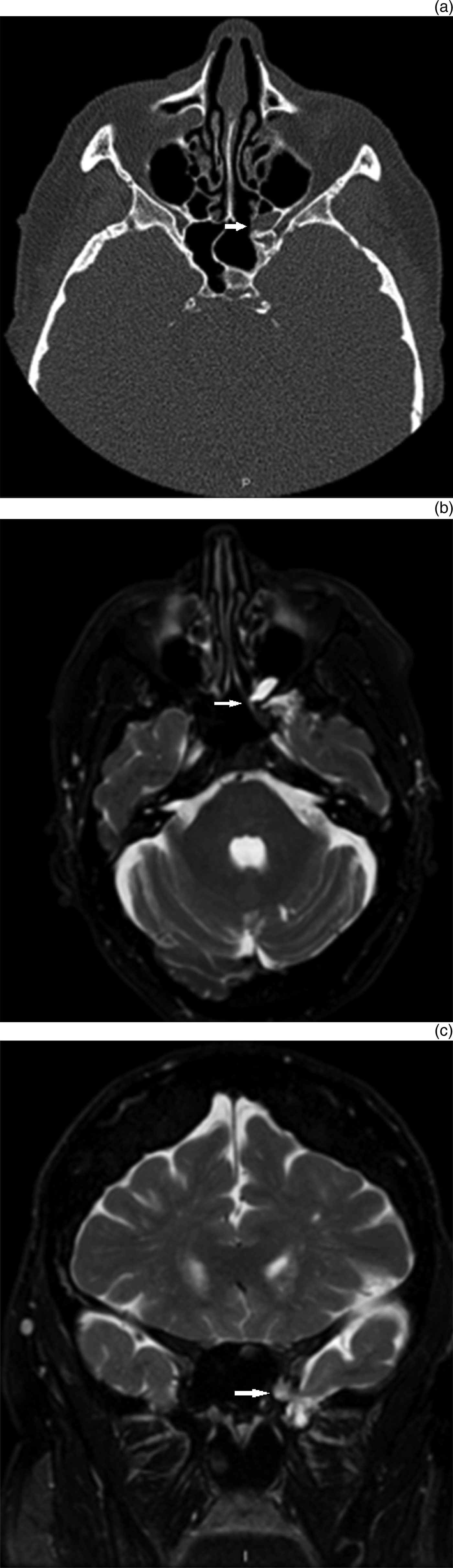
A review of the patient's pre-operative CT scans revealed a bony defect in the inferomedial aspect of the left temporal fossa communicating with the left sphenoid sinus that was not documented on the initial report. Clinical examination did not reveal an obvious leak or polyp recurrence, and the sphenoid had a normal appearance on sphenoidotomy. The patient subsequently underwent CT and magnetic resonance imaging (MRI) (Figure 3), and was scheduled for a repair of the sphenoid cerebrospinal fluid (CSF) leak.
(a) Post-operative axial computed tomography scan at the level of the left sphenoid sinus bony defect (arrow) (P = posterior). (b) Axial, T2-weighted magnetic resonance imaging (MRI) scan at the level of the bony defect, showing high signal intensity in the bony defect (arrow). (c) Coronal, T2-weighted MRI scan at the level of the bony defect (arrow) (I = inferior).
Discussion
Nasal glial heterotopia, historically referred to as nasal gliomas, are part of a family of congenital midline masses that include encephaloceles and dermoid cysts. They are a collection of benign, mature brain glial tissues that present in the nose or paranasal regions. Nasal glioma cases usually present perinatally with an external mass, nasal obstruction and/or feeding difficulties, and occur in 1 in 20 000–40 000 live births.Reference Harley1
There is no recognised familial aetiology, malignant potential or sex predilection. Sixty per cent of nasal gliomas present externally around the nasal region, 30 per cent present intranasally, and 10 per cent have both internal and external components.Reference Hengerer2 External nasal gliomas are often found at the level of the glabella, while intranasal gliomas usually arise along the lateral nasal cavity wall, at or above the level of the middle turbinate,Reference Black and Smith3 as in the case reported here.
Nasal gliomas are differentiated from encephaloceles in that they lack anatomical communication with central nervous system tissue.Reference Martinez-Lage, Garcia-Contreras, Ferri-Niguez and Sola4, Reference Patterson, Kapur and Chandra5 This gave rise to the Furstenberg test, which is typically positive when the ipsilateral internal jugular vein is compressed, and the mass enlarges and becomes pulsatile, thus differentiating encephaloceles from nasal gliomas.
Theories for nasal glioma include a failure of anterior neuropore fusion, extracranial separation of embryonal neuroglia, neurological cells in the nasal mucosa that grow through the fronto-ethmoidal suture and encephaloceles in which the intracranial connection has been lost.Reference Morgan and Evans6–Reference Karma, Rasanen and Karaja9
Ten to 15 per cent of gliomas have a dural connection, with a fibrous stalk extending towards a defect in the skull base.Reference Frodel, Larrabee and Raisis10–Reference Smith, Schwartz, Luse and Ogura13 The likelihood of a dural connection is two to three times higher for intranasal gliomas as compared with their external counterparts.Reference Harley1 Occasionally, they can arise from the septum.
A search on PubMed, Embase and Medline was undertaken on 30 March 2014 using the following combinations of search terms: ‘nasal’ and ‘glioma’; ‘heterotrophic’ and ‘cerebral’; ‘cerebral’ and ‘heterotopia’; or ‘heterotrophic’ and ‘glial’. The articles searched were limited to those written in the English language and conducted on human subjects. Articles referenced in the papers that were identified using this search method were also included in the review. Studies of nasal gliomas presenting in patients younger than 16 years of age were excluded.
There were 14 case reports of nasal gliomas in adults, with presentation as late as 81 years.Reference Smith, Schwartz, Luse and Ogura13–Reference Katz and Lewis26 Most patients were asymptomatic, or had symptoms of nasal obstruction or clear rhinorrhoea. Diagnosis was usually made following an incidental finding of heterotrophic glial tissue after incisional biopsy of a nasal polyp. Other presentations were acute or recurrent meningitis,Reference Smith, Schwartz, Luse and Ogura13, Reference Penner and Thompson16 meningoencephalitis,Reference Chau, Hopkins and McGilligan21 and visual loss.Reference Majithia, Liyanage, Hewitt and Grant17, Reference Jain, Shukla, Aryya, Srivastava and Kumar18
Histologically, nasal gliomas have no meningeal envelope, and are covered by skin or nasal respiratory mucosa. They are composed of vascularised, dense fibrous connective tissue, with mature astrocytic and oligodendrocytic glial cells, and sometimes contain neurons and ependymal cells. Secondary changes of fibrogliosis or gemistocytic alteration of glial cells are often reported, probably due to compression and infarction. These can make the differentiation between encephaloceles histologically difficult, and the distinction may have to be made based on radiological or surgical findings.Reference Jain, Shukla, Aryya, Srivastava and Kumar18, Reference Yeoh, Bale and de Silva27, Reference Patterson, Kapur and Chandra28 In terms of radiological investigations, CT can be used to identify bony defects, while MRI both offers better soft tissue definition, and can be used to identify the site and extent of the tumour and any intracranial extension. However, radiographic reports do not always exclude a bony defect or an associated intracranial connection, as was the case in our experience and others.Reference Bradley and Singh12, Reference Penner and Thompson16, Reference Chau, Hopkins and McGilligan21
The management of nasal gliomas in children is generally accepted as being surgical excision, in order to prevent the lesion from affecting developing facial structures or increasing the risk of meningitis.Reference Rahbar, Resto, Robson, Perez-Atayde, Goumnerova and McGill29 Surgical excision is approached either endoscopically or via an external lateral rhinotomy and/or craniotomy if there is a significant intracranial connection. The risk of recurrence following excision is between 4 and 10 per cent.Reference Katz and Lewis26
The management in asymptomatic or mildly symptomatic adults is more contentious, as nasal gliomas are slow-growing and there is no known malignant potential. If diagnosis is based on an incisional biopsy of a nasal polyp, further assessment is required to ensure a CSF leak has not been caused post-operatively (a repair should ensue if a CSF leak is evident). If an incisional biopsy has not been undertaken, we would recommend a multidisciplinary discussion, involving otorhinolaryngologists, radiologists, neurosurgeons and ophthalmologists, before embarking on excision. This is because resection carries the risk of CSF leak and meningitis, and must be balanced against functional and cosmetic outcomes.
• A literature review identified 14 cases of nasal glioma presenting in adults; these were all intranasal entities
• Diagnosis is usually incidental following biopsy of a presumed nasal polyp; other presentations are meningitis, meningoencephalitis and visual loss
• Histologically, they have no meningeal envelope, are covered by skin or respiratory mucosa, and contain fibrous connective tissue, with mature astrocytic and oligodendrocytic glial cells
• Excision of these entities in asymptomatic adults should be weighed up against post-operative risks of cerebrospinal fluid leak and meningitis
In conclusion, nasal gliomas presenting in adulthood are rare. The literature review revealed 14 case reports. For obvious reasons, adult presentations thus far have been intranasal entities. Nevertheless, pre-operative awareness of these rare cases is important. Incisional biopsies of nasal masses conducted before careful scrutiny of pre-operative imaging investigations may lead to an inadvertent post-operative CSF leak or meningitis, as reported in this case report and in the literature.
Acknowledgement
We would like to thank Dr Chee Yeong (consultant radiologist) for his advice and expertise.