Introduction
Despite severe losses during the great extinction event at the Permian–Triassic boundary, brachiopods were still a major component of the faunas of the latter period (Kummel, Reference Kummel1979). The recovery of brachiopods began in the mid- to late Induan (early Early Triassic) and pre-extinction diversity levels are reported to have been attained in the Anisian (early Middle Triassic; Chen et al., Reference Chen, Kaiho and George2005, Reference Chen, Chen and Tong2010), much later than for ammonoids (late Induan; Zakharov and Popov, Reference Zakharov and Popov2014). Early Triassic brachiopod faunas are globally poor (19 genera all over the world after Vörös, Reference Vörös2010; according to Clapham et al., Reference Clapham, Fraiser, Marenco and Shen2013, 45 genera are present already in the Induan but even this number is quite low if compared with Permian faunas, cf. infra), while Middle Triassic ones are distinctly richer (84 Anisian and 91 Ladinian genera; Vörös, Reference Vörös2010); the acme of Triassic brachiopod diversity was in the Norian (133 genera). However, unlike Anisian (Sun et al., Reference Sun, Hao, Sun and Jiang2009) and Late Triassic brachiopods, Ladinian (late Middle Triassic) brachiopod faunas are poorly known (Sun, Reference Sun1980, p. 1181; Dagys, Reference Dagys1993, p. 82). The complete absence of Ladinian brachiopods in the Caucasus sections in which both Anisian and Carnian representatives of this phylum are known and with neither major lithological shifts nor changes in abundance of other faunal groups led Ruban (Reference Ruban2006a, Reference Rubanb) to postulate that an “enigmatic Ladinian crisis” took place during the aforementioned stage. Ladinian brachiopod faunas in South China are also distinctly less diverse than Anisian ones but it is uncertain whether this is due to global or local causes (Shen et al., Reference Shen, Zhang, Li, Mu and Xie2006).
The aims of the present study are: (1) to document a hitherto poorly known Ladinian brachiopod fauna from Mt. Svilaja (Dinarides, Croatia), including the description of one new genus and two new species, as well as the redescription of an inadequately described genus and species, all three unknown outside the studied locality; (2) to show that a sparsely recorded evolutionary line, a representative of which is present in the studied material, may be traced from the Permian to Recent times, including reinterpretation of systematic position of one Permian and one Recent brachiopod genus as well as a formal description of a new suborder of the Terebratulida.
History of research
Ladinian brachiopods from the Svilaja range were first mentioned by von Kerner (Reference von Kerner1908, p. 283) who reported two unpublished identifications by Schloenbach, ‘Retzia (?) quadricostata’ and ‘Spiriferina hirsuta’, and two (also unpublished) by Bittner, ‘Spirigera cf. trigonella’ and ‘Rhynchonella vivida var. dalmatina’. This material, however, came from other outcrops in the same mountain range. Silicified faunas from the section at Svilaja dealt with herein were first described by Chorowicz and Termier (Reference Chorowicz and Termier1975); however, only a small fraction of the total diversity (e.g., a single brachiopod species compared to five described herein, a single anthozoan compared to nine present in the fauna, and two gastropod taxa compared to several dozen known in the locality; J. Stolarski and A. Kaim, unpublished data) was revealed. Silicified brachiopods from Svilaja were also the subject of a preliminary communication by Jaecks et al. (Reference Jaecks, Leighton, Kolar-Jurkovšek, Jurkovšek and Jelaska2003); however, his interpretation of the brachiopod described here as Thecocyrtella dagysii n. sp. as a post-Paleozoic productide is rejected by the present authors.
Geologic setting
In terms of present geography, the study area (Fig. 1.1) is situated in the Dinarides, a mountain belt belonging to the southern branch of the Alpine chain (Ager, Reference Ager1980), occupying the northeastern coast of the Adriatic Sea. More precisely, Svilaja is an isolated mountain range in central Dalmatia, situated ca. 30 km north from the city of Split; it is separated from the main chain of the Dinarides by the valley of Cetina with the Peruča Lake (Peručko jezero). The Mt. Svilaja section is located along the Mijići–Zelovo Sutinsko road at approximately 43°42’51” N 16°32’8” E (between the towns of Muć and Sinj; Fig. 1.2).
Geological setting of the studied fauna. (1) General map showing the position of the Svilaja range within the Dinarides. The rectangle shows the area enlarged in (2). (2) Hypsometric map showing the position of the studied section in the Svilaja range. Mountain ranges in full capitals; Towns in Roman typeface; rivers and lakes italicized. (3) Global paleogeography in the Middle Triassic. The polygon shows the area enlarged in (4). (4) Paleogeographical reconstruction of the western edge of the Tethys in the Late Anisian (supposed not much different to the situation in the Ladinian). Simplified and redrawn after Jawecka et al. (Reference Jawecka, Zakrzewska and Słodkowska1987), Dercourt et al. (Reference Dercourt, Ricou and Vrielynck1993). AM=Armorican Massif; BM=Bohemian Massif, CAP=Central Apennine Plateau, E=Ebro Massif, IM=Iberian Massif, MC=Massif Central, VH=Vindelician High. Minuscules a–k denote approximate localizations of Ladinian brachiopod faunas all over the world; see text for explanation.

In terms of geology, the Dinarides represent the collision chain between the northeastern edge of the Apulia (=Adria) microplate and Europe (Tari, Reference Tari2002). The south-western part of this mountain range, called Outer (or External) Dinarides, represents a complex set of carbonate platforms referred to collectively as the Adriatic-Dinaridic Carbonate Platform (Vlahović et al., Reference Vlahović, Tišliar, Velić and Matičec2002). In the Middle Triassic this area was an epicontinental sea situated at the western edge of the Tethys (Adriatic continental margin; Fig. 1.4) subject to rifting processes without, however, any apparition of oceanic crust until the Norian (middle Late Triassic) (Pamić et al., Reference Pamić, Gušić and Jelaska1998; but see Sudar et al., Reference Sudar, Gawlick, Lein, Missoni, Kovacs and Jovanovic2013). The considered area belongs to a geological unit called the Dalmatian-Herzegovinan Composite Terrane (Karamata, Reference Karamata2006) and in the Middle Triassic was situated in the equatorial zone (15° N paleolatitude according to Dercourt et al., Reference Dercourt, Ricou and Vrielynck1993 and Stampfli et al., Reference Stampfli, Borel, Marchant and Mosar2002; 5° N according to Szulc, Reference Szulc2000, and Golonka et al., Reference Golonka, Krobicki, Pająk, Van Giang and Zuchiewicz2006; 10° S paleolatitude according to Karamata, Reference Karamata2006 and references therein; Fig. 1.3). Details of paleogeographic reconstructions of the area considered are still debated (e.g., Channell and Kozur, Reference Channell and Kozur1997 and references therein; compare also slight differences in paleogeographic maps in Fig. 1.3 and 1.4).
The Middle Triassic of the studied section at Mt. Svilaja starts with Anisian dolomitic Otarnik Breccia. Late Anisian to early Ladinian vitrocrystallic tuffs (‘pietra verde’) are conformably overlain by cherty limestones and dolomites (the horizon with the fauna studied) with abundant calcareous algae [the most common species being Diplopora annulata Schafhäutl and Teutloporella herculea (Stoppani) Pia (Grgasović et al., Reference Grgasović, Kolar-Jurkovšek and Jurkovšek2007)], foraminifers, radiolarians, serpulids, gastropods, bivalves, ammonoids, brachiopods, crinoids, and conodonts. The ammonoid assemblage is composed of both leiostraca (Proarcestes sp.) and trachyostraca: Alkaites dinaricus Balini, Jurkovšek and Kolar-Jurkovšek, Reference Balini, Jurkovšek and Kolar-Jurkovšek2006, Detoniceras svilajanus Balini, Jurkovšek and Kolar-Jurkovšek, Reference Balini, Jurkovšek and Kolar-Jurkovšek2006, and Argolites trinodosus Balini, Jurkovšek and Kolar-Jurkovšek, Reference Balini, Jurkovšek and Kolar-Jurkovšek2006 (Balini et al., Reference Balini, Jurkovšek and Kolar-Jurkovšek2006). Based on conodont fauna this interval is attributed to the Ladinian hungaricus Assemblage Zone (Jelaska et al., Reference Jelaska, Kolar-Jurkovšek, Jurkovšek and Gušic2003). This interval is followed by well bedded pelagic limestone with sporadic marl laminae which are attributed to mungoensis (Ladinian) and murchianus (upper Ladinian to ?lower Carnian) conodont zones. A paleokarstified emersion surface can be observed at the top of these limestones (Bucković and Martinuš, Reference Bucković and Martinuš2010).
Material and methods
Preservation and taphonomy
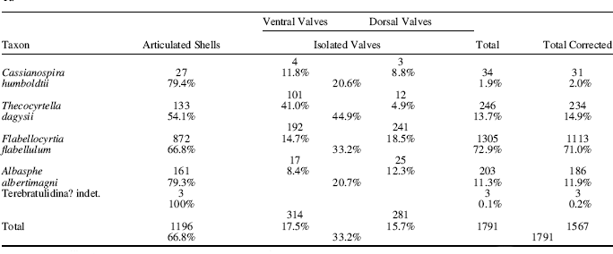
The described fauna is silicified and has been extracted from limestone through etching in acetic acid. The faunal content of the rock is quite rich; brachiopods and gastropods are the most abundant, corals and algae are abundant, echinoderms are frequent, whereas bivalves and ammonoids are rare. The present sample (1791 specimens; see Table 1) has been obtained after dissolving ca. 100 kg of rock; a minor part thereof consists of specimens naturally weathered from the rock.
Numerical characteristics of the material studied.
“Total corrected” stands for the sum of articulated shells and of those of either isolated valves which are more numerous; the percentages referred to the “total” (next to last column) are then these in the studied material, whereas those referred to the “total corrected” are an approximation (minimum number of individuals) of the original biocoenosis.
The silicification process does not affect all parts of the shell equally. It was observed that shells of Flabellocyrtia flabellulum and of Thecocyrtella dagysii sometimes possess a deltidium and sometimes not (compare Figs 4.12, 4.24 and 4.14, 4.17, 4.25–4.29, 4.34 as well as 6.1, 6.2, 6.5, 6.7 and 6.3, 6.13, 6.18, 6.32, 6.34–6.37; specimens lacking the deltidium are more frequent). This is best interpreted as resulting from selective silicification; a very similar situation (pseudodeltidium lacking or more seldom present due to selective silicification) was described by Pope (Reference Pope1982, p. 4; see also Holdaway and Clayton, Reference Holdaway and Clayton1982). Hollow spaces appearing inside the shell are also an artifact explained by the silicification either proceeding from the external surfaces towards the internal layers and often remaining incomplete (for an example of a list of artifacts caused by silicification, see Caruthers and Stanley, Reference Caruthers and Stanley2008, p. 155) or occurring preferentially along certain layers of the shell (Crowley, Reference Crowley1988; Carson, Reference Carson1991, p. 462). In some cases, concentric rings (beekite) typical of silicification obscure the original surface of the valves (Fig. 4.31). Although most of original microstructure gets indeed lost during the silicification process (Brenchley and Harper, Reference Brenchley and Harper1998, p. 78), by no means should it be understood that all microstructural information is lost during diagenesis. In several cases, punctae are preserved (Thecocyrtella: Fig. 4.4, Flabellocyrtia: Fig. 4.3, Albasphe: Fig. 4.2) and sometimes the same may be said even about the axial cavity of the spines of the spiralium in Flabellocyrtia. Overall, however, the silicification in the material studied may be described as intermediate in character between two extreme types referred to in a description of silicified Carboniferous brachiopods by Sun and Baliński (Reference Sun and Baliński2008, p. 491), namely preserving very fine details including some information on microstructure on one hand and obliteration of original patterns up to the apparition of a secondary “spongy” texture on another hand.
The percentage of disarticulated valves in the material amounts to about 33% (see Table 1 for details). Referring to a paleoecological analysis of Middle Triassic brachiopod assemblages with different percentages of disarticulated valves (Pálfy, Reference Pálfy1991), the value for Svilaja assemblage corresponds approximately to “reasonable ‘background’ disarticulation under average circumstances,” i.e., without much postmortem transport. However, the ratios of ventral versus dorsal valves are somewhat different from the value 1:1 expected if there is no postmortem bias (Chen et al., Reference Chen, Chen and Tong2010 and references therein). This is especially the case for the spiriferinide Thecocyrtella dagysii in which ventral valves are over eight times more numerous than dorsal ones; this, however, is best explained as expressing their different preservational qualities (Alexander, Reference Alexander1986, Reference Alexander1990)—the ventral valve is massive, whereas the dorsal one is fragile—or perhaps some kind of differential sorting. For the other species the value of the discussed ratio is between 1.26 and 1.46; this seems to indicate a moderate bias. The assemblage is thus interpreted as parautochthonous (perhaps even autochthonous).
The sample is of low diversity (Shannon index=0.88) and relatively medium dominance (Simpson D index=0.54). Rarefaction analysis (Fig. 2) suggests that the present collection is fully saturated and additional sampling is rather unlikely to result in a discovery of additional species. Very small differences between percentages of species referred to the total material and corrected values (minimum number of individuals; Zuschin et al., Reference Zuschin, Stachowitsch and Stanton2003 and references therein) allow to consider that the biocoenosis structure is reflected more or less accurately in the material.
Rarefaction curve for the brachiopod sample from Svilaja.
Methods
Internal features of described brachiopods have been investigated by X-ray microtomography (Zeiss XRadia MicroXCT-200) at the NanoFun laboratory at the Institute of Paleobiology; this method was applied to single specimens of Flabellocyrtia flabellulum, Thecocyrtella dagysii, and Albasphe albertimagni. Results concerning these three species were not the same. In F. flabellulum and T. dagysii the optical densities of silicified shell and silicified matrix fragments are quite different (on CT images shell is grey and rock is white), wherefore sections are easy to interpret, with the obvious reservation that absence of an anatomic structure in the selectively silicified material (see above) does not disprove its presence in vivo. On the contrary, in the analyzed specimen of A. albertimagni the optical densities of various parts of the shell are not so different, hence there was no clear limit between the rock matrix and the shell (on CT scans, shell showed a continuous intergrading of grey to white half-tones and in some cases it was impossible to decide if a white spot corresponds to the brachiopod or to the matrix).
The self-evident advantages of the X-ray tomography method, above all, its non-destructive character and the possibility of obtaining sections along several series of planes from a single specimen (Figs 5, 7, and 10), have been widely discussed in a number of contributions (Hamada et al., Reference Hamada, Tateno and Suzuki1991; Błażejowski et al., Reference Błażejowski, Binkowski, Bitner and Gieszcz2011; Zapalski and Dohnalik, Reference Zapalski and Dohnalik2013; and references therein). A disadvantage of this method is, however, that it produces only images based on difference in density between shell and the surrounding rock (or the air in case of our etched material). The classic method of obtaining sections from acetate peels allows visualising the internal structure of the shell with growth lines and crystallite orientation, which is not possible using X-ray microtomography. For example, Kozłowski (Reference Kozłowski1929) who studied Devonian and Triassic representatives of Cyrtinidina (Cyrtina and Hirsutella) by means of serial sections and acetate peels was able to show that in both the median septum was composed of three distinct lamellae. This character could not be checked in Flabellocyrtia (Bittnerulidae, Cyrtinoidea). Nonetheless, in the case of our material the X-ray microtomography is the only way to investigate the internal structures as it is impossible to make acetate peels from silicified material.
Due to the small size of the studied brachiopods, photographs were taken mostly under a scanning electron microscope. Selected specimens were mounted on stubs, coated with platinum, and examined using a Philips XL-20 microscope at the Institute of Paleobiology in Warsaw. Larger brachiopods (Figs 3.12–3.16, 4.30–4.34, 6.4–6.7, 6.9–6.18, 8.22–8.31) were photographed in visible light after coating with ammonium chloride, although some shell regions were refractory to coating, probably due to their extraction from the rock by acid etching. Medium-sized specimens were photographed under Nikon SMZ 1500 binocular equipped with Nikon D5200 digital camera; some photographs were assembled from a series of up to five frames using the Helicon Focus programme. The largest ones were photographed with a Nikon D-1X digital camera with a Nikkor micro 55/2.8 lens.
Cassianospira humboldtii (von Klipstein, 1845). (1–6,11) Six articulated shells BJ 2281, 2292, 3057, 2293, 2382, ZPAL Bp 72/4/1 (6, 11) in dorsal views (1–6) and an enlargement of the interarea (11). (7, 8) Ventral valves BJ 2383, 2439. (9, 10) Dorsal valves BJ 2440, 3054. (12–16) Articulated shell ZPAL Bp 72/4/1 in dorsal, ventral, lateral, posterior, and anterior views. (17–21) Terebratulidina? gen. et sp. indet. (17, 18) juvenile articulated shell BJ 3056 in dorsal and lateral views. (19–21) Fragment of an adult shell BJ 3055 in dorsal, ventral, and posterior views. (1–11, 17–21) SEM micrographs; (12–16) images taken in visible light.
The main collection is housed at the Jurkovšek Palaeontological Collection that has been registered with the Slovenian National History Museum in Ljubljana and abbreviated BJ. Representative collections of species studied is also kept at the Institute of Paleobiology of the Polish Academy of Sciences in Warsaw (collection number ZPAL Bp 72).
Systematic paleontology
Phylum Brachiopoda Duméril, Reference Duméril1805
Remarks
The classification framework and the authorship of the names follow the revised Treatise on Invertebrate Paleontology, Part H (Williams et al. Reference Williams, Carlson and Brunton2000), except for the superfamily Zeillerioidea, the authorship of which is to be attributed to Schuchert (in Schuchert and LeVene, Reference Schuchert and LeVene1929, p. 24) who introduced the subfamily Zeilleriinae by Latinising the vernacular (invalid) name “Zeilleriidés” used by Rollier (Reference Rollier1915). Allan (Reference Allan1940), credited with the authorship in the Treatise, was in fact only the first to use the name at family level. The first use at superfamily level was by Kyansep (Reference Kyansep1961) (personal communication, M. Manceñido, 2014).
Synonymy lists are established with short comments by means of special signs following the system described by Halamski (Reference Halamski2009, p. 46–47; further references therein). For species published in monographs issued during a several years’ period, the full citation follows the model proposed by Halamski and Baliński (Reference Halamski and Baliński2013, p. 265).
Subphylum Rhynchonelliformea Williams et al., Reference Williams, Carlson, Brunton, Holmer and Popov1996
Class Rhynchonellata Williams et al., Reference Williams, Carlson, Brunton, Holmer and Popov1996
Order Athyridida Boucot, Johnson and Staton, Reference Boucot, Johnson and Staton1964
Suborder Retziidina Boucot, Johnson and Staton, Reference Boucot, Johnson and Staton1964
Superfamily Retzioidea Waagen, Reference Waagen1883
Family Neoretziidae Dagys, Reference Dagys1972
Genus Cassianospira Dagys, Reference Dagys1972
Type species
Retzia loczyi Bittner, Reference Bittner1900; Carnian.
Remarks
This genus has been reported from the Upper Triassic (Carnian and Norian) up to now (Dagys, Reference Dagys1974, p. 288; Alvarez and Rong, Reference Álvarez and Rong2002, p. 1591). This is the first report from the Middle Triassic (Ladinian).
Cassianospira humboldtii (von Klipstein, 1845 in 1843–45 [‘1843’])
*1845 Spirifer Humboldtii von Klipstein, p. 233; pl. 15, fig. 17.
1853 Waldheimia? Humboldtii; Gray, p. 85.
1889 Retzia Humboldtii Klipst. sp.; Bittner, p. 168.
1890 Retzia Humboldtii Klipst. spec.; Bittner, p. 88; pl. 2, fig. 33.
Type material
Articulated shell from the Carnian of San Cassian (southern Alps) figured by Klipstein (1845, pl. 15, fig. 17) NHMUK PI OR 30635, holotype by monotypy.
Description
Shell teardrop-shaped, wider than long when young, about as wide as long in larger individuals due to the growth of the ventral umbo, markedly to very strongly ventribiconvex, up to 4.1 mm in width; maximal width about the midlength of the dorsal valve or slightly anteriorly. Hinge line and anterior commissure straight. Ventral valve high; interarea apsacline, nearly flat near the hinge line, markedly incurved in umbonal region; a rather narrow delthyrium closed by a symphytium bordered by low rounded rims (Fig. 3.11); foramen submesothyrid. Dorsal valve weakly convex.
Ornamentation of stout, high, rounded plicae, beginning at umbo, separated by narrower furrows; up to five pairs on the ventral valve, a median one and up to four pairs of lateral ones on the dorsal valve. Growth lines not observed.
Dorsal interior: cardinal flange thick, supported by a high and thin (Fig. 3.10) or moderately thick (Fig. 3.9) median septum, the length of the septum equaling approximately two-thirds of that of the valve; spiralium not preserved. Ventral interior (Fig. 3.7, 3.8) poorly preserved.
Material
Twenty-seven mostly subcomplete articulated shells, four ventral, and three dorsal valves.
Dimensions
ZPAL Bp 72/4/1: length 2.7 mm; width 2.5 mm; thickness 1.8 mm.
Remarks
The publication date of Cassianospira humboldtii requires a comment. The Beiträge zur geologischen Kenntniss der östlichen Alpen by A. v. Klipstein are dated 1843 at the title page (in the reference list of the Treatise on Invertebrate Paleontology, Part H, Revised, confounded with Beiträge zur geologischen und topographischen Kenntnis der östlichen Alpen by the same author printed in three parts between 1871 and 1883). However, “Spirifer humboldtii” (our Cassianospira humboldtii; not to be confounded with Spirifer humboldtii d’Orbigny, 1842, which comes from the Silurian of Bolivia; Schuchert, Reference Schuchert1897, p. 287; Bassler, Reference Bassler1915, p. 892) is dated 1844 by Gray (Reference Gray1853, p. 85), whereas Bittner (Reference Bittner1889, p. 159) and Sherborne (Reference Sherborne1927, p. 3053) give the date 1845. As often in the nineteenth century, the monograph by Klipstein was apparently printed during several years; the date given by the Index Animalium (Sherborne, Reference Sherborne1927) is accepted here.
Cassianospira humboldtii is known from the Carnian of the Alps. It differs from Cassianospira loczyi (Bittner, Reference Bittner1900) in having the median plica on the dorsal valve of the same height as the admedian pair of plicae (lower than the lateral ones in C. loczyi). Cassianospira klipsteinii (Bittner, Reference Bittner1889) is another similar species differing in higher interarea (Bittner Reference Bittner1890, pl. 2, figs 30–31; Zardini, Reference Zardini1988, pl. 10, fig. 13)
Order Spiriferidina Ivanova, Reference Ivanova1972
Suborder Cyrtinidina Carter and Johnson in Carter et al., Reference Carter, Johnson, Gourvennec and Hou1994
Superfamily Suessioidea Waagen, Reference Waagen1883
Family Bittnerulidae Schuchert in Schuchert and LeVene, Reference Schuchert and LeVene1929
Subfamily Bittnerulinae Schuchert in Schuchert and LeVene, Reference Schuchert and LeVene1929
Genus Thecocyrtella Bittner, Reference Bittner1892
Type species
Cyrtotheca ampezzana Bittner, Reference Bittner1890; Cortina d’Ampezzo, Alps; Carnian.
Remarks
Carter (Reference Carter2006, p. 1889) included absence of fold and sulcus among diagnostic characters of Thecocyrtella. T. dagysii is similar to T. ampezzana in internal characters but possesses a ventral sulcus; we consider that this is not a character justifying separation at genus level but rather a reason for the emendation of the diagnosis; the discussed fragment should run “sulcus absent or present”.
Thecocyrtella dagysii new species
Figures 4.1, 4–34; 5
(1, 4–34) Thecocyrtella dagysii Halamski, Bitner, Kaim, Kolar-Jurkovšek, and Jurkovšek n. sp. (1) A poorly preserved shell BJ 3081 attached to a fragment of a thallus of a (dasyclad?) alga. (4) Punctae (seen from inside) of the shell ZPAL Bp 72/3/1. (5, 10) Dorsal interior BJ 3084, general view and enlargement of the cardinalia. (6, 7) Dorsal interior BJ 3085 in oblique and ventral views. (8, 9) Juvenile articulated shell BJ 3098, dorsal view and enlargement of the interarea. (11) Dorsal interior BJ 3104 showing crura. (12, 13) Ventral valve BJ 3087, dorsal view and enlargement of the deltidium. (14–18) Four ventral valves ZPAL Bp 72/3/1, BJ 3095, 3094, and 3093 in dorsal views and an enlargement of the interarea. (19, 24) Articulated shell BJ 3086 in postero-dorsal view and enlargement of the interarea. (20–23) Four ventral valves 3089, 3092, 3102, and 3103 in ventral views. (25–29) Five articulated shells BJ 3096, 3101, 3099, 3100, and 3097 in postero-dorsal views. (30–34) Holotype BJ 3083 in dorsal, ventral, lateral, anterior, and posterior views. (2) Albasphe albertimagni Halamski, Bitner, Kaim, Kolar-Jurkovšek, and Jurkovšek n. gen. n. sp.; punctae (seen from inside) of the shell ZPAL Bp 72/2/6. (3) Flabellocyrtia flabellulum Chorowicz and Termier, Reference Chorowicz and Termier1975; punctae (seen from inside) of the shell BJ 3082. (1–29) SEM micrographs; (30–34) images taken in visible light.
Thecocyrtella dagysii Halamski, Bitner, Kaim, Kolar-Jurkovšek, and Jurkovšek n. sp.: X-ray microtomograph sections of the holotype BJ 3083. (1) Sections parallel to the delthyrium, distances (in mm) measured from front; (2) sections parallel to the commissural plane, distances (in mm) measured from the tip of the ventral umbo; (3) sections parallel to the symmetry plane, distances (in mm) measured either side from the latter. J=jugum, s=septum, sn=spondylium, sp=spiralium.
Type material
Holotype, BJ 3083.0; illustrated paratypes, BJ 3081, 3084–3101; ZPAL Bp 72/3/1; type locality, Svilaja, Dinarides; cherty limestones and dolomites, Ladinian, Middle Triassic.
Diagnosis
Thecocyrtella with a deep ventral sulcus.
Dimensions
Holotype BJ 3083: length 2.7 mm; width 2.1 mm; thickness 2.2 mm.
Description
Shell very strongly ventribiconvex. Dorsal valve medially indented, nearly flat, hinge line straight, postero-lateral extremities auriculate; maximal width at hinge line; a strong median rib present in some specimens (Fig. 4.28–4.30), sometimes situated in the middle of a faint sulcus (Fig. 4.30). Ventral valve pyramidal, beak incurved, with a narrow, deep V-shaped median groove originating at umbo; lateral slopes mostly smooth, seldom with faint plication. Interarea very high, apsacline, concave; delthyrium narrow (0.11–0.16 of the area width), with subparallel edges, covered by a wider (0.28–0.37 of the area width) deltidium (Fig. 4.9, 4.13, 4.19; rarely preserved, cf. supra). Shell substance punctate (Fig. 4.4).
Dorsal interior: crura subparallel (Fig. 4.7) or divergent (Fig. 4.5, 4.10), then coalescing to form a jugum (Figs 4.11; 5.1, section at 0.918 mm; 5.2, section at 1.557 mm); spiralium continuing quite far towards the ventral beak but less than one entire whorl is preserved (Fig. 5.3). Ventral interior: median septum thin, moderately high, not very long; spondylium (Fig. 5.1, sections at 2.150 and 2.296 mm) present in umbonal region (0.4 mm long in a specimen 2.4 mm long); teeth small.
Etymology
In honor of Algirdas Dagys (30.08.1932–7.01.2000), Lithuanian paleontologist, in recognition of his contribution to the study of Triassic brachiopods (name Latinised as Dagysius, then genitive Dagysii).
Material
One hundred and thirty-three articulated shells, 101 ventral valves, and 12 dorsal valves (paratypes).
Remarks
The described material is included in Thecocyrtella Bittner, Reference Bittner1892 on account on shape, minute size, (near) lack of radial ornamentation, and possession of a ventral septum and a spondylium. Dagys (Reference Dagys1974, p. 149) showed that Bittner (Reference Bittner1890) misinterpreted an incomplete beak of the type species T. ampezzana (Bittner, Reference Bittner1890) as representing an attachment scar (a similar condition is shown in Fig. 4.19); as a matter of fact, there is no such structure in the discussed genus. For the terminological question of deltidium vs. symphytium, cf. infra, remarks on Flabellocyrtia flabellulum.
T. dagysii n. sp. differs from T. ampezzana and from T. orientalis Ivanova in Dagys, Reference Dagys1965 [this species was sometimes cited as “T. orientalis Ivanova, Reference Ivanova1960” but the original publication (Ivanova, Reference Ivanova1960, p. 279, text-fig. 407, pl. 64: 9) was without either description or diagnosis; the first to provide a diagnosis was Dagys (Reference Dagys1965, p. 107), so the name must be considered as validly introduced only in the latter publication] in having a deep ventral sulcus. Bittnerula zitteli (Bittner, Reference Bittner1890) (syn. Spirifer buchii von Klipstein, 1843; Dagys, Reference Dagys1974 considered Bittnerula as a synonym of Thecocyrtella) has a weaker ventral sulcus and a much higher interarea. Thecocyrtelloidea tubulosa Yang and Xu, 1966 from the Anisian of Guizhou has no spondylium and its spiralium has at least five whorls (Yang and Xu, 1966).
The interpretation of brachiopods described herein under Thecocyrtella dagysii n. sp. as post-Paleozoic productides was proposed in a preliminary communication by Jaecks et al. (Reference Jaecks, Leighton, Kolar-Jurkovšek, Jurkovšek and Jelaska2003) without knowledge of the internal structures, which are crucial for correct identification. Moreover, Jaecks et al. (Reference Jaecks, Leighton, Kolar-Jurkovšek, Jurkovšek and Jelaska2003) misinterpreted some poorly preserved unidentified epizoans in the umbonal region of some specimens as alleged productide spines.
Subfamily Hirsutellinae Xu and Liu, Reference Xu and Liu1983
Genus Flabellocyrtia Chorowicz and Termier, Reference Chorowicz and Termier1975
Type species
Flabellocyrtia flabellulum Chorowicz and Termier, Reference Chorowicz and Termier1975; Svilaja; Ladinian.
Flabellocyrtia flabellulum Chorowicz and Termier, Reference Chorowicz and Termier1975
Flabellocyrtia flabellulum Chorowicz and Termier, Reference Chorowicz and Termier1975: external and internal morphology of shells. (1–3) articulated shells BJ 3105, 3070, and 3072 showing different preservation of the deltidium (preserved in 1, 2; not preserved in 3). (4–7) Articulated shells ZPAL Bp 72/1/3 and 72/1/4 in dorsal and posterior views. (8) Fragmentary dorsal valve ZPAL Bp 72/1/5 with an almost entire crus. (9–18) Articulated shells ZPAL Bp 72/1/1 and 72/1/2 in dorsal, ventral, lateral, anterior, and posterior views. (19–30) Dorsal interiors BJ 3062 (19), 3061 (20, 24, 28), 3060 (21, 25, 29), 3059 (22, 26, 30), and 3058 (23, 27), general views (19–23), enlargements of cardinalia (24–27), and of marginal regions (28–30). (31, 36) Fragments of spiralium BJ 3068 and 3069 showing spines. (32, 37) Fragmentary ventral valves BJ 3063 and 3064 (apical regions preserved) in antero-lateral view showing different shapes of the median septum. (33) Ventral valve BJ 3065 showing spiralium. (34–35) Ventral valves BJ 3066 and 3067 in postero-dorsal views. (1–3, 8, 19–37) SEM micrographs; (4–7, 9–18) images taken in visible light.
Flabellocyrtia flabellulum Chorowicz and Termier, Reference Chorowicz and Termier1975: X-ray microtomograph sections of the articulated shell BJ 3106. (1) Sections parallel to the delthyrium, distances (in mm) measured from the tip of the ventral umbo; (2) sections parallel to the commissural plane, distances as above; (3) sections parallel to the symmetry plane, distances (in mm) measured either side from the latter. S=septum, sp=spiralium, t=tichorhinum.
*1975 Flabellocyrtia flabellulum Chorowicz and Termier, p. 235–237; text-fig. 4; pl. 20, figs 5–13.
Type material
Specimens figured by Chorowicz and Termier (Reference Chorowicz and Termier1975: text-fig. 4, pl. 20, figs 5–13), syntypes; repository unknown [not traced in UPMC, MNHN, FLS, and UCBL].
Description
Shell up to 8.6 mm in width, wider than long, very strongly ventribiconvex. Anterior commissure weakly unisulcate. Dorsal valve weakly convex with a wide, triangular median sulcus to more seldom weakly concave; maximal width at hinge line or about the posterior third; hinge line straight; interarea sublinear. Ventral valve: interarea approximately catacline, high, incurved; delthyrium occupying about one sixth to one fifth of the interarea width, closed by a seldom preserved deltidium (Fig. 6.1, 6.2, 6.5, 6.7; incomplete in the umbonal region in all studied specimens). Ornamentation of strong, acute plicae separated by V-shaped furrows about as wide as the plicae; a median plica and 3(–4) lateral ones per flank on the dorsal valve; ventrally up to five pairs of plicae. Shell substance punctate (Fig. 4.3).
Dorsal interior: External plications reflected on the internal side on the valve. Cardinal process (ctenophoridium) multilobate (Fig. 6.25, 6.26), situated on a wide platform (Fig. 6.24–6.27) extending through the median third of the valve width, giving rise laterally to crura (Fig. 6.8) and narrow, posteriorly directed hinge plates (Fig. 6.25). Crura stout, wide, anteriorly directed (Figs 6.8; 7.2, section at 3.578 mm), diverging at 40°–70°, long, extending nearly to the antero-lateral margin. Radial traces of narrow mantle canals present in marginal region (Fig.6.28–6.30). Spiralium of about four whorls (Fig. 6.33; 7.1, section at 3.377 mm); hollow spines can sometimes be observed on external sides of the first whorl (Fig. 6.31, 6.36; 7.2, section at 3.226 mm, enlargement; 7.3, section at 0.924 mm, enlargement).
Ventral interior: Teeth flat, relatively wide (Fig. 6.34, 6.35). Median septum posteriorly with a tichorhinum (Fig. 7.2, section at 0.465 mm), the canal thereof without a septum; anteriorly septum high, blade-like (Fig. 6.32, 6.37).
Material
Eight hundred seventy-two articulated shells, 192 ventral valves, and 241 dorsal valves; collection numbers BJ 3058–3072, 3082, 3105–3106; ZPAL Bp 72/1.
Dimensions (in mm)
ZPAL Bp 72/1/3: length (l) 6.8, length of the dorsal valve (d) 5.3, width (w) 6.3, thickness (t) 5.7; ZPAL Bp 72/1/4: l 8.5, d 5.7, w 6.5, t 5.4; ZPAL Bp 72/1/1: l 6.0, d 4.8, w 7.3, t 5.4; ZPAL Bp 72/1/4: l 8.4, d 6.4, w 8.0, t 6.9.
Remarks
The dorsal cardinalia were interpreted by Chorowicz and Termier (Reference Chorowicz and Termier1975, p. 236) as consisting of a large cardinal process occupying one third of the valve length linked to short crura. As a matter of fact, the cardinal process (ctenophoridium, similarly as in Hirsutella, the type genus of the subfamily to which Flabellocyrtia belongs) is of usual size but situated on a wide platform and the crura are long (see description above). The structures closing the delthyrium of Flabellocyrtia and Thecocyrtella are called deltidium by Carter (Reference Carter2006); in the material studied no median line of junction could be observed, wherefore, following Williams and Brunton (Reference Williams and Brunton1996), the term ‘symphytium’ would theoretically be more appropriate; the present authors decided to follow the general usage reflecting the fact that such a junction line is seldom preserved in the fossil material (R. Gourvennec, pers. comm., March 2014).
Order Terebratulida Waagen, Reference Waagen1883
Suborder Gwyniidina Halamski and Bitner, new
Diagnosis
Terebratulida with secondarily lost loop; lophophore supported by submarginal ridges in some representatives.
Remarks
Representatives of the order Terebratulida are characterized by the lophophore being supported by a loop (Cloud, Reference Cloud1942; Williams and Wright, Reference Williams and Wright1961; Cooper, Reference Cooper1983; Boucot and Wilson, Reference Boucot and Wilson1994; MacKinnon and Lee, Reference MacKinnon and Lee2006), originating either alone (suborder Terebratulidina) or in conjunction with the septal pillar (suborder Terebratellidina) (MacKinnon and Lee, Reference MacKinnon and Lee2006). The identification of an early-branching (Permian to Recent) lineage of terebratulides lacking a loop, even if such a simplification be not a strictly unique case among over six hundred genera belonging to this order (cf. infra, Gwynioidea, remarks), justifies the erection of a new suborder. Such a proposal is supported by results of molecular studies that place Recent Gwynia in an isolated position, namely as a basal branch of the terebratellidines (Cohen, Reference Cohen2007), although with relatively weak support (personal communication, B. Cohen, 2013).
Superfamily Gwynioidea MacKinnon, Reference MacKinnon2006
Diagnosis
Shell minute, punctate, rounded to medially indented in outline. Loop absent. Submarginal ridges supporting the lophophore present in some representatives. Cardinal process weakly developed or absent. Dental plates absent. Septa with internal cavities, either ventral or dorsal, present in some representatives.
Families included
Dispheniidae Grant, Reference Grant1988; Gwyniidae MacKinnon, Reference MacKinnon2006 (nomen translatum ex Gwynioidea MacKinnon, Reference MacKinnon2006; herein).
Remarks
The superfamily Gwynioidea MacKinnon, Reference MacKinnon2006 was introduced (MacKinnon Reference MacKinnon2006) for two related (Logan et al., Reference Logan, MacKinnon and Phorson1997) genera of minute brachiopods characterized by presence of submarginal ridges supporting the lophophore, Recent Gwynia (Logan et al., Reference Logan, MacKinnon and Phorson1997; Simon and Willems, Reference Simon and Willems1999; Álvarez and Emig, Reference Álvarez and Emig2005) and Jurassic Zellania (Baker, Reference Baker1970). The latter is closely related to Triassic Albasphe n. gen. described herein, which in its turn appears to be an evolutionary descendant of Permian Disphenia. Bitner et al. (Reference Bitner, Melnik and Zezina2013) described the Recent genus Simpliciforma in which the diagnostic submarginal ridges have been lost secondarily. The absence of submarginal ridges in the specimen described by Lüter (Reference Lüter2008) as Gwynia macrodentata allows to transfer this species to the genus Simpliciforma (see also comments in Bitner et al., Reference Bitner, Melnik and Zezina2013, p. 287). The links between each pair of stratigraphically successive genera are clear but the entire evolutionary line defined in this way is much less easy to characterize. The superfamily Gwynioidea is redefined herein to include two families, Paleozoic to Mesozoic Dispheniidae Grant, Reference Grant1988 and Pleistocene to Recent Gwyniidae MacKinnon, Reference MacKinnon2006 (for nomenclatural issue related to such a treatment, see Appendix). Baker (Reference Baker1970) convincingly showed that probable affinities of Zellania were with primitive terebratulides belonging to the superfamily Stringocephaloidea King, Reference King1850; nonetheless, he refrained from formally proposing such a conclusion on account of the stratigraphic gap between Devonian Stringocephaloidea and Middle Jurassic Zellania. Discovery of Permian and Triassic predecessors of the latter genus eliminates this objection and the superfamily Gwynioidea can be interpreted as descending from a representative of the suborder Terebratulidina Waagen, Reference Waagen1883 (incerti subordinis in MacKinnon, Reference MacKinnon2006). The comparison of all five genera belonging to the superfamily Gwynioidea is given in Table 2. A new formal classification of the superfamily Gwynioidea is given in the Appendix.
Comparison of Permian to Recent representatives of Gwyniidina new suborder

Grant (Reference Grant1988) suggested (without giving any reason) that Dispheniidae might be related to the superfamily Zeillerioidea Schuchert in Schuchert & LeVene, Reference Schuchert and LeVene1929. Such a classification was probably inspired by shape similarity (indented anterior margin) between Disphenia and well-known Liassic representatives of Zeilleria like Z. cor (Delance, Reference Delance1974). However, zeillerioids are quite different, having a well-developed loop and a mesothyrid or permesothyrid foramen (Baker, Reference Baker2006). Generally speaking, shape alone is a poor taxonomic character in brachiopods. For example, Halamski and Segit (Reference Halamski and Segit2006) disproved an evolutionary scheme among Paleozoic terebratulides based on external shapes on account of micro-ornamentation. Such a criterion is unfortunately not utilisable in the lineage studied: Disphenia and Albasphe are known solely from silicified specimens, so the information is missing; Zellania, Gwynia, and Simpliciforma are entirely devoid of micro-ornamentation. Weakly developed cardinal process in Disphenia myiodes may also have been an argument for a relationship with the Zeillerioidea in which a cardinal process is most often absent (Baker, Reference Baker2006). However, among the Terebratulida cardinal process presence or absence does not seem to be such an important taxonomic criterion as, for example, in Rhynchonellida. Among nine species of the genus Xenobrochus (Dyscolioidea, Terebratulidina) most possess a cardinal process but three species have lost it secondarily (Bitner, Reference Bitner2011). Grant’s (Reference Grant1988) suggestion is therefore rejected herein and the family Dispheniidae is accommodated within the Gwynioidea (see diagnosis above).
In a preliminary communication Savage (Reference Savage2005) suggested that Disphenia and Cardiarina Cooper, Reference Cooper1956 (the latter originally described as a possible rhynchonellide) may be related and both included into the Terebratulida; however, in the definitive treatment of the same matter (Savage, Reference Savage2006) Cardiarina has been considered as incertae sedis and Disphenia was omitted. Cardiarina seems unrelated to Disphenia given lack of septa in both valves and different microstructure (impunctate in the former, punctate in the latter); it has been interpreted as a possible representative of the Lambdarinidae by Baliński and Sun (Reference Baliński and Sun2008).
The Recent species Simplicithyris kurilensis Zezina, Reference Zezina1976, lacking both crura and loop, has strong dental plates (Zezina, Reference Zezina1976); this character distinguishes it from all representatives of the Gwynioidea (cf. supra, diagnosis). The systematic position of another Recent terebratulide species lacking a loop, Oceanothyris juveniformis Bitner and Zezina in Bitner et al., Reference Bitner, Melnik and Zezina2013, requires further investigation. Despite the similarity of name reflecting external homeomorphy, the Paleocene genus Gwyniella Johansen, Reference Johansen1987 is unrelated to Gwynia; it possesses a poorly developed brachidium and was tentatively assigned to the family Megathyrididae by Johansen (Reference Johansen1987, p. 26; superfamily and family uncertain in Lee et al., Reference Lee, MacKinnon and Smirnova2007).
Family Dispheniidae Grant, Reference Grant1988
Diagnosis
Gwynioidea possessing either a dorsal or a ventral septum with an intra-septal cavity.
Subfamily Albasphinae Halamski and Bitner, new
Diagnosis
Dispheniidae with submarginal ridges and the dorsal septum with a cavity.
Genera included
Albasphe n. gen., Ladinian (Middle Triassic); Zellania Moore, Reference Moore1855, Early to Middle Jurassic (discussion on stratigraphic range of this genus is reported to the Appendix).
Genus Albasphe new genus
Type species
Albasphe albertimagni n. gen. n. sp., as below.
Other species
Type species only.
Diagnosis
Shell minute, rounded to bilobate in outline. Ventral valve: accessory ridges absent; median septum absent, sometimes a low myophragm present. Dorsal valve: delthyrium open; median septum high, with an intra-septal cavity; submarginal ridges present.
Etymology
An arbitrary combination of letters consisting of alb, for Albertus and of a, without and sphe from Greek sphen, wedge; the first part from the specific epithet, the second and the third to recall that the new genus has not the wedge-shaped outline unlike its probable evolutionary predecessor Disphenia. The genus name is feminine.
Remarks
Albasphe n. gen. differs from the Jurassic Zellania in the anatomy of the ventral valve, namely in lack of accessory ridges (present in the latter) and weak development of the septum (only a short and low ridge vs. a high and long blade-like ventral septum in Zellania), as well as in larger size.
Albasphe albertimagni new species
Albasphe albertimagni Halamski, Bitner, Kaim, Kolar-Jurkovšek, and Jurkovšek n. gen. n. sp. (1–2, 8–9, 13, 14) Ventral valves ZPAL Bp 72/2/6, 72/2/5, BJ 3079, and 3080 in oblique (1, 8) and dorsal (2, 9, 13, 14) views. (3–7, 10, 11–12, 15–16, 20–21) Dorsal valves ZPAL Bp 72/2/7, BJ 3078, 3076, 3077, 3075, ZPAL Bp 72/2/4, and 72/2/1 in ventral (3, 4, 6, 10, 11, 15, 20) and oblique (5, 7, 12, 16, 21) views. (17–19) Articulated shells ZPAL Bp 72/2/2 and 72/2/3 in dorsal views (17, 19) and enlargement of the delthyrium (18). (22–31) Articulated shells BP 3074 and 3073 (holotype) in dorsal, ventral, lateral, posterior, and anterior views. (1–21) SEM micrographs. (22–31) images taken in visible light.
Albasphe albertimagni Halamski, Bitner, Kaim, Kolar-Jurkovšek, and Jurkovšek n. gen. n. sp. (1) Transverse section through the shell (based on the section at 1.422 mm in Fig. 10.1). (2) Reconstructions of internal structures showing both valves in oblique views (not to scale).
Albasphe albertimagni Halamski, Bitner, Kaim, Kolar-Jurkovšek, and Jurkovšek n. gen. n. sp.: X-ray microtomograph sections of the holotype BJ 3073. (1) Transversal sections, distances (in mm) measured from the tip of the ventral umbo; (2) sections parallel to the commissural plane, distances (in mm) measured from the highest point of the dorsal valve; (3) Sections parallel to the symmetry plane, distances (in mm) measured either side from the latter.
Type material
Holotype, BJ 3073; illustrated paratypes, BJ 3073–3080, ZPAL Bp 72/2/1–7; type locality, Svilaja, Dinarides; cherty limestones and dolomites, Ladinian, Middle Triassic.
Diagnosis
As for the genus.
Description
Shell teardrop-shaped and rounded (Fig. 8.17, 8.22) to transversely subtriangular in outline (Fig. 8.19, 8.27), width to length ratio 0.90–1.33 (mean 1.08; N=20); in the latter case bilobed through a median indentation of the anterior commissure, the length of the indentation not exceeding one sixth of that of the dorsal valve; weakly to moderately ventribiconvex. Maximal width of the shell at about ⅔ of its length. Anterior commissure gently unisulcate. Dorsal valve moderately convex, in transverse individuals with a V-shaped sulcus; beak acute. Ventral valve convex either with a weak sublinear median sulcus (Fig. 8.28, 8.31) or regularly convex (Fig. 8.23, 8.26); beak nearly straight; delthyrium open (Fig. 8.18), symphytium absent. Shell smooth; shell substance punctate (Fig. 4.2); micro-ornamentation, if any, not preserved.
Dorsal interior: median septum beginning about the posterior fourth of the valve length, rising abruptly and arching backwards, its posterior extremity thus consisting in lateral profile of a rounded sinus in the lower part and of an overhanging upper part (Fig. 8.7, 8.12, 8.16), nearly reaching the ventral valve floor (Fig. 9.1), then bifurcating about the midlength of the valve, thus forming an internal cavity, open anteriorly in form of an isosceles concave-sided triangle (Fig. 8.6, 8.11, 8.15). Submarginal ridges situated in the immediate proximity of the margin, the angular length of each ridge ca. 90° (Fig. 8.10). Inner socket ridges either of the same thickness as the submarginal ridges of which they form a mere continuation (Figs 8.12, 9.2) or much thicker (Fig. 8.3).
Ventral interior: a short and low myophragm is present in some specimens (Fig. 8.1, 8.13); faint muscle scars on its either scars are visible in a single shell (Fig. 8.13); accessory ridges absent; teeth rounded, stout but rather small (Fig. 8.14).
Etymology
In honor of Albertus Magnus (ca. 1200–15.11.1280), naturalist.
Material
One hundred and sixty-one articulated shells, 17 ventral valves, and 25 dorsal valves.
Dimensions
Holotype BJ 3073: length 2.1 mm; width 2.6 mm; thickness 1.0 mm. Paratype BJ 3074: length 2.3 mm; width 2.1 mm; thickness 1.2 mm.
Remarks
Albasphe albertimagni n. gen. n. sp. shows important shape variability. In a single sample both rounded and bilobate individuals are found, the former resembling the inferred evolutionary predecessor Disphenia, the latter similar to the inferred descendant Zellania. This variability cannot be explained by ontogenetic differences alone, as rounded individuals (“morphological juveniles”) attain a size comparable to that of bilobed individuals (compare Fig. 8.22 and 8.27 representing shells 2.3 and 2.1 mm long, respectively).
A. albertimagni is externally homoeomorphic with representatives of the family Lambdarinidae (Rhynchonellida, Late Devonian to Permian) from which it is distinguished by shell microstructure and internal features. In the Lambdarinidae such a shape difference as seen in the studied sample is indicative of a distinction at genus level (bilobed: Lambdarina; rounded: Dacryrina; Baliński and Sun, Reference Baliński and Sun2008).
Suborder Terebratulidina Waagen, Reference Waagen1883
Superfamily and family unknown
Terebratulidina? gen. et sp. indet.
Material
A single fragmentary adult specimen and two complete articulated shells of juvenile specimens.
Remarks
The material is very limited and poorly preserved, precluding any precise determination. Nevertheless, a smooth, biconvex shell with astrophic hinge line, a large, labiate permesothyrid foramen, and a partly visible symphytium strongly suggest the suborder Terebratulidina; similar morphologies, however, do occur among representatives of the order Athyridida, so in absence of diagnostic interior characters such an assignment cannot be excluded either (even if judged much less probable by the authors).
Functional interpretation of intra-septal cavities in Gwyniidae
The intra-septal cavities in septa of Disphenia, Albasphe, and Zellania are an unusual feature as they have no analogue in any of over 4,000 described brachiopod genera. Neither Baker (Reference Baker1970) describing Zellania nor Grant (Reference Grant1988) dealing with Disphenia proposed a functional interpretation thereof.
The most elementary explanation would be that such a cavity might have contained a peculiar anatomic structure. This, however, seems quite implausible. In effect, the opening of those cavities is located at the anterior margin of the valve, so they could have been filled only by the edge of the mantle. It is improbable that any special organ be situated in such a position. It might be hypothesized that this opening served as a brood pouch, a specialised excrescence of the mantle (Lüter, Reference Lüter2007). This, however, seems unlikely as well, as brood pouches are usually situated in the posterior region of the shell (Rudwick, Reference Rudwick1964, Reference Rudwick1966; Hoffmann and Lüter, Reference Hoffmann and Lüter2009; Seidel et al., Reference Seidel, Hoffmann, Kaulfuss and Lüter2012; Kaulfuss et al., Reference Kaulfuss, Seidel and Lüter2013) and, even if in rare cases they may be situated anteriorly (Michalík, Reference Michalík1996), they are paired structures unlikely to be located in a median position.
It is more promising to try explaining function of this structure in terms of constructional morphology. The role of an anterior widening of the septum is evidently a support of the lophophore; quite different structures having the same role and position may be observed in the Devonian orthide Phragmophora (Cooper, Reference Cooper1955; Biernat, Reference Biernat1959; Halamski, Reference Halamski2009). A solid structure of the same width as the septum with an internal cavity would be very heavy. A large open delthyrium suggests that a functional pedicle was present in Albasphe; a burden at the anterior end of the shell would therefore cause an unstable situation. A hollow septum works as well as the lophophore support without having the above-mentioned inconvenience.
It follows that the function of the intra-septal cavities may be interpreted as a simple way of reducing the weight of a broad-base septum.
Evolution of the Gwynioidea
A sparsely recorded evolutionary line represented by five monospecific genera, formally described herein as the suborder Gwyniidina, is postulated to have existed since the Permian. The oldest two representatives, Permian Disphenia and Triassic Albasphe, are related to similar shallow water biotopes and quite similar in external morphology.
The phylogenetic relation between Disphenia to Albasphe is intriguing as the bifurcated septum, present in the ventral valve of Disphenia, is found in the dorsal valve of Albasphe.
The morphological difference between Albasphe and Zellania is accompanied by some size diminution (Albasphe is up to ca. 2.5 mm long, while Zellania is ca. 1.5 mm long; maximal recorded length of Disphenia is 3.3 mm). This might be related to different habitats of these taxa: Albasphe is interpreted to have dwelt in submarine meadows, whereas Zellania lived in cryptic habitats (Baker, Reference Baker1970). Nonetheless, internal structures of Zellania are not simplified in respect of those of Albasphe; in fact, quite the opposite. The evolutionary line is presented (Fig. 11) as if stratigraphic succession reflected directly the phylogenetic order.
Stratophenetic intepretation of the terebratulide suborder Gwyniidina. Ventral valves on the left, dorsal ones on the right. Disphenia redrawn after Grant (Reference Grant1988), Zellania after Baker (Reference Baker1970), Gwynia after Simon and Willems (Reference Simon and Willems1999), the dorsal valve of Simpliciforma profunda after Bitner et al. (Reference Bitner, Melnik and Zezina2013), the ventral valve of S. macrodentata after Lüter (Reference Lüter2008). Drawings by B. Waksmundzki.
The Recent representatives of Gwyniidina are pedomorphic and simplified, a fact probably related to their abyssal habitat (Bitner et al., Reference Bitner, Melnik and Zezina2013). Simpliciforma macrodentata (Lüter, Reference Lüter2008) has still a low ventral median septum, Gwynia capsula has none, and S. profunda Bitner and Zezina in Bitner et al., Reference Bitner, Melnik and Zezina2013 has lost even the ridges supporting the lophophore. It may be added that representatives of Zellania were interpreted by Rollier (Reference Rollier1917, p. 183) as probably juvenile shells.
The lophophore of Zellania is interpreted as schizolophe (Baker, Reference Baker1970) and similarity of internal structures allows to extend this interpretation to Albasphe. In contemporary Gwynia the lophophore is simpler, trocholophe, as described by Emig (Reference Emig1992) who subsequently argued that this condition is plesiomorphic for all Brachiopoda. In this case, however, it is merely a simplification of a more advanced lophophore (see also Carlson in Williams and Carlson, Reference Williams and Carlson2007, p. 2870, who argued that the ancestral condition of the adult lophophore in brachiopods is spirolophe).
The origin of the order Terebratulida is interpreted as pedomorphic (Jin and Chatterton, Reference Jin and Chatterton1996) and pedomorphosis played an important role in the evolution of the terebratulides (e.g., MacKinnon, Reference MacKinnon2001; Pérez-Huerta, Reference Pérez-Huerta2004). The representatives of the suborder Gwyniidina characterized by small size and lack of the loop (simplification of previously existing structures) may be interpreted in their turn as pedomorphic descendants of the Stringocephaloidea (as suggested for Zellania already by Baker, Reference Baker1970, on account of the shell microstructure and the ontogeny of dorsal cardinalia). The evolutionary trends in the Gwyniidina include size diminution and simplification of internal structures, once again features of pedomorphosis.
Paleoecology
The fauna at Svilaja is extremely rich by means of the abundance but in some groups also in terms of diversity. Characteristic are abundant occurrences of algae, corals and sponges, which seemingly constitute the framework of the community. Apparently the most diversified is motile epibenthos represented mainly by gastropods and echinoderms. Infaunal animals are rare and represented mostly by bivalves. Sessile animals are not much diversified but may reach large numbers as it is in the case of the brachiopods studied herein. The brachiopods in Svilaja occur abundantly but are of low diversity. All five species identified in the brachiopod assemblage are small-sized and were probably tiering above the sea bottom. Most likely they lived attached to the dasycladalean algae which served as the only suitable substrate in this environment. Such an interpretation is supported by a specimen of Thecocyrtella dagysii found in situ on an algal thallus (Fig. 4.1). All in all, the environment at Svilaja might be interpreted as a submarine “meadow” composed of representatives of at least five species of Dasycladales (Chorowicz and Termier, Reference Chorowicz and Termier1975; Grgasović et al., Reference Grgasović, Kolar-Jurkovšek and Jurkovšek2007). This kind of biocoenosis was common in the Anisian (Termier and Termier, Reference Termier and Termier1968, p. 153–156; Kotański, Reference Kotański2013) but much rarer in the Ladinian (Flügel, Reference Flügel1991; Bassi and Fugagnoli, Reference Bassi and Fugagnoli2005).
Comparison
The brachiopod fauna at Svilaja displays high degree of endemicity. Out of four named taxa described in the present paper, only one species is known from elsewhere. The athyridide Cassianospira humboldtii has been originally described from southern Alps (von Klipstein, 1843–Reference von Klipstein45). Although the full taxonomic treatment of bivalves from Svilaja is not yet assessed, Chorowicz and Termier (Reference Chorowicz and Termier1975) suggested similar level of endemicity also in this group. The three named species of ammonites reported from Svilaja by Balini et al. (Reference Balini, Jurkovšek and Kolar-Jurkovšek2006) are also unknown outside the study area. On the contrary, anthozoans from Svilaja are similar to those from Carnian deposits of the Dolomites, at least at genus level (J. Stolarski, unpublished data).
As it would be difficult to compare the brachiopod fauna from Svilaja with the other faunas on the specific, generic, or even familial level (because of the low number of species in particular faunas), a preliminary comparison on the ordinal level has been attempted. The following Ladinian brachiopod faunas have been taken into account (in order of decreasing number of species; species grouped by orders except for the Terebratulida, in which suborders are distinguished; see Fig. 12 for taxonomic composition): the fauna from Guizhou Province (southern China: c. 24 taxa, possibly less due to uncertain taxonomy; Yang and Xu, 1966; supplemented after data in Enos et al., Reference Enos, Lehrmann, Jiayong, Youyi, Jiafei, Chaikin, Minzoni, Berry and Montgomery2006 and references therein; Fig. 1.3: a); the fauna from southern Alps (17 species; Torti and Angiolini, Reference Torti and Angiolini1997; Fig. 1.3: b); the fauna from Ostrý vrch (Carpathians, southern Slovakia: 12 species; Kochanová and Pevný, Reference Kochanová and Pevný1982; Fig. 1.3: c); the fauna from the Tulcea Zone in Dobrogea (Romania: 10 species; Iordan, Reference Iordan1993; Fig. 1.3: d), consisting of 10 species; the fauna from Siberia (8 species; Dagys, Reference Dagys1965; Fig. 1.3: e); the fauna from western Bulgaria (4 species; Benatov, Reference Benatov2001; Fig. 1.3: f).
Results of cluster analysis; Morisita similarity measure with paired group algorithm: (1) two-way cluster analysis of seven Ladinian (Middle Triassic) brachiopod faunas and four Permian faunas; (2) cluster of the same faunas after excluding Dictyonellida, Productida, Orthotetida, Orthida, and Spiriferida (orders that became extinct before the Ladinian) and Terebratellina (appeared after the Permian). Analyzes performed with PAST v.2.17b software (Hammer et al., Reference Hammer, Harper and Ryan2001).
Single Ladinian brachiopod taxa were also reported from Iran (Siblík Reference Siblík1991; Fig. 1.3: g), Israel (Feldman, Reference Feldman2002, Reference Feldman2013; Fig. 1.4: h), Spain (Márquez-Aliaga et al., Reference Márquez-Aliaga, Emig and Brito1999, Reference Márquez-Aliaga, Emig and López-Gómez2007; Fig. 1.4: k), and British Columbia (Sandy and Blodgett, Reference Sandy and Blodgett2011; Fig. 1.3: i). A richer late Ladinian fauna from Nevada remains undescribed (Balini, Reference Balini2008; Bonuso and Hernandez, Reference Bonuso and Hernandez2013; Fig. 1.3: j). These data are enumerated here for sake of completeness but disregarded in further comparison.
The presence of Gwyniidina at Svilaja, which are otherwise known only from the Permian, gave an impression that the fauna might be more similar to Permian brachiopod faunas than to the Triassic ones. Therefore, we decided to include additionally four Permian brachiopod faunas to our analysis, and namely: the fauna from the Glass Mountains in Texas (1026 species after Cooper and Grant, Reference Cooper and Grant1977); the Artinskian fauna from southern Thailand (109 species after Grant, Reference Grant1976); the Kungurian to Wordian fauna of the Chios (Khios) island in Greece (18 species after Grant, Reference Grant1993b; Angiolini et al., Reference Angiolini, Carabelli and Gaetani2005); and the Lopingian fauna of the Hydra island also in Greece (37 species after Grant, Reference Grant1972, Reference Grant1988, Reference Grant1993a, Reference Grant1995; Shen and Clapham, Reference Shen and Clapham2009).
The cluster analysis (Fig. 12.1) shows that fauna at Svilaja stands out of all the other Ladinian faunas worldwide, still however, is separate in respect to any of the Permian faunas. The latter might be, however, biased by the absence of Dictyonellida, Productida, Orthotetida, Orthida, and Spiriferida in the Ladinian faunas (became extinct before the Ladinian) and the Terebratellidina in the Permian faunas (appeared after the Permian). After removal of these groups from the data matrix we re-ran the analysis and it appears that the fauna from Svilaja again stands out of all Ladinian and Permian faunas (Fig. 12.2). This is probably related to presence of spiriferinides and athyridides in the Svilaja assemblage and absence of rhynchonellides and terebratellidines that became increasingly important in the Mesozoic faunas. Most likely this composition reflects the unusual environment of algal meadows where the brachiopods are small pendent animals living attached at some distance above the sea floor while rhynchonellides and terebratellidines are usually living directly on the sea floor. The environment recorded in the silicified assemblage from Svilaja is rarely so well preserved in the fossil record and perhaps that is the reason why it does not fit into the scheme of Mesozoic brachiopod biofacies established by Ager (Reference Ager1965, Reference Ager1993).
Conclusions
Ladinian deposits at Mt. Svilaja in Dalmatia (Outer Dinarides, Croatia) yielded an abundant brachiopod fauna of low diversity. It consists of five species and the most common is Flabellocyrtia flabellulum Chorowicz and Termier, Reference Chorowicz and Termier1975 making over 70% of the material. The remaining species are Cassianospira humboldtii (von Klipstein, 1845) (the only one known elsewhere, i.e. from the Anisian of Southern Alps), Thecocyrtella dagysii n. sp., Albasphe albertimagni n. gen. n. sp., and poorly preserved Terebratulidina? gen. et sp. indet. The assemblage is interpreted as parautochthonous and representing the ecosystem of a dasycladacean submarine meadow.
Thecocyrtella dagysii n. sp. differs from the other representatives of the genus in possessing a ventral sulcus. Dorsal cardinalia of Flabellocyrtia flabellulum Chorowicz and Termier, Reference Chorowicz and Termier1975 are reinterpreted in comparison with the original description and shown to consist of a small cardinal process situated on a large platform.
Albasphe albertimagni n. gen. n. sp. possesses a dorsal septum with an intra-septal cavity and dorsal submarginal ridges, both features in common with Aalenian Zellania davidsonii Moore, Reference Moore1855 from which it differs in lack of the ventral septum and of ventral submarginal ridges.
Permian Disphenia, Triassic Albasphe, and Jurassic Zellania are interpreted as members of a sparsely recorded pedomorphic evolutionary line of terebratulides with secondarily lost loop, described formally as Gwyniidina n. subordo, consisting of the single superfamily Gwynioidea MacKinnon, Reference MacKinnon2006, and subdivided into newly emended Dispheniidae Grant, Reference Grant1988 (Dispheniinae Grant, Reference Grant1988 with the single genus Disphenia and Albasphinae n. subfam. with Albasphe and Zellania) and Gwyniidae Grant, Reference Grant1988 (nomen translatum herein; including Recent Gwynia and Simpliciforma).
We consider the trocholophe lophophore of Gwynia as secondarily simplified, not a plesiomorphic condition as interpreted by Emig (Reference Emig1992).
Acknowledgments
Zuoyu Sun (Peking University, PR China) and Michał Krobicki (Polish Geological Institute, Sosnowiec, Poland) provided copies of hard-to-find publications. Cyprian Kulicki and Katarzyna Janiszewska (both Institute of Paleobiology, Warsaw) helped in SEM and MicroCT techniques respectively. Bogusław Waksmundzki (Warsaw University) prepared the pictorial reconstructions (Fig. 10). CT analyzes were done in the NanoFun laboratories co-financed by the European Regional Development Fund within the Innovation Economy Operational Programme POIG.02.02.00-00-025/09 operated by Katarzyna Janiszewska. Andrzej Baliński, Jarosław Stolarski (both Institute of Paleobiology) and Rémy Gourvennec (Université de Bretagne occidentale, Brest, Brittany, France) discussed some matters related to the text. The field work in August 2004 (BJ and TKJ) was possible through the Slovenia-Croatia Cooperation in Science and Technology. The study was partly supported by the Slovenian Research Agency (programme number P1-0011). The research of Andrzej Kaim was completed during the tenure of the Fulbright Fellowship at the American Museum of Natural History, New York, USA. The text benefited from the reviews by Miguel Manceñido (La Plata Natural Sciences Museum, Argentina) and Fernando Álvarez (Universidad de Oviedo, Spain). All the above-mentioned persons and institutions are gratefully acknowledged.
Appendix. Classification of the suborder Gwyniidina
The terebratulidine superfamily Gwynioidea was introduced (MacKinnon, Reference MacKinnon2006) for two genera without a formal description of a family. As emended by the present authors, the superfamily contains as many as five genera, wherefore a few nomenclatural acts are necessary. Given that the concerned material is not dealt with in the systematic part, the newly characterized family group taxa Gwyniidae and Dispheniinae are diagnosed in the present Appendix.
Suborder Gwyniidina Halamski and Bitner, new
Diagnosis.—See above.
Superfamily Gwynioidea MacKinnon, Reference MacKinnon2006
Diagnosis.—See above.
Remarks.—The newly redefined superfamily Gwynioidea MacKinnon, Reference MacKinnon2006 includes the family Dispheniidae Grant, Reference Grant1988. If priority and coordination principles (Art. 36.1 of the ICZN) were to be strictly applied, the name of the superfamily would have to be derived from the older family name (therefore, “Disphenioidea Grant, Reference Grant1988”). However, on virtue of the Art. 35.5 of the ICZN (“If after 1999 a name in use for a family-group taxon is found to be older than a name in prevailing usage for a taxon at higher rank in the same family-group taxon, the older name is not to displace the younger name”), the name in current usage is maintained.
Family Dispheniidae Grant, Reference Grant1988
Diagnosis.—See above.
Subfamily Dispheniinae Grant, Reference Grant1988
Diagnosis.—Dispheniidae without submarginal ridges and having a ventral septum with a cavity.
Remarks
Given the introduction of Albasphinae subfam. nov., it is necessary to erect another subfamily of the Dispheniidae containing the type genus. The name Dispheniinae is a nomen translatum ex Dispheniidae Grant, Reference Grant1988.
Genera included
Disphenia Grant, Reference Grant1988.
Range
Permian.
Subfamily Albasphinae Halamski and Bitner, new
Diagnosis, genera included, range
See above.
Genus Albasphe, new
Remarks
Our interpretation of Zellania is based on the detailed redescription of the type species Z. davidsonii from the Upper Aalenian of the Cotswolds, South England (Baker, Reference Baker1970). Concerning that taxon, it may be observed that the original spelling of the epithet was davidsonii (Moore, Reference Moore1855, p. 111, 112), so the variant davidsoni used by Moore himself (Reference Moore1860, p. 444), Davidson (Reference Davidson1876, p. 112) and Baker (Reference Baker1970) must be treated as an incorrect subsequent spelling (Art. 33.4 of the ICZN). More interestingly, it should also be noted that several other species have been reported under that generic name, namely:
Zellania laboucherei Moore, Reference Moore1855; Dundry, England; inferior oolite, Aalenian. Said by Moore (Reference Moore1855, p. 113) to differ from Z. davidsonii by “shape, the absence of radiating striae, and by the constant presence of lines of growth”. However, as Baker (Reference Baker1970) failed to find any striae in Z. davidsonii, the two co-occurring species may be hypothesised to be synonymous.
Zellania liasiana Moore, Reference Moore1855; Ilminster, England; Upper Lias (=Toarcian?). The interior shows clear submarginal ridges (Moore, Reference Moore1855, pl. 1, fig. 8), so the generic identification seems secure.
Zellania globata Moore, Reference Moore1860. Hampton, oolite (=Aalenian?). Reported also from the Fuller’s Earth (=Bajocian) at Bath and the Coral Rag (=Oxfordian) at Lyneham, Wiltshire (Davidson, Reference Davidson1876, p. 112). “The interior of the dorsal valve possesses a well defined circular ridge, entirely encircling the inner portion of the shell. In this species I have been unable to observe any trace of a central septum, which in those previously figured is well defined.” (Moore, Reference Moore1860, p. 444). Requires restudy. Davidson’s material is lost (Muir-Wood, Reference Muir-Wood1936, p. 105).
Zellania oolitica Moore, Reference Moore1860. Same locality and stratum as Z. davidsonii and Z. laboucherei. The characters reported by Moore (Reference Moore1860, p. 444) do not seem to warrant a specific distinction. However, Davidson (1872, p. 113) notes: “I have not seen any good interior, or indication of the presence of a septum in either valve.” Requires restudy.
Zellania obesa Moore, Reference Moore1867; Stout’s Hill, lower Lias (=Hettangian to Sinemurian). Known from a single specimen (Moore, Reference Moore1867, p. 540, pl. 16, fig. 28; Davidson, Reference Davidson1876, p. 303, pl. 11, fig. 22). Interior unknown.
Zellania pictolithica (Manceñido and Damborenea, 1991); Piedra Pintada, Neuquén Province, Argentina; Pliensbachian, Lower Jurassic (=Scumulus? pictolithicus Manceñido and Damborenea, 1991; see Manceñido and Damborenea, 1991 for description and Manceñido, Reference Manceñido2010 for reinterpretation). Interior not figured.
Family Gwyniidae MacKinnon, Reference MacKinnon2006
Diagnosis
Gwynioidea not having any septum with an intra-septal cavity.
Remarks
The superfamily Gwynioidea MacKinnon, Reference MacKinnon2006 was introduced (MacKinnon Reference MacKinnon2006) without any subordinate family-group taxon. Given that Dispheniidae Grant, Reference Grant1988 are now included, it is necessary to erect a family. The name Gwyniidae is a nomen translatum ex Gwynioidea MacKinnon, Reference MacKinnon2006.
Genera included
Gwynia King, 1859, Pleistocene to Recent; Simpliciforma Bitner and Zezina in Bitner et al., Reference Bitner, Melnik and Zezina2013, Recent.
Range
Pleistocene to Recent.