Premature constriction and/or closure of the ductus arteriosus is a rare entity in which the majority of events occur secondary to medication or structural lesions.Reference Schierz, Giuffrè, Piro, La Placa and Corsello 1 – Reference Momma and Takao 3 Premature closure/constriction of the ductus arteriosus has been well known to lead to pulmonary hypertension, tricuspid and pulmonary regurgitation, and right ventricular dysfunction.Reference Schierz, Giuffrè, Piro, La Placa and Corsello 1 – Reference Momma and Takao 3 Recently, Schierz et al.Reference Schierz, Giuffrè, Piro, La Placa and Corsello 1 reported a case of hypertrophic cardiomyopathy due to premature closure/constriction of the ductus arteriosus. This article showed that premature closure/constriction of the ductus arteriosus could also lead to left ventricular dysfunction.Reference Schierz, Giuffrè, Piro, La Placa and Corsello 1 Here, we present a term newborn with dilated cardiomyopathy due to premature closure/constriction of the ductus arteriosus. The patient first showed severe left ventricular dysfunction, but he had fully recovered with a complete resolution of his cardiomyopathy within 6 months of age.
Case report
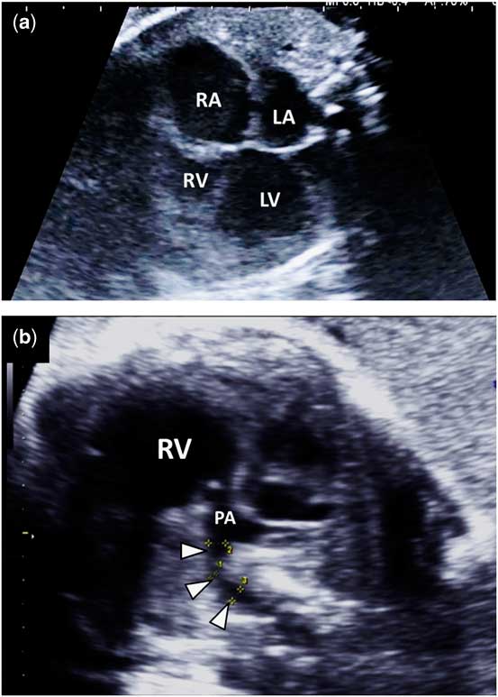
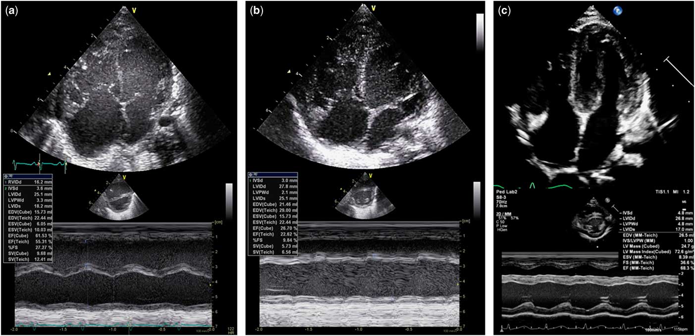
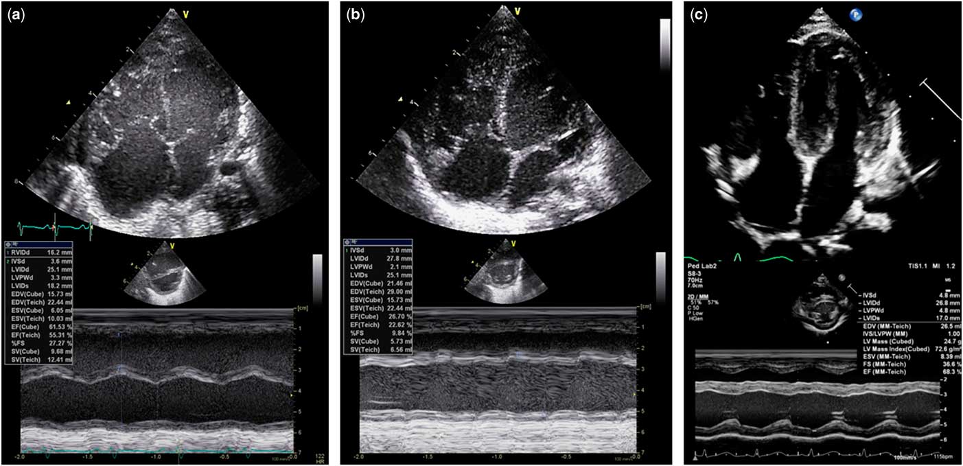
A 27-year-old woman, gravida 1, was referred at 36 weeks and 5 days of gestation because of rapidly progressive fetal cardiomegaly. She had neither a personal nor familial history of CHD. She had taken no non-steroidal anti-inflammatory drugs or prostaglandin inhibitors during her pregnancy. Despite maternal regular checkups, fetal cardiomegaly had not been pointed out until 35 weeks of gestation. A detailed fetal echocardiography showed a cardiothoracic area ratio of 0.58, a dilated left ventricle, and a hypertrophic right ventricle (Fig 1a). The right atrium was also dilated because of severe tricuspid regurgitation with a peak systolic velocity of 3.4 m/second. There was no structural cardiac anomaly. The pulmonary valve was patent and an antegrade flow in the pulmonary trunk was observed. However, the ductus arteriosus was narrowed and colour Doppler flow mapping could not identify blood flow through the ductus (Fig 1b). These findings were consistent with premature closure/constriction of the ductus arteriosus. On follow-up, a cardiotocogram showed non-reassuring fetal status with a sinusoidal pattern. Subsequently, a 3050-g male neonate was born by emergent Cesarean delivery at 36 weeks and 6 days. The Apgar score was 2 at 1 minute and the patient was intubated immediately owing to severe dyspnoea. Haematological parameters of venous blood were as follows: pH 7.25; pCO2, 39.9 mmHg; pO2, 44.7 mmHg; HCO3 −, 17.3 mmoL/L; base excess, −9.5 mmoL/L; aspartate aminotransferase, 63 IU/L; alanine aminotransferase, 8 IU/L; lactase dehydrogenase, 632 IU/L; creatine kinase, 326 IU/L; creatine kinase, MB 70 IU/L; and cardiac troponin, T 0.226 ng/mL (reference range:⩽0.10). Neonatal echocardiography revealed a left ventricular dilatation concomitant with a hypertrophic right ventricle and moderate tricuspid regurgitation. Neither aortic nor mitral regurgitation was evident. The left ventricular end-diastolic diameter and left ventricular ejection fraction were 25.1 mm (z score 3.4) and 62%, respectively (Fig 2a and Supplementary video S1). The ductus arteriosus was almost closed. The patient required respiratory support and inotropic agent and diuretic administration. The left ventricular function was transiently worse the day after birth. Echocardiography showed that the left ventricular end-diastolic diameter was dilated to 27.8 mm (z score 4.4) and ejection fraction was decreased to 27% with diffuse hypokinesis. There was neither pericardial effusion nor non-compacted myocardium. These findings of the left ventricular dilation and systolic dysfunction were compatible with dilated cardiomyopathy.Reference Jefferies and Towbin 4 However, both ventricular functions gradually improved day by day. The patient was extubated on the 14th day after birth and discharged on the 47th day. Echocardiography at the time of discharge showed a persistent mild left ventricular dilatation. At 6 months of age, the cardiomyopathy had fully resolved (Fig 2c and Supplementary video S1). The possibility of metabolic and infectious cardiomyopathies was excluded by metabolic screening and viral isolation.
Fetal echocardiographic findings during the initial visit. ( a ) A four-chamber view at 36 weeks and 5 days shows a dilated right atrium (RA) and left ventricle (LV), and a hypertrophic right ventricle (RV). The cardiothoracic ratio at that time was 0.58. ( b ) The narrowed ductus arteriosus (arrow heads). LA=left atrium; PA=pulmonary artery.

Sequential echocardiographic findings after birth. Figures show four-chamber view (upper part) and M-mode measurement of the left ventricle (lower part). ( a ) At birth: left ventricular end-diastolic diameter and left ventricular ejection fraction are 25.1 mm (z score 3.4) and 62%, respectively. ( b ) On the day after birth: left ventricular end-diastolic diameter and left ventricular ejection fraction are 27.8 mm (z score 4.4) and 27%, respectively. ( c ) At 6 months of age: left ventricular end-diastolic diameter and left ventricular ejection fraction are 26.8 mm (z score 1.3) and 75%, respectively.
Discussion
We demonstrated a case of dilated cardiomyopathy due to premature closure/constriction of the ductus arteriosus for the first time. Although the patient showed left ventricular dilatation concomitant with severe systolic dysfunction soon after birth, the cardiomyopathy was gradually improved by the medical treatments.
Mechanical constriction of the ductus arteriosus can cause an increase in fetal pulmonary blood flow, which leads to hypertrophy of the pulmonary artery smooth muscle and an increase in right ventricular afterload.Reference Enzensberger, Wienhard and Weichert 2 , Reference Momma and Takao 3 In the fetal circulation, however, premature closure/constriction of the ductus arteriosus can also increase blood flow through the foramen ovale and increase pulmonary blood flow after an increase in left ventricular preload.Reference Momma and Takao 3 In a fetal rat, the constriction of the ductus arteriosus after the administration of indomethacin soon led to a dilated left ventricle and a hypertrophic right ventricle,Reference Momma and Takao 3 which was a finding similar to the present case. When the ductus constricts rapidly, a rapid increase in left ventricular preload may lead to left ventricular dilated cardiomyopathy as an uncommon manifestation in premature closure/constriction of the ductus arteriosus. This case suggested that clinical manifestations of premature closure/constriction of the ductus arteriosus depend on the timing and speed of constriction of the ductus.
Supplementary material
To view supplementary material for this article, please visit https://doi.org/10.1017/S1047951118001002
Acknowledgements
The authors thank Dr Takeshi Kawakami and Dr Mika Ozaki (Department of Obstetrics and Gynecology, Japan Community Healthcare Organization, Kyushu Hospital), and Dr Mamie Watanabe, Dr Yuichiro Sugitani, Dr Chiaki Iida, Dr Hiromitsu Shirouzu, and Dr Naoki Kawaguchi (Department of Pediatrics, Japan Community Healthcare Organization, Kyushu Hospital) for assistance of data collection. Author’s contributions: Dr S.O. led conceptualisation and design of the analysis, analysed and interpreted data, and drafted the initial manuscript; Dr J.M. supervised data collection, reviewed the manuscript critically, and approved the final manuscript as submitted and Dr Y.I. contributed to collection of the data, reviewed the manuscript critically, and approved the final manuscript as submitted.
Financial Support
This research received no specific grant from any funding agency or from commercial or not-for-profit sectors.
Conflicts of Interest>
None.
Ethical Standards
The parents of the study participant provided informed consent for publication.