Neurophysiology techniques can provide objective information in movement disorders, a subspecialty practice area that traditionally relies on clinical judgment and experience for diagnosis. While expert clinical assessment remains fundamental, it can be limited by phenotypic overlap and subjective interpretation. Given the growing global burden of movement disorders, there is an increasing need to improve access to accurate diagnosis through more objective tools, such as neurophysiological assessment. In Canada, the variability in access to specialized expertise may further challenge timely and accurate diagnosis, highlighting the need for objective assessments.
With refinements in technique and advances in technology, the clinical utility and impact of neurophysiology in movement disorders practice have been demonstrated across a wide range of clinical contexts, with neurophysiological tests leading to significant gains in diagnostic accuracy and certainty.
Reference Grippe and Chen1
This is most commonly the case in functional movement disorders, which benefit greatly from objective measures.
Reference Kassavetis, Chen and Ganos2
Neurophysiology also aids in distinguishing tremor from myoclonus, characterizing tremor and identifying the myoclonus generator, among other applications that may be difficult to address through clinical assessment alone.
Reference Schwingenschuh, Van der Stouwe and Pandey3,Reference Latorre, Ganos and Hamada4
For example, data from the Toronto center showed that among referrals for functional movement disorders, 13% of patients had a change in diagnosis following neurophysiological assessment. In patients referred for jerky movements, 27% were ultimately found not to have myoclonus, and tremor was not confirmed in 20% of cases. These findings illustrate the added value of neurophysiological testing in clarifying phenomenology and refining clinical diagnoses.
Reference Grippe and Chen1,Reference Kassavetis, Chen and Ganos2
Clinical neurophysiological studies for the assessment of movement disorders are well established in many centers worldwide, with broader dissemination in Europe, followed by Asia and North America.
Reference Kassavetis, Chen and Ganos2
Although one center in Canada has performed these evaluations for more than 20 years,
Reference Grippe and Chen1
access to this specialized testing in Canada is limited and lacks standardized approaches within the communities of movement disorders and clinical neurophysiologists.
Historical View
The application of neurophysiological techniques for the assessment of movement disorders dates to 1946, when they were first used in the evaluation of non-epileptic myoclonus. One of the earliest findings was giant somatosensory evoked potentials (SEP), followed by the description of a cortical transient preceding a myoclonic jerk, and the cortical (C) reflex, as methods to distinguish cortical from subcortical origins of myoclonus.
Reference Latorre, Ganos and Hamada4
In 1991, the brainstem reflex myoclonus was localized to the brainstem based on a characteristic pattern of muscle activation. In the same year, the startle response was recognized as pathological when non-habituating in patients with hyperekplexia. Subsequent work expanded the neurophysiological characterization of myoclonus with the identification of the spinal cord and peripheral nerve or nerve root as generators of myoclonus.
Reference Latorre, Ganos and Hamada4
The Bereitschaftspotential (BP), a slow, negative electroencephalogram (EEG) marker of volitional action obtained using the EEG–electromyography (EMG) back-averaging technique, was described in 1964 by Kornhuber and Deecke as the first report of EEG activity preceding volitional movement. Since then, it has been used to study different aspects of movement, including the relation to the unconscious initiation of a freely voluntary act or free will. Most recently, it has been applied in brain–computer interfaces as a signal to control movement and has a wide range of possible applications in the rehabilitation scenario.
Reference Pitt, Brumberg, Burnison, Mehta and Kidwai5
In the movement disorders clinical practice, the BP was shown to precede functional jerky movements, thereby emerging as a neurophysiological marker of functional etiology in this movement phenotype.
Reference Terada, Ikeda and Van Ness6
Neurophysiological techniques have also become increasingly important in the evaluation of tremor. Orthostatic tremor was the first tremor subtype in which characteristic surface electromyography (EMG) findings were identified and incorporated in the diagnosis. Gradually, neurophysiology assumed a broader role in the assessment of other tremor syndromes, such as essential tremor and enhanced physiological tremor. Subsequently, specific neurophysiological criteria were established for the diagnosis of functional tremor.
Reference Schwingenschuh, Van der Stouwe and Pandey3
Neurophysiology is also considered a key diagnostic tool in disorders characterized by motor system hyperexcitability, such as stiff person spectrum disorders.
Reference Grippe and Chen1
Technical Summary
Recent publications have increasingly contributed to the standardization of neurophysiological protocols. However, knowledge of these assessments remains limited outside the movement disorders community, underscoring the need for broader implementation, further protocol standardization and improved integration with clinical neurophysiology laboratory settings.
Reference Kassavetis, Chen and Ganos2
In the following section, we outline the principal tests that comprise this emerging subspecialty, define the minimum equipment required and discuss the training necessary to conduct and interpret these assessments effectively.
The techniques used are summarized in Figure 1. The main indications for movement disorders neurophysiology testing include the assessment of jerky movements to differentiate between tremor, myoclonus and tics, identification of the myoclonus and myoclonus generator, classification of tremor subtypes, evaluation of functional etiology in patients with jerky movements or tremor, assessment of patients with instability while standing to investigate orthostatic tremor or orthostatic myoclonus and evaluation of patients with stiff person syndrome. The minimal proposed protocol for each indication is detailed in the supplementary material.
Figure 1.
Summary of common clinical neurophysiology techniques used in studies of movement disorders. Adapted from: Grippe T, Callister M, Vial F, Shabbir M, Chen R. Chapter 17: Clinical neurophysiology studies in the diagnosis of movement disorders. In: Handbook of Clinical Neurology (under review). BP = Bereitschaftspotential; CNS = Central nervous system; EEG = electroencephalogram; EMG = electromyography; JLBA = Jerk-locked back-averaging; LLR = Long-latency reflex; R= Right; SEP = somatosensory evoked potentials.
Figure 1. Long description
A diagram illustrating common clinical neurophysiology techniques used in studies of movement disorders. Panel 1: Polymography with accelerometry. The panel shows surface electromyography recordings from the right extensor carpi radialis and flexor carpi ulnaris muscles, along with an accelerometer channel. The graph displays two surface EMG channels and an accelerometer channel of an alternating tremor with a mean frequency of 4.5 Hz. Panel 2A: JLBA Bereitschaftspotential. The panel shows an example of EEG and a surface EMG channel with an average of 116 epochs processed with jerk-locked back-averaging showing BP before EMG contraction by 1.9 seconds. Panel 2B: JLBA cortical transient. The panel shows seven EEG channels and a surface EMG channel processed with jerk-locked back-averaging in a patient with cortical myoclonus. Panel 3: Somatosensory evoked potentials. The panel shows an example of a giant SEP with N20-P25 component of 20 microvolts with stimulation of the right median nerve in a patient with cortical myoclonus. Panel 4: Long-latency reflex/C-reflex. The panel shows an example of C-reflex recorded at the right abductor pollicis brevis after stimulation of the left median nerve in a patient with cortical myoclonus. Panel 5: Exteroceptive reflex. The panel shows exaggerated exteroceptive reflexes following stimulation of the posterior tibial nerve in a patient with Stiff person syndrome, initial short-latency EMG burst 30-80 milliseconds, followed by a period of tonic activity with a gradual tonic discharge beginning after 100 milliseconds.
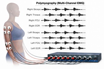
To perform these assessments, a minimum set of equipment is required. The most important component is polymyography (Figure 2), which is fundamental to all analyses. At least four channels are required to perform surface EMG, which may be used in combination with accelerometers, for the initial characterization of the movements. Systems with up to 16 channels may be used to allow more efficient and comprehensive assessments. Surface EMG electrodes are placed over the affected muscles and enable initial characterization of jerky movements, including differentiation between tremor and myoclonus. For tremor assessments, EMG and accelerometry are generally sufficient. Additional simple tools, such as a metronome to pace voluntary movements, are required for assessment of specific diagnostic criteria such as in functional tremor or essential tremor. Video recording synchronized with the neurophysiological data is desirable, as correlation between video and EMG findings can be crucial for accurate interpretation. EEG is often required for the evaluation of myoclonus and functional jerky movements. In these cases, EEG–EMG back-averaging techniques are used to identify the BP or cortical transients that precede myoclonus. Recording of SEP is also useful. The standard electrode setups for these studies are illustrated in Figure S1. The minimum EEG setup includes 10 electrodes and an electrical stimulator for peripheral nerve stimulation.
Figure 2.
Schematic representation of an eight-channel upper limbs polymyography for tremor study. EMG = electromyography; ECR = extensor carpi radialis; FCU = flexor carpi ulnaris. Figure generated using AI.
Some protocols require sensory stimulation. Electrical stimulation is particularly useful for the evaluation of reflex myoclonus and exaggerated reflexes, including blink reflexes and exteroceptive reflexes. Auditory stimulation may be used to trigger the startle reflex. Therefore, a sound generator and headphones are necessary for this assessment. Other forms of sensory stimulation, such as tapping with a reflex hammer or stimulation with a 128 Hz tuning fork, are also used, but no special equipment is required, since these are part of the standard neurological examination. The use of needle EMG is limited to specific protocols, such as muscle assessment in stiff person syndrome,
Reference Grippe and Chen1
assessment of deep muscles such as the diaphragm and, in selected cases, power spectral analysis in patients with dystonia.
Reference Latorre, Ganos and Hamada4
Its use is therefore considered optional, and these assessments can be performed in collaboration with a neuromuscular EMG team.
Movement Neurophysiology in Canada
There is a growing global effort to expand the integration of movement disorder clinical neurophysiology tools into routine clinical practice. Key barriers to wider implementation include limited availability of structured training, resource constraints and the absence of standardized protocols across centers.
Reference Kassavetis, Chen and Ganos2
In Canada, these assessments are currently routinely performed only at two centers, with a third center in development.
The sole fellowship training site in Canada is based at the Toronto Western Hospital. Trainees are expected to have a prior background in movement disorders or clinical neurophysiology, preferably both. A training period of 6 months to 1 year, or completion of approximately 50 supervised assessments, is suggested prior to independent practice. A minimum of 6-month duration would generally be appropriate only if the trainee is concurrently pursuing subspecialty training in clinical neurophysiology or movement disorders. To increase the impact of movement disorders neurophysiology to benefit more Canadians, we foresee a need for standardization of techniques alongside recognition of this subspecialty practice area by the Canadian Society of Clinical Neurophysiologists.
There is a need for a national network to support ongoing clinical research, improve evidence for existing and emerging applications, develop new training opportunities and increase accessibility of these techniques to reach more Canadians.








 You have
Access
Neurophysiology techniques can provide objective information in movement disorders, a subspecialty practice area that traditionally relies on clinical judgment and experience for diagnosis. While expert clinical assessment remains fundamental, it can be limited by phenotypic overlap and subjective interpretation. Given the growing global burden of movement disorders, there is an increasing need to improve access to accurate diagnosis through more objective tools, such as neurophysiological assessment. In Canada, the variability in access to specialized expertise may further challenge timely and accurate diagnosis, highlighting the need for objective assessments.
With refinements in technique and advances in technology, the clinical utility and impact of neurophysiology in movement disorders practice have been demonstrated across a wide range of clinical contexts, with neurophysiological tests leading to significant gains in diagnostic accuracy and certainty. Reference Grippe and Chen1 This is most commonly the case in functional movement disorders, which benefit greatly from objective measures. Reference Kassavetis, Chen and Ganos2 Neurophysiology also aids in distinguishing tremor from myoclonus, characterizing tremor and identifying the myoclonus generator, among other applications that may be difficult to address through clinical assessment alone. Reference Schwingenschuh, Van der Stouwe and Pandey3,Reference Latorre, Ganos and Hamada4 For example, data from the Toronto center showed that among referrals for functional movement disorders, 13% of patients had a change in diagnosis following neurophysiological assessment. In patients referred for jerky movements, 27% were ultimately found not to have myoclonus, and tremor was not confirmed in 20% of cases. These findings illustrate the added value of neurophysiological testing in clarifying phenomenology and refining clinical diagnoses. Reference Grippe and Chen1,Reference Kassavetis, Chen and Ganos2
Clinical neurophysiological studies for the assessment of movement disorders are well established in many centers worldwide, with broader dissemination in Europe, followed by Asia and North America. Reference Kassavetis, Chen and Ganos2 Although one center in Canada has performed these evaluations for more than 20 years, Reference Grippe and Chen1 access to this specialized testing in Canada is limited and lacks standardized approaches within the communities of movement disorders and clinical neurophysiologists.
Historical View
The application of neurophysiological techniques for the assessment of movement disorders dates to 1946, when they were first used in the evaluation of non-epileptic myoclonus. One of the earliest findings was giant somatosensory evoked potentials (SEP), followed by the description of a cortical transient preceding a myoclonic jerk, and the cortical (C) reflex, as methods to distinguish cortical from subcortical origins of myoclonus. Reference Latorre, Ganos and Hamada4 In 1991, the brainstem reflex myoclonus was localized to the brainstem based on a characteristic pattern of muscle activation. In the same year, the startle response was recognized as pathological when non-habituating in patients with hyperekplexia. Subsequent work expanded the neurophysiological characterization of myoclonus with the identification of the spinal cord and peripheral nerve or nerve root as generators of myoclonus. Reference Latorre, Ganos and Hamada4
The Bereitschaftspotential (BP), a slow, negative electroencephalogram (EEG) marker of volitional action obtained using the EEG–electromyography (EMG) back-averaging technique, was described in 1964 by Kornhuber and Deecke as the first report of EEG activity preceding volitional movement. Since then, it has been used to study different aspects of movement, including the relation to the unconscious initiation of a freely voluntary act or free will. Most recently, it has been applied in brain–computer interfaces as a signal to control movement and has a wide range of possible applications in the rehabilitation scenario. Reference Pitt, Brumberg, Burnison, Mehta and Kidwai5 In the movement disorders clinical practice, the BP was shown to precede functional jerky movements, thereby emerging as a neurophysiological marker of functional etiology in this movement phenotype. Reference Terada, Ikeda and Van Ness6
Neurophysiological techniques have also become increasingly important in the evaluation of tremor. Orthostatic tremor was the first tremor subtype in which characteristic surface electromyography (EMG) findings were identified and incorporated in the diagnosis. Gradually, neurophysiology assumed a broader role in the assessment of other tremor syndromes, such as essential tremor and enhanced physiological tremor. Subsequently, specific neurophysiological criteria were established for the diagnosis of functional tremor. Reference Schwingenschuh, Van der Stouwe and Pandey3 Neurophysiology is also considered a key diagnostic tool in disorders characterized by motor system hyperexcitability, such as stiff person spectrum disorders. Reference Grippe and Chen1
Technical Summary
Recent publications have increasingly contributed to the standardization of neurophysiological protocols. However, knowledge of these assessments remains limited outside the movement disorders community, underscoring the need for broader implementation, further protocol standardization and improved integration with clinical neurophysiology laboratory settings. Reference Kassavetis, Chen and Ganos2 In the following section, we outline the principal tests that comprise this emerging subspecialty, define the minimum equipment required and discuss the training necessary to conduct and interpret these assessments effectively.
The techniques used are summarized in Figure 1. The main indications for movement disorders neurophysiology testing include the assessment of jerky movements to differentiate between tremor, myoclonus and tics, identification of the myoclonus and myoclonus generator, classification of tremor subtypes, evaluation of functional etiology in patients with jerky movements or tremor, assessment of patients with instability while standing to investigate orthostatic tremor or orthostatic myoclonus and evaluation of patients with stiff person syndrome. The minimal proposed protocol for each indication is detailed in the supplementary material.
Summary of common clinical neurophysiology techniques used in studies of movement disorders. Adapted from: Grippe T, Callister M, Vial F, Shabbir M, Chen R. Chapter 17: Clinical neurophysiology studies in the diagnosis of movement disorders. In: Handbook of Clinical Neurology (under review). BP = Bereitschaftspotential; CNS = Central nervous system; EEG = electroencephalogram; EMG = electromyography; JLBA = Jerk-locked back-averaging; LLR = Long-latency reflex; R= Right; SEP = somatosensory evoked potentials.
Figure 1. Long description
A diagram illustrating common clinical neurophysiology techniques used in studies of movement disorders. Panel 1: Polymography with accelerometry. The panel shows surface electromyography recordings from the right extensor carpi radialis and flexor carpi ulnaris muscles, along with an accelerometer channel. The graph displays two surface EMG channels and an accelerometer channel of an alternating tremor with a mean frequency of 4.5 Hz. Panel 2A: JLBA Bereitschaftspotential. The panel shows an example of EEG and a surface EMG channel with an average of 116 epochs processed with jerk-locked back-averaging showing BP before EMG contraction by 1.9 seconds. Panel 2B: JLBA cortical transient. The panel shows seven EEG channels and a surface EMG channel processed with jerk-locked back-averaging in a patient with cortical myoclonus. Panel 3: Somatosensory evoked potentials. The panel shows an example of a giant SEP with N20-P25 component of 20 microvolts with stimulation of the right median nerve in a patient with cortical myoclonus. Panel 4: Long-latency reflex/C-reflex. The panel shows an example of C-reflex recorded at the right abductor pollicis brevis after stimulation of the left median nerve in a patient with cortical myoclonus. Panel 5: Exteroceptive reflex. The panel shows exaggerated exteroceptive reflexes following stimulation of the posterior tibial nerve in a patient with Stiff person syndrome, initial short-latency EMG burst 30-80 milliseconds, followed by a period of tonic activity with a gradual tonic discharge beginning after 100 milliseconds.
To perform these assessments, a minimum set of equipment is required. The most important component is polymyography (Figure 2), which is fundamental to all analyses. At least four channels are required to perform surface EMG, which may be used in combination with accelerometers, for the initial characterization of the movements. Systems with up to 16 channels may be used to allow more efficient and comprehensive assessments. Surface EMG electrodes are placed over the affected muscles and enable initial characterization of jerky movements, including differentiation between tremor and myoclonus. For tremor assessments, EMG and accelerometry are generally sufficient. Additional simple tools, such as a metronome to pace voluntary movements, are required for assessment of specific diagnostic criteria such as in functional tremor or essential tremor. Video recording synchronized with the neurophysiological data is desirable, as correlation between video and EMG findings can be crucial for accurate interpretation. EEG is often required for the evaluation of myoclonus and functional jerky movements. In these cases, EEG–EMG back-averaging techniques are used to identify the BP or cortical transients that precede myoclonus. Recording of SEP is also useful. The standard electrode setups for these studies are illustrated in Figure S1. The minimum EEG setup includes 10 electrodes and an electrical stimulator for peripheral nerve stimulation.
Schematic representation of an eight-channel upper limbs polymyography for tremor study. EMG = electromyography; ECR = extensor carpi radialis; FCU = flexor carpi ulnaris. Figure generated using AI.
Some protocols require sensory stimulation. Electrical stimulation is particularly useful for the evaluation of reflex myoclonus and exaggerated reflexes, including blink reflexes and exteroceptive reflexes. Auditory stimulation may be used to trigger the startle reflex. Therefore, a sound generator and headphones are necessary for this assessment. Other forms of sensory stimulation, such as tapping with a reflex hammer or stimulation with a 128 Hz tuning fork, are also used, but no special equipment is required, since these are part of the standard neurological examination. The use of needle EMG is limited to specific protocols, such as muscle assessment in stiff person syndrome, Reference Grippe and Chen1 assessment of deep muscles such as the diaphragm and, in selected cases, power spectral analysis in patients with dystonia. Reference Latorre, Ganos and Hamada4 Its use is therefore considered optional, and these assessments can be performed in collaboration with a neuromuscular EMG team.
Movement Neurophysiology in Canada
There is a growing global effort to expand the integration of movement disorder clinical neurophysiology tools into routine clinical practice. Key barriers to wider implementation include limited availability of structured training, resource constraints and the absence of standardized protocols across centers. Reference Kassavetis, Chen and Ganos2 In Canada, these assessments are currently routinely performed only at two centers, with a third center in development.
The sole fellowship training site in Canada is based at the Toronto Western Hospital. Trainees are expected to have a prior background in movement disorders or clinical neurophysiology, preferably both. A training period of 6 months to 1 year, or completion of approximately 50 supervised assessments, is suggested prior to independent practice. A minimum of 6-month duration would generally be appropriate only if the trainee is concurrently pursuing subspecialty training in clinical neurophysiology or movement disorders. To increase the impact of movement disorders neurophysiology to benefit more Canadians, we foresee a need for standardization of techniques alongside recognition of this subspecialty practice area by the Canadian Society of Clinical Neurophysiologists.
There is a need for a national network to support ongoing clinical research, improve evidence for existing and emerging applications, develop new training opportunities and increase accessibility of these techniques to reach more Canadians.
Supplementary material
The supplementary material for this article can be found at https://doi.org/10.1017/cjn.2026.10635.
Author contributions
TG contributed to the conception of the manuscript and drafted the initial version. DW contributed to manuscript conception and critical review of the manuscript. AL contributed to manuscript conception and critical review. MN contributed to the critical review of the manuscript. RC contributed to manuscript conception and critical review. All authors reviewed and approved the final manuscript for submission.
Funding statement
The authors declare no specific funding for this work.
Competing interests
No conflicts to declare.