Introduction
Advances in high-throughput DNA sequencing and genomics have impacted on parasitological research by allowing the rapid sequencing of parasite genomes, providing opportunities for increased understanding of parasite biology. The genomes of more than twenty helminths have been determined (Zarowiecki and Berriman, Reference Zarowiecki and Berriman2015), providing insights into parasite evolution and genetic diversity. These genomes could also provide a potentially valuable resource for the research community to guide investigations into diagnosis, treatment and control of parasitic diseases.
The genome sequence of the cestode parasite that causes cystic echinococcosis, Echinococcus granulosus, has been published by two independent teams of researchers. A draft genome of E. granulosus was first published in 2013 together with the reference genome of the closely related species, Echinococcus multilocularis, as well as draft genomes of the tapeworm parasites Taenia solium and Hymenolepis microstoma (Tsai et al. Reference Tsai, Zarowiecki, Holroyd, Garciarrubio, Sanchez-Flores, Brooks, Tracey, Bobes, Fragoso, Sciutto, Aslett, Beasley, Bennett, Cai, Camicia, Clark, Cucher, De Silva, Day, Deplazes, Estrada, Fernandez, Holland, Hou, Hu, Huckvale, Hung, Kamenetzky, Keane and Kiss2013). A second draft genome of E. granulosus was described later in the same year (Zheng et al. Reference Zheng, Zhang, Zhang, Zhang, Li, Lu, Zhu, Wang, Huang, Liu, Kang, Chen, Wang, Chen, Yu, Gao, Jin, Gu, Wang, Zhao, Shi, Wen, Lin, Jones, Brejova, Vinar, Zhao, McManus, Chen and Zhou2013). The published genomes of E. granulosus are potentially valuable resources for research into echinococcosis. The availability of the E. granulosus genome has been described as providing ‘a new dawn for improved diagnosis, treatment and control of echinococcosis’ (Zhang et al. Reference Zhang, Wang and McManus2014). Similarly, it has been suggested that data mining of the genomes of E. granulosus and other parasites offers a new starting point for parasitology research (Lv et al. Reference Lv, Wu, Zhang, Ji, Cai, Luo, Wang and Li2015).
An antigen termed EG95 was cloned from mRNA obtained from the early larval stage (oncosphere) of E. granulosus (Lightowlers et al. Reference Lightowlers, Lawrence, Gauci, Young, Ralston, Maas and Health1996). EG95 expressed in Escherichia coli has been shown to confer a high level of protection against E. granulosus infection in livestock animals in several vaccine trials (Lightowlers et al. Reference Lightowlers, Jensen, Fernandez, Iriarte, Woollard, Gauci, Jenkins and Heath1999; Lightowlers, Reference Lightowlers2006). EG95 has been found to be encoded by a gene which is a member of a small family of closely related genes. The gene family was characterized by cloning of E. granulosus genomic DNA fragments and sequencing of the fragments using Sanger-based sequencing methods (Chow et al. Reference Chow, Gauci, Cowman and Lightowlers2001). Seven cloned genomic fragments were characterized, six of which encompass eg95 gene family members (designated eg95-1 to eg95-6), which encode three closely related proteins (EG95-1, EG95-5 and EG95-6), with eg95-1 encoding the cDNA from which the EG95 vaccine antigen has been expressed. Echinococcus granulosus is known to be genetically heterogeneous and the eg95 gene family has been characterized (by gene cloning) from two genotypes of E. granulosus: G1 (Chow et al. Reference Chow, Gauci, Cowman and Lightowlers2001) and G6 (Alvarez Rojas et al. Reference Alvarez Rojas, Gauci, Nolan, Harandi and Lightowlers2012). The G1 and G6 genotype groupings are known to cause the great majority of all reported cases of cystic echinococcosis in humans (Alvarez Rojas et al. Reference Alvarez Rojas, Romig and Lightowlers2014). Other studies have aimed to characterize the eg95-related genes in E. granulosus using polymerase chain reaction (PCR)-based methods (Zhang et al. Reference Zhang, Li, You, Zhang, Turson, Loukas and McManus2003; Haag et al. Reference Haag, Gottstein and Ayala2009); however, there have been concerns raised about the creation of PCR-related artefacts in the sequences resulting from these investigations, due to the likelihood of template switching among very closely related target sequences amplified using non gene-specific PCR primers (Alvarez Rojas et al. Reference Alvarez Rojas, Gauci, Nolan, Harandi and Lightowlers2012).
The approach used to sequence the eg95 family in the G1 and G6 genotypes of E. granulosus utilized the cloning of gDNA fragments having sizes which corresponded to fragments detected on Southern blots probed with labelled EG95 cDNA. This method is unable to unequivocally identify all the fragments that may contain closely related gDNA sequences because of the potential for co-migration of different fragments containing the eg95-related sequences. The method is also unable to differentiate alleles from different genes per se. The availability of two draft genomes of E. granulosus provided us with the opportunity to clarify the number and genomic organization of eg95-related genes.
Materials and methods
The E. granulosus genome data published by Tsai et al. (Reference Tsai, Zarowiecki, Holroyd, Garciarrubio, Sanchez-Flores, Brooks, Tracey, Bobes, Fragoso, Sciutto, Aslett, Beasley, Bennett, Cai, Camicia, Clark, Cucher, De Silva, Day, Deplazes, Estrada, Fernandez, Holland, Hou, Hu, Huckvale, Hung, Kamenetzky, Keane and Kiss2013) was available for analysis from the Sanger Institute (http://www.sanger.ac.uk/resources/downloads/helminths). The genome data of E. granulosus published by Zheng et al. (Reference Zheng, Zhang, Zhang, Zhang, Li, Lu, Zhu, Wang, Huang, Liu, Kang, Chen, Wang, Chen, Yu, Gao, Jin, Gu, Wang, Zhao, Shi, Wen, Lin, Jones, Brejova, Vinar, Zhao, McManus, Chen and Zhou2013) was available through the National Centre for Biotechnology Information (NCBI, http://www.ncbi.nlm.nih.gov/assembly/GCA_000524195.1, dated 28/01/2014).
Echinococcus granulosus genome data were analysed for eg95-related genes using two strategies. At the nucleotide level, the DNA sequences were extracted corresponding to those genes identified in Tsai et al. (Reference Tsai, Zarowiecki, Holroyd, Garciarrubio, Sanchez-Flores, Brooks, Tracey, Bobes, Fragoso, Sciutto, Aslett, Beasley, Bennett, Cai, Camicia, Clark, Cucher, De Silva, Day, Deplazes, Estrada, Fernandez, Holland, Hou, Hu, Huckvale, Hung, Kamenetzky, Keane and Kiss2013) and Zheng et al. (Reference Zheng, Zhang, Zhang, Zhang, Li, Lu, Zhu, Wang, Huang, Liu, Kang, Chen, Wang, Chen, Yu, Gao, Jin, Gu, Wang, Zhao, Shi, Wen, Lin, Jones, Brejova, Vinar, Zhao, McManus, Chen and Zhou2013) that were listed as being related to eg95. Tsai et al. (Reference Tsai, Zarowiecki, Holroyd, Garciarrubio, Sanchez-Flores, Brooks, Tracey, Bobes, Fragoso, Sciutto, Aslett, Beasley, Bennett, Cai, Camicia, Clark, Cucher, De Silva, Day, Deplazes, Estrada, Fernandez, Holland, Hou, Hu, Huckvale, Hung, Kamenetzky, Keane and Kiss2013) reported five eg95-related genes, designated EgrG_000328500.1, EgrG_000375200.1, EgrG_000710400.1, EgrG_000554700.1 and EgrG_002001600.1 (Supplementary table S12.4 and from ftp site at the Sanger Institute, file ‘Egranulosus_v3_2014_05_27.products’, modified 6/2/14, accessed March 2017). Zheng et al. (Reference Zheng, Zhang, Zhang, Zhang, Li, Lu, Zhu, Wang, Huang, Liu, Kang, Chen, Wang, Chen, Yu, Gao, Jin, Gu, Wang, Zhao, Shi, Wen, Lin, Jones, Brejova, Vinar, Zhao, McManus, Chen and Zhou2013) reported seven eg95-related genes (Supplementary Table 47) designated: EGR_05614, EGR_08805, EGR_10541, EGR_06928, EGR_11122, EGR_06751 and EGR_10281.
To confirm the presence or absence of other eg95-related genes, each database was also interrogated using BLAST, with the E. granulosus BLAST server provided at the Sanger Institute, as well as the GeneDB website used for the E. granulosus genome published by Tsai et al. (Reference Tsai, Zarowiecki, Holroyd, Garciarrubio, Sanchez-Flores, Brooks, Tracey, Bobes, Fragoso, Sciutto, Aslett, Beasley, Bennett, Cai, Camicia, Clark, Cucher, De Silva, Day, Deplazes, Estrada, Fernandez, Holland, Hou, Hu, Huckvale, Hung, Kamenetzky, Keane and Kiss2013). The sequences of the eg95 gene family members originally described by Chow et al. (Reference Chow, Gauci, Cowman and Lightowlers2001) were used as input: eg95-1 (Genbank accession AF134378), eg95-2 (AF199352), eg95-3 (AF199353), eg95-4 (AF199349), eg95-5 (AF199350), eg95-6 (AF199347) and eg95-7 (AF199348). The eg95-related gene sequences with the highest similarity found using BLAST were identified and used in alignments (ClustalW) and in comparative analyses with the previously described eg95-related genes from E. granulosus.
At the protein level, the amino acid sequences of proteins encoded by eg95-related genes identified in both genome projects were compared with the three proteins encoded by the eg95 gene family (EG95-1, EG95-5, EG95-6) from E. granulosus G1 strain (Chow et al. Reference Chow, Gauci, Cowman and Lightowlers2001), with the percentage amino acid identity determined using ClustalW alignment.
Results
Analysis of eg95 related genes in the E. granulosus genome assembled by Tsai et al.
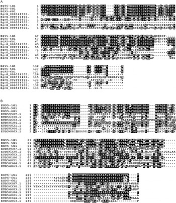
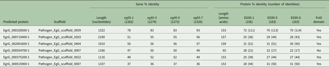
At the nucleotide level, the six eg95-related genes identified from the genome described by Tsai et al. (Reference Tsai, Zarowiecki, Holroyd, Garciarrubio, Sanchez-Flores, Brooks, Tracey, Bobes, Fragoso, Sciutto, Aslett, Beasley, Bennett, Cai, Camicia, Clark, Cucher, De Silva, Day, Deplazes, Estrada, Fernandez, Holland, Hou, Hu, Huckvale, Hung, Kamenetzky, Keane and Kiss2013) have different lengths, ranging from 1116 to 2190 nucleotides (total length of gene from the start to the stop codon), which differed when compared with the genes reported by Chow et al. (Reference Chow, Gauci, Cowman and Lightowlers2001) who reported 1262 bp for eg95-1 and 1274 bp for eg95-5 and eg95-6. Table 1 shows the nucleotide sequence identity between the two groups, with the gene encoding the predicted protein EgrG_000328500.1 showing greatest nucleotide identity to one of the genes described by Chow et al. (93% homology to the eg95-7 gene, reported to be a pseudogene). The predicted protein from this genomic fragment contains an ATA triplet replacing the initiator codon (Fig. 1A), as is the case for the eg95-7 pseudogene described by Chow et al. (Reference Chow, Gauci, Cowman and Lightowlers2001). None of the remaining genes identified by Tsai et al. (Reference Tsai, Zarowiecki, Holroyd, Garciarrubio, Sanchez-Flores, Brooks, Tracey, Bobes, Fragoso, Sciutto, Aslett, Beasley, Bennett, Cai, Camicia, Clark, Cucher, De Silva, Day, Deplazes, Estrada, Fernandez, Holland, Hou, Hu, Huckvale, Hung, Kamenetzky, Keane and Kiss2013) showed more than 57% nucleotide sequence identity with any of the eg95 gene family members described by Chow et al. (Reference Chow, Gauci, Cowman and Lightowlers2001). The lack of homology, between eg95-related genes identified in the genome data prepared by Tsai et al. (Reference Tsai, Zarowiecki, Holroyd, Garciarrubio, Sanchez-Flores, Brooks, Tracey, Bobes, Fragoso, Sciutto, Aslett, Beasley, Bennett, Cai, Camicia, Clark, Cucher, De Silva, Day, Deplazes, Estrada, Fernandez, Holland, Hou, Hu, Huckvale, Hung, Kamenetzky, Keane and Kiss2013) and the eg95 gene family described by Chow et al. (Reference Chow, Gauci, Cowman and Lightowlers2001), was reflected in low levels of predicted amino acid sequence identity between the corresponding proteins (Table 1). Amino acid sequence identity ranged from 22 to 70% between the two groups and the length of the predicted proteins ranged between 65 and 159 amino acids, with none matching the 156 amino acid length of the vaccine antigen EG95-1.
(A) Amino acid sequence alignment of proteins predicted from genes identified to be eg95-related and described in the genome of E. granulosus (Tsai et al. Reference Tsai, Zarowiecki, Holroyd, Garciarrubio, Sanchez-Flores, Brooks, Tracey, Bobes, Fragoso, Sciutto, Aslett, Beasley, Bennett, Cai, Camicia, Clark, Cucher, De Silva, Day, Deplazes, Estrada, Fernandez, Holland, Hou, Hu, Huckvale, Hung, Kamenetzky, Keane and Kiss2013) compared with proteins encoded by the eg95 gene family members described by Chow et al. (Reference Chow, Gauci, Cowman and Lightowlers2001). (B) Amino acid sequence alignment of proteins predicted from genes identified to be eg95-related and described in the genome of E. granulosus (Zheng et al. Reference Zheng, Zhang, Zhang, Zhang, Li, Lu, Zhu, Wang, Huang, Liu, Kang, Chen, Wang, Chen, Yu, Gao, Jin, Gu, Wang, Zhao, Shi, Wen, Lin, Jones, Brejova, Vinar, Zhao, McManus, Chen and Zhou2013) compared with proteins encoded by the eg95 gene family members described by Chow et al. (Reference Chow, Gauci, Cowman and Lightowlers2001). EG95-1G1, EG95-5G1 and EG95-6G1 refer to the proteins described in Chow et al. (Reference Chow, Gauci, Cowman and Lightowlers2001), with EG95-1G1 representing the protective vaccine antigen.

Homology of proteins predicted from genes identified to be eg95-related and described in the genome of E. granulosus (Tsai et al. Reference Tsai, Zarowiecki, Holroyd, Garciarrubio, Sanchez-Flores, Brooks, Tracey, Bobes, Fragoso, Sciutto, Aslett, Beasley, Bennett, Cai, Camicia, Clark, Cucher, De Silva, Day, Deplazes, Estrada, Fernandez, Holland, Hou, Hu, Huckvale, Hung, Kamenetzky, Keane and Kiss2013) compared with proteins encoded by the eg95 gene family members described by Chow et al. (Reference Chow, Gauci, Cowman and Lightowlers2001)

Analysis of eg95 related genes in the E. granulosus genome assembled by Zheng et al.
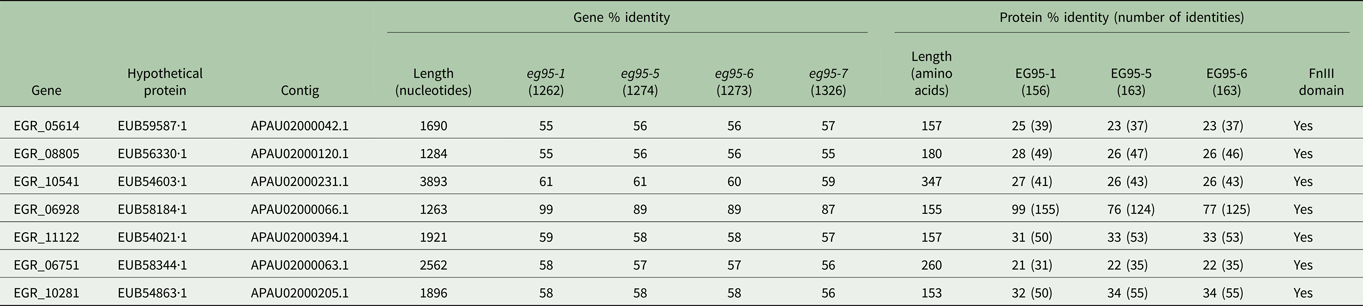
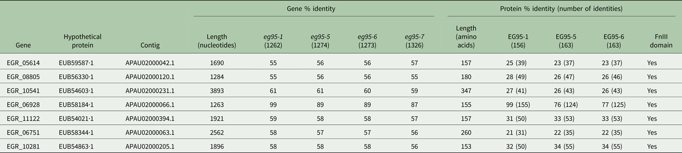
The E. granulosus genome data published by Zheng et al. (Reference Zheng, Zhang, Zhang, Zhang, Li, Lu, Zhu, Wang, Huang, Liu, Kang, Chen, Wang, Chen, Yu, Gao, Jin, Gu, Wang, Zhao, Shi, Wen, Lin, Jones, Brejova, Vinar, Zhao, McManus, Chen and Zhou2013) reported the presence of seven eg95-related genes, which were originally designated EG_05614, EG_08805, EG_10541, EG_06928, EG_11122, EG_06751 and EG_10281. In the most recently updated version of the genome, the same genes contain the prefix EGR. These genes have lengths ranging from 1263 to 3893 nucleotides. The gene with the highest homology to any of the eg95 genes reported by Chow et al. (Reference Chow, Gauci, Cowman and Lightowlers2001) is EGR_06928, having 99% nucleotide sequence identity with eg95-1. The other six genes have nucleotide sequence homology with eg95-1 from 55 to 61%, and between 56 and 61% with either eg95-5 or eg95-6 (Table 2). At the amino acid level, the gene EGR_06928 encodes the hypothetical protein EUB58184.1, which has 99% homology with the vaccine antigen EG95-1 (Fig. 1B). This protein contains 155 amino acids compared with 156 in the EG95-1 vaccine antigen. The length of the other six predicted proteins encoded by the genes identified by Zheng et al. (Reference Zheng, Zhang, Zhang, Zhang, Li, Lu, Zhu, Wang, Huang, Liu, Kang, Chen, Wang, Chen, Yu, Gao, Jin, Gu, Wang, Zhao, Shi, Wen, Lin, Jones, Brejova, Vinar, Zhao, McManus, Chen and Zhou2013) varies from 153 to 347 amino acids and their homology with the vaccine antigen is between 21 and 31% (Table 2). Homology of these proteins with either EG95-5 or EG95-6, described by Chow et al. (Reference Chow, Gauci, Cowman and Lightowlers2001), ranges between 22 and 34%. All of the predicted proteins contain conserved amino acids, which are characteristic of a fibronectin type III (FnIII) domain. Although the gene EGR_06928 listed in Table 2 and identified by Zheng et al. (Reference Zheng, Zhang, Zhang, Zhang, Li, Lu, Zhu, Wang, Huang, Liu, Kang, Chen, Wang, Chen, Yu, Gao, Jin, Gu, Wang, Zhao, Shi, Wen, Lin, Jones, Brejova, Vinar, Zhao, McManus, Chen and Zhou2013) encodes a protein that is almost identical to the EG95 vaccine antigen, it was not possible to determine which of the four fragments characterized by Chow et al. (Reference Chow, Gauci, Cowman and Lightowlers2001) that encode the EG95-1 protein correspond to EGR_06928. The EG95-related sequence contained within EGR_06928 contains 15 nucleotides within the predicted two introns and 3′UTR, which indicate that the sequence appears to be a mosaic of all four genes.
Homology of proteins predicted from genes identified to be eg95-related and described in the genome of E. granulosus (Zheng et al. Reference Zheng, Zhang, Zhang, Zhang, Li, Lu, Zhu, Wang, Huang, Liu, Kang, Chen, Wang, Chen, Yu, Gao, Jin, Gu, Wang, Zhao, Shi, Wen, Lin, Jones, Brejova, Vinar, Zhao, McManus, Chen and Zhou2013) compared with proteins encoded by the eg95 gene family members described by Chow et al. (Reference Chow, Gauci, Cowman and Lightowlers2001)
Discussion
Our analyses of the eg95 gene family in the two published genome databases of E. granulosus highlight limitations in the current E. granulosus genome assemblies when applied to gene family studies. It was not possible to map the eg95 gene family to the two published draft E. granulosus genomes. None of the genes/proteins listed as EG95 in the genome annotation by Tsai et al. (Reference Tsai, Zarowiecki, Holroyd, Garciarrubio, Sanchez-Flores, Brooks, Tracey, Bobes, Fragoso, Sciutto, Aslett, Beasley, Bennett, Cai, Camicia, Clark, Cucher, De Silva, Day, Deplazes, Estrada, Fernandez, Holland, Hou, Hu, Huckvale, Hung, Kamenetzky, Keane and Kiss2013) were identical to any member of the previously described eg95 genes, nor did they have close similarity to the eg95-1 gene encoding the EG95 vaccine antigen sequence (Table 1), which had been determined previously by cloning gDNA restriction fragments (Chow et al. Reference Chow, Gauci, Cowman and Lightowlers2001) and also deduced from cloned E. granulosus mRNA (Lightowlers et al. Reference Lightowlers, Lawrence, Gauci, Young, Ralston, Maas and Health1996). A single gene (EGR_06928, Table 2) identified in the E. granulosus genome annotation prepared by Zheng et al. (Reference Zheng, Zhang, Zhang, Zhang, Li, Lu, Zhu, Wang, Huang, Liu, Kang, Chen, Wang, Chen, Yu, Gao, Jin, Gu, Wang, Zhao, Shi, Wen, Lin, Jones, Brejova, Vinar, Zhao, McManus, Chen and Zhou2013) having a relatively high degree of homology with known eg95-related genes was found to be a mosaic of four different gene fragments (eg95-1 to eg95-4) previously described from sequencing of both strands of cloned genomic DNA (Chow et al. Reference Chow, Gauci, Cowman and Lightowlers2001). The eg95-5 and eg95-6 genes identified by Chow et al. (Reference Chow, Gauci, Cowman and Lightowlers2001), encoding the related proteins EG95-5 and EG95-6, were found in neither of the E. granulosus genome databases.
Second-generation sequencing technologies are being used to generate large quantities of sequence data and numerous associated publications. However, major challenges remain which relate to assembly and interpretation of the data generated. Holroyd and Sanchez-Flores (Reference Holroyd and Sanchez-Flores2012) suggested that the genome assembly process is one of the most challenging steps in a genome project, since reconstructing the original genome sequence from short fragments is made more difficult by the possibility of sequences occurring in multiple positions within a genome. Although second-generation sequencing technologies have provided opportunities to investigate the genome of a species in great detail, it is well recognized that the assembly of short sequence reads into genes provides significant computational challenges (Koren and Phillippy, Reference Koren and Phillippy2015). For the purposes that we sought to use the two published E. granulosus genomic datasets, the data were found to have limited utility.
In addition to the genes previously annotated by Tsai et al. (Reference Tsai, Zarowiecki, Holroyd, Garciarrubio, Sanchez-Flores, Brooks, Tracey, Bobes, Fragoso, Sciutto, Aslett, Beasley, Bennett, Cai, Camicia, Clark, Cucher, De Silva, Day, Deplazes, Estrada, Fernandez, Holland, Hou, Hu, Huckvale, Hung, Kamenetzky, Keane and Kiss2013) and Zheng et al. (Reference Zheng, Zhang, Zhang, Zhang, Li, Lu, Zhu, Wang, Huang, Liu, Kang, Chen, Wang, Chen, Yu, Gao, Jin, Gu, Wang, Zhao, Shi, Wen, Lin, Jones, Brejova, Vinar, Zhao, McManus, Chen and Zhou2013) as being eg95-related (Tables 1 and 2), BLAST assessment using the original eg95 genes from Chow et al. (Reference Chow, Gauci, Cowman and Lightowlers2001) as the query to search the two genome databases also identified several other genes encoding related proteins. BLAST identified one gene, located on EgG_scaffold_0007 (encoding the predicted protein EgrG_000515900.1, Table 1), as eg95-related because it contained an FnIII domain, while the same protein was annotated by Tsai et al. (Reference Tsai, Zarowiecki, Holroyd, Garciarrubio, Sanchez-Flores, Brooks, Tracey, Bobes, Fragoso, Sciutto, Aslett, Beasley, Bennett, Cai, Camicia, Clark, Cucher, De Silva, Day, Deplazes, Estrada, Fernandez, Holland, Hou, Hu, Huckvale, Hung, Kamenetzky, Keane and Kiss2013) as a gene encoding a member of the EMY162 antigen family in tapeworms. Similarly, other genes were misidentified in the BLAST searches of the two E. granulosus genome databases as EG95-related (data not shown) and were likely to have been selected because they encoded proteins that have an FnIII domain in common with EG95 (Tables 1 and 2). These proteins showed much lower homology to the EG95-1 vaccine antigen (<21% amino acid sequence identity), likely due only to the presence of characteristic amino acid signature residues in the FnIII domains. The FnIII domain is a common domain found in many proteins from diverse animal species (Doolittle and Bork, Reference Doolittle and Bork1993). The domain has been identified in several cestode parasite vaccine antigens (Lightowlers et al. Reference Lightowlers, Flisser, Gauci, Heath, Jensen and Rolfe2000).
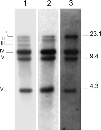
Both E. granulosus genome sequencing projects were undertaken using the G1 genotype, which has been more recently assigned as E. granulosus sensu stricto. This is the same genotype that was used for the initial cloning of the EG95 mRNA from a New Zealand isolate of E. granulosus and also the same genotype used for the gDNA fragment cloning and Sanger sequencing conducted by Chow et al. (Reference Chow, Gauci, Cowman and Lightowlers2001) from Australian E. granulosus isolates. Although the same genotype was used in both the genomic sequencing project undertaken by Tsai et al. (Reference Tsai, Zarowiecki, Holroyd, Garciarrubio, Sanchez-Flores, Brooks, Tracey, Bobes, Fragoso, Sciutto, Aslett, Beasley, Bennett, Cai, Camicia, Clark, Cucher, De Silva, Day, Deplazes, Estrada, Fernandez, Holland, Hou, Hu, Huckvale, Hung, Kamenetzky, Keane and Kiss2013) and the project undertaken by Zheng et al. (Reference Zheng, Zhang, Zhang, Zhang, Li, Lu, Zhu, Wang, Huang, Liu, Kang, Chen, Wang, Chen, Yu, Gao, Jin, Gu, Wang, Zhao, Shi, Wen, Lin, Jones, Brejova, Vinar, Zhao, McManus, Chen and Zhou2013), it was not possible to accurately map any of the eg95 genes described by Chow et al. (Reference Chow, Gauci, Cowman and Lightowlers2001) in either of the published genomes. Chow et al. (Reference Chow, Gauci, Cowman and Lightowlers2001) characterized four genomic DNA fragments (eg95-1, eg95-2, eg95-3 and eg95-4) encoding the same EG95 vaccine antigen (EG95-1), an additional two fragments encoding two other related proteins (EG95-5 and EG95-6) and a seventh encoding a pseudogene (eg95-7). These were identified from a pool of several individual parasite isolates. Southern blot hybridization analysis of genomic DNA obtained from various isolates of E. granulosus with the same G1 genotype identified differences in the eg95 DNA fragment hybridization pattern between isolates (Fig. 2). Despite the absence of bands II or III (each comprising eg95-2) in some isolates, all seven eg95 genomic fragments have been confirmed (by gene-specific PCR) to be present in all three isolates (Chow, Reference Chow2002). The Southern blot differences appear to be due to an EcoRI site polymorphism in the DNA fragment upstream from eg95-2. Smyth (Reference Smyth1962) and Spakulova et al. (Reference Spakulova, Orosova and Mackiewicz2011) presented evidence suggesting that E. granulosus cells were diploid and this is consistent with information from the E. multilocularis reference genome, which indicates that E. multilocularis is diploid (Tsai et al. Reference Tsai, Zarowiecki, Holroyd, Garciarrubio, Sanchez-Flores, Brooks, Tracey, Bobes, Fragoso, Sciutto, Aslett, Beasley, Bennett, Cai, Camicia, Clark, Cucher, De Silva, Day, Deplazes, Estrada, Fernandez, Holland, Hou, Hu, Huckvale, Hung, Kamenetzky, Keane and Kiss2013). The four fragments described by Chow et al. (Reference Chow, Gauci, Cowman and Lightowlers2001), eg95-1, eg95-2, eg95-3 and eg95-4, all encode the same protein and may include alleles of a single or more limited number of genes. However, the current draft versions of the E. granulosus genome are insufficiently complete to be able to confirm this. Assessment of the two E. granulosus genomes using the Core Eukaryotic Genes Mapping Approach (CEGMA) (Parra et al. Reference Parra, Bradnam, Ning, Keane and Korf2009) suggests that the genome sequenced by Tsai et al. (Reference Tsai, Zarowiecki, Holroyd, Garciarrubio, Sanchez-Flores, Brooks, Tracey, Bobes, Fragoso, Sciutto, Aslett, Beasley, Bennett, Cai, Camicia, Clark, Cucher, De Silva, Day, Deplazes, Estrada, Fernandez, Holland, Hou, Hu, Huckvale, Hung, Kamenetzky, Keane and Kiss2013) is 92·74% complete, while the CEGMA completeness score for the genome of Zheng et al. (Reference Zheng, Zhang, Zhang, Zhang, Li, Lu, Zhu, Wang, Huang, Liu, Kang, Chen, Wang, Chen, Yu, Gao, Jin, Gu, Wang, Zhao, Shi, Wen, Lin, Jones, Brejova, Vinar, Zhao, McManus, Chen and Zhou2013) is 89·11%.
Southern blot hybridization analysis of genomic DNA obtained from different isolates of E. granulosus. Parasite DNA was digested with EcoRI and probed with eg95 cDNA in Southern hybridization as described in Chow et al. (2001). Panel 1; 5 µg genomic DNA pooled from adult worms of a New Zealand E. granulosus isolate. Panel 2; 6 µg genomic DNA of protoscolesces obtained from an individual hydatid cyst from a naturally infected Australian sheep. Panel 3; 4 µg genomic DNA of protoscolesces from an individual cyst collected from a naturally infected sheep in Australia but from a different property to that from which the sample used in panel 2 was obtained. The six bands showing homology to eg95 cDNA are marked on the left and numbered I – VI. Size estimates of the bands are 23·1, 19·0, 17·0, 13·0, 9·4 and 4·0 kb, respectively. The positions of molecular size markers (kb) are denoted on the right.

A high number of heterozygous single-nucleotide polymorphisms (SNPs, 79 804) were observed in E. granulosus protoscoleces (Tsai et al. Reference Tsai, Zarowiecki, Holroyd, Garciarrubio, Sanchez-Flores, Brooks, Tracey, Bobes, Fragoso, Sciutto, Aslett, Beasley, Bennett, Cai, Camicia, Clark, Cucher, De Silva, Day, Deplazes, Estrada, Fernandez, Holland, Hou, Hu, Huckvale, Hung, Kamenetzky, Keane and Kiss2013) compared with the E. multilocularis reference genome, which was found to contain at least 40-fold fewer heterozygous SNPs (168–1855 SNPs for six isolates). The difficulties encountered in mapping eg95 genes to the E. granulosus genomes may be associated in part with the high number of SNPs in the draft genomes. The genome sequenced by Tsai et al. (Reference Tsai, Zarowiecki, Holroyd, Garciarrubio, Sanchez-Flores, Brooks, Tracey, Bobes, Fragoso, Sciutto, Aslett, Beasley, Bennett, Cai, Camicia, Clark, Cucher, De Silva, Day, Deplazes, Estrada, Fernandez, Holland, Hou, Hu, Huckvale, Hung, Kamenetzky, Keane and Kiss2013) was derived from two hydatid cysts of the G1 genotype (E. granulosus sensu stricto) recovered from bovines in Uruguay. Zheng et al. (Reference Zheng, Zhang, Zhang, Zhang, Li, Lu, Zhu, Wang, Huang, Liu, Kang, Chen, Wang, Chen, Yu, Gao, Jin, Gu, Wang, Zhao, Shi, Wen, Lin, Jones, Brejova, Vinar, Zhao, McManus, Chen and Zhou2013) found 145 534 SNP sites in their E. granulosus genome, sequenced with DNA from a single ovine liver cyst (originating from a single egg and thus representing a clone). The four genomic fragments (eg95-1 to eg95-4) described by Chow et al. (Reference Chow, Gauci, Cowman and Lightowlers2001) as encoding a single, identical EG95-1 protein may well consist of one gene and three alleles. However, we were unable to identify the other genes described by Chow et al. (Reference Chow, Gauci, Cowman and Lightowlers2001), eg95-5 and eg95-6, in either of the genome databases, raising doubts about the accuracy of the available genome data to support any hypothesis concerning the number of genes encoding the EG95-1 protein in the genome.
The accuracy of eg95 gene prediction from the E. granulosus draft genomes may have been improved by alignment of transcriptome reads from the oncosphere stage of the parasite to the genome sequence. Expression of eg95 is up-regulated in the oncosphere life cycle stage and either absent or expressed at a very low level in other stages (Chow et al. Reference Chow, Gauci, Cowman and Lightowlers2004). For this reason, eg95-1 cDNAs may not have been present in transcriptomes of the E. granulosus life-cycle stages that were used to generate the genomic sequences. Although Tsai et al. (Reference Tsai, Zarowiecki, Holroyd, Garciarrubio, Sanchez-Flores, Brooks, Tracey, Bobes, Fragoso, Sciutto, Aslett, Beasley, Bennett, Cai, Camicia, Clark, Cucher, De Silva, Day, Deplazes, Estrada, Fernandez, Holland, Hou, Hu, Huckvale, Hung, Kamenetzky, Keane and Kiss2013) provided transcriptomic data for E. granulosus protoscoleces (extracted from a single porcine liver cyst), transcriptomic data from oncospheres of E. granulosus were not used to aid gene prediction. Zheng et al. (Reference Zheng, Zhang, Zhang, Zhang, Li, Lu, Zhu, Wang, Huang, Liu, Kang, Chen, Wang, Chen, Yu, Gao, Jin, Gu, Wang, Zhao, Shi, Wen, Lin, Jones, Brejova, Vinar, Zhao, McManus, Chen and Zhou2013) undertook transcriptome data analysis from four different life-cycle stages of E. granulosus (including oncospheres) that could be used to validate the expression of predicted genes, although it is unclear whether transcriptomic data specific to EG95 were used to improve the accuracy of eg95 gene prediction. The expressed sequence tags (EST) used by Zheng et al. (Reference Zheng, Zhang, Zhang, Zhang, Li, Lu, Zhu, Wang, Huang, Liu, Kang, Chen, Wang, Chen, Yu, Gao, Jin, Gu, Wang, Zhao, Shi, Wen, Lin, Jones, Brejova, Vinar, Zhao, McManus, Chen and Zhou2013) had an average length of 300 bp. ESTs of this length, derived from a group of longer, very closely related genes (eg95, 1262–1326 nucleotides) would be expected to have limited utility in assembly and mapping of eg95-related sequences to that genome.
We were unable to map the previously characterized eg95 genes to the recently published E. granulosus genome data. Similar limitations to the interpretation of genome data have been observed by others. Schatz et al. (Reference Schatz, Witkowski and McCombie2012) noted that families of nearly identical genes are notoriously difficult to assemble and that they can be mis-assembled into a mosaic sequence without necessarily representing any member of the family. Fragmented and mis-assembled genome data may have an impact on gene and SNP annotation that can lead to erroneous biological observations. For example, Florea et al. (Reference Florea, Souvorov, Kalbfleisch and Salzberg2011) analysed two Bos taurus genome assemblies, built from the same data, and observed large changes in the assemblies, which led to significant differences in gene annotation. They found that a remarkably high number (40%) of the bovine genes (>9500) varied significantly between the two genome assemblies, mainly due to genome mis-assembly and local sequence variations. Their findings and those of others (Denton et al. Reference Denton, Lugo-Martinez, Tucker, Schrider, Warren and Hahn2014) have highlighted the effects of low-quality genome assembly on gene annotation and provide an argument for the need for continued improvement of draft genomes, appealing to the scientific community to develop systematic methods to measure the accuracy of genome assembly and annotation. Goodswen et al. (Reference Goodswen, Barratt, Kennedy and Ellis2015) noted inconsistencies when using genomic data to predict potential vaccine antigens against the parasite Neospora caninum, highlighting annotation inaccuracies in genome data that can lead to erroneously predicted vaccine candidates by bioinformatics programs.
Similar appeals to the scientific community have been provided by Alkan et al. (Reference Alkan, Sajjadian and Eichler2011) who noted that although the genomes of many species are now accessible, the proportion of each genome that can be reliably accessed has diminished substantially (<80%), which may have a significant impact on genome analyses. Although a potential solution to inaccuracies caused by the reduced quality of genome data may include new sequencing technologies, which increase read length or sequence library size, other factors influencing genome assembly include the cost and effort required to experimentally validate genome assemblies prior to publication. Emerging third-generation DNA sequencing technologies, such as long-read sequencing developed by Pacific Biosciences and Oxford Nanopore, have the potential to overcome many of the limitations researchers face with short read technologies (Rhoads and Au, Reference Rhoads and Au2015; Lu et al. Reference Lu, Giordano and Ning2016). Long reads may improve de novo sequence assembly and transcriptome analyses, having the advantage of allowing genome assembly that includes large stretches of repetitive sequence regions. The E. granulosus genome was estimated by Zheng et al. (Reference Zheng, Zhang, Zhang, Zhang, Li, Lu, Zhu, Wang, Huang, Liu, Kang, Chen, Wang, Chen, Yu, Gao, Jin, Gu, Wang, Zhao, Shi, Wen, Lin, Jones, Brejova, Vinar, Zhao, McManus, Chen and Zhou2013) as being 151·61 Mb in size, including 45·86 Mb of repeats (22, 340 contigs consisting of 13,158 repeats). The apparently high proportion of repeats present in the E. granulosus genome indicates that these repeat regions would have been difficult to assemble from data prepared using second-generation sequencing technologies alone.
The presence of the FnIII domain in the EG95 vaccine antigen and other protective cestode antigens, as well as in multi-domain proteins and diverse animal species, highlights the importance of annotation in producing genomic resources useful to the scientific community. Structural and functional gene annotation is possible using computational annotation methods; however, accurately ascribing biological function to genes remains a challenge (Bolger et al. Reference Bolger, Arsova and Usadel2017). The ability to provide curated gene annotations based on additional experimental data for E. granulosus would contribute significantly more value to E. granulosus genome resources. The E. granulosus draft genomes, like those of other organisms, have been released as new resources for the wider parasitological research community as a first step in the development of a valuable resource. Contribution to the manual curation and validation of these genomes by the wider research community would significantly enhance the utility of the draft genomes. On this basis, we suggest that there is a need for substantial investment in validating the existing draft genomes and to provide resources to help improve their reliability and utility. Whether that investment in time is likely to receive financial support remains to be seen, particularly as the ‘genomes’ are already published in high-profile journals.
The current genome assemblies of E. granulosus have the potential to be useful resources for future investigations into the parasite's biology. While the benefits of genomic data for parasitic disease research are obvious, improvements in the accuracy of some draft genomes are warranted and would contribute significantly to enhancing the scientific value of the datasets for future scientific investigations.
Financial support
This work was supported by the Australian National Health and Medical Research Council, Grants 1003546 and 1043327 (ML).