Implications
Iron deficiency is the most common nutritional deficiency around the world and it is considered a major public health problem. The mechanism and control of iron uptake by the gut has puzzled investigators in both in vivo and in vitro studies. A variety of experimental models have been used in studies related to iron, however, in vitro systems or animal models features different iron metabolism results compared with humans. This review covers the main experimental models commonly used in metabolic and nutritional studies of iron homeostasis, including their advantages and disadvantages.
Introduction
Iron deficiency is the most common nutritional disorder around the world and it is considered a public health problem (McLean et al., Reference McLean, Egli, de Benoist, Wojdyla and Cogswell2007). Because of iron's insolubility and potential toxicity under physiological conditions, special molecules have evolved for its acquisition, transport and storage in soluble, nontoxic forms (Kolachala et al., Reference Kolachala, Sesikeran and Nair2007). The mechanism and control of iron uptake by the gut has puzzled investigators in both in vivo and in vitro studies (Latunde-Dada et al., Reference Latunde-Dada, Pires-Bianchi and Dutra-Oliveira1998; Morgan and Oates, Reference Morgan and Oates2002; Latunde-Dada, Reference Latunde-Dada2009). Studies about the effect of iron are focused mainly in two ways; basic studies to elucidate iron metabolism, and nutritional studies to evaluate the efficacy of iron supplementation to prevent or treat iron deficiency and anemia (Lynch and Stoltzfus, Reference Lynch and Stoltzfus2003; Nadadur et al., Reference Nadadur, Srirama and Mudipalli2008; Theurl et al., Reference Theurl, Aigner, Theurl, Nairz, Seifert, Schroll, Sonnweber, Eberwein, Witcher, Murphy, Wroblewski, Wurz, Datz and Weiss2008; West and Oates, Reference West and Oates2008; Lönnerdal, Reference Lönnerdal2009). There are many studies available in the scientific literature about iron absorption and regulation. Iron supplementation studies determine factors that affect iron absorption and oxidative damage (Beach et al., Reference Beach, Hansen, Bukhave, Jensen, Sorensen, Kristensen, Purslow, Skibsted and Sandstrom2003; Casanueva and Viteri, Reference Casanueva and Viteri2003; Troost et al., Reference Troost, Saris, Haenen, Bast and Brummer2003; Lonnerdal et al., Reference Lonnerdal, Bryant, Liu and Theil2006; Nagababu et al., Reference Nagababu, Gulyani, Earley, Cutler, Mattson and Rifkind2008; Jin et al., Reference Jin, Frohman, Thannhauser, Welch, Glahn and Jin2009).
A variety of experimental models have been used in studies related to iron through the time (Latunde-Dada et al., Reference Latunde-Dada, Pires-Bianchi and Dutra-Oliveira1998; Srigiridhar and Nair, Reference Srigiridhar and Nair2000; Morgan and Oates, Reference Morgan and Oates2002; Troost et al., Reference Troost, Saris, Haenen, Bast and Brummer2003; Nagababu et al., Reference Nagababu, Gulyani, Earley, Cutler, Mattson and Rifkind2008; Quintero et al., Reference Quintero, González and Sánchez2008; Jin et al., Reference Jin, Frohman, Thannhauser, Welch, Glahn and Jin2009; Latunde-Dada, Reference Latunde-Dada2009). Nevertheless, in vitro systems or animal models have different iron metabolism behaviour when compared with humans (Latunde-Dada et al., Reference Latunde-Dada, Pires-Bianchi and Dutra-Oliveira1998; Vaghefi et al., Reference Vaghefi, Nedjaoum, Guillochon, Bureau, Arhan and Bougle2005; Patterson et al., Reference Patterson, Lei and Millar2008; Quintero et al., Reference Quintero, González and Sánchez2008). Studies related to iron involve different procedures including isotopic techniques, simulated enzymatic gastrointestinal digestion or hemoglobin (Hb) depletion–repletion (Srigiridhar and Nair, Reference Srigiridhar and Nair2000; Beach et al., Reference Beach, Hansen, Bukhave, Jensen, Sorensen, Kristensen, Purslow, Skibsted and Sandstrom2003; Vaghefi et al., Reference Vaghefi, Nedjaoum, Guillochon, Bureau, Arhan and Bougle2005; Kolachala et al., Reference Kolachala, Sesikeran and Nair2007; Quintero et al., Reference Quintero, González and Sánchez2008). This paper reviews the main experimental models commonly used in iron metabolic and nutritional studies, including their advantages and disadvantages.
Iron metabolism
Iron is complexed in the food, and the nature of the foodstuff determines its bioavailability. In most cases, dietary iron has to be dissociated and made soluble in order for the iron to be absorbed. The usually low pH of the stomach serves this function, releasing iron and maintaining it in Fe3+ state (Bothwell et al., Reference Bothwell, Charlton, Cook and Finch1979; Andrews, Reference Andrews1999). Once released, free iron comes into contact with absorptive cells of the proximal small intestine.
Iron absorption takes place in the duodenum and the proximal jejunum (Nadadur et al., Reference Nadadur, Srirama and Mudipalli2008). Nonheme iron is rendered soluble in gastric secretion and remains soluble in the upper small intestine; its absorption is mediated by divalent metal transporter 1 (DMT1). Heme iron from Hb or myoglobin is transported by a protein namely PCFT/HCP1 and FLVCR, that acts also as a folate transporter and this seems to be its main role. This transporter protein appears to function independently of the putative heme receptor and receptor-mediated endocytosis and it acts as a direct heme iron transfer process across plasma membranes (Qiu et al., Reference Qiu, Jansen, Sakaris, Min, Chattopadhyay, Tsai, Sandoval, Zhao, Akabas and Goldman2006).
Ferroportin (FPN) plays a role in iron transport enterocytes and in iron release from hepatocytes and macrophages. The diffusion of Fe2+ across the basolateral membrane is facilitated by FPN and Hephaestin, a membrane-bound protein that promotes oxidation of Fe2+ to Fe3+ prior to its release from transporter molecule. The cellular iron uptake, storage and efflux depend on the functional demands of the different cell types. In the majority of eukaryotic cells, iron uptake occurs primarily by transferrin receptor (TFR)-mediated endocytic pathway. There are two TFRs, namely TFR-1 and TFR-2. The TFR-1 features a high affinity binding the complex transferrin-Fe2+, and it has a key role involved on iron uptake in the majority of cells, while TFR-2 is expressed primarily in liver and binds the complex transferrin-Fe3+ with lower affinity (Nadadur et al., Reference Nadadur, Srirama and Mudipalli2008). In addition to the cellular acquisition of iron by the classic transferrin-dependent pathway, there is another pathway, the uptake of nontransferrin-bound iron (NTBI) that requires iron reduction and subsequent cellular uptake of Fe2+ by DMT1 (Lane and Lawen, Reference Lane and Lawen2008). The iron reduction of NTBI and uptake is mediated by mucosal ferric reductases such as Duodenal Cytochrome b (Dcytb; Krause et al., Reference Krause, Neitz, Magert, Schulz, Forssmann, Schulz-Knappe and Adermann2000). In presence of catalytic concentrations of ascorbate, Dcytb may catalyze electron transfer from intracellular ascorbate to extracellular ascorbyl free radical to generate ascorbate, which could then directly donate a single electron to Fe3+ or Cu2+. These results are supported by a novel model of NTBI reduction and uptake pathway in K562 erythroleukemia cells (Lane and Lawen, Reference Lane and Lawen2008). Figure 1 summarizes iron absorption pathways in the intestinal enterocyte.
Summary diagram of iron absorption pathways in the intestinal enterocyte. Nonheme iron: All nonheme iron is ultimately taken up from the lumen by divalent metal transporter (DMT1) situated on the microvillus membrane, before joining the labile iron pool in the cytoplasm. Ferric iron must first be reduced to the ferrous form by Dcytb before uptake. Ferrous iron in the labile iron pool is then transferred to the circulation by ferroportin (FPN), which requires hephaestin for oxidation to the ferric form in order to bind to circulating transferrin. Heme iron: Heme iron is taken up by receptor-mediated endocytosis. Internalized heme iron is degraded by Heme-oxigenase, releasing nonheme iron. The nonheme iron is then transported to the cytoplasm, joined to the labile iron pool and is transferred to the bloodstream by FPN in the same manner as nonheme iron.

The stored iron accounts for 20% to 30% of body iron and the majority is bound to ubiquitin and the highly conserved iron binding protein, ferritin (Nadadur et al., Reference Nadadur, Srirama and Mudipalli2008). Absorption is regulated according to the body's needs by hepcidin, a small cysteine-rich cationic peptide currently considered as the most important factor controlling iron absorption (Lynch, Reference Lynch2007). A study using human biopsies and rats concluded that the intestine iron uptake takes place through a sequential transfer involving interaction of luminal transferrin, transferrin–TFR and ferritin (Kolachala et al., Reference Kolachala, Sesikeran and Nair2007).
In addition, levels of key proteins in the absorptive enterocyte are influenced by oxygen tension in the cell, which, in turn, affects the transcription factor hypoxia-inducible factor 2α (HIF-2α) (Mastrogiannaki et al., Reference Mastrogiannaki, Matak, Keith, Simon, Vaulont and Peyssonnaux2009). This fact leads to subsequent changes in the transcription of DMT1 and FPN. In addition, the content of iron within the enterocyte regulates iron absorption through its effects on iron regulatory proteins (IRP) types 1 and 2 and their subsequent effect on mRNAs encoding DMT1, FPN, ferritin and HIF-2α. (Galy et al., Reference Galy, Ferring-Appel, Kaden, Grone and Hentze2008). The IRPs bind to specific sequences (iron-responsive elements ) that influence mRNA translation (linked with FPN, ferritin and HIF-2α) or stability (linked with TfR-1 and DMT1; Sánchez et al., Reference Sanchez, Galy, Muckenthaler and Hentze2007). Consequently, presence of hypoxia or cellular iron deficiency, DMT1 and FPN are upregulated, promoting iron absorption from the diet.
Hepcidin is secreted into the circulation by hepatocytes and play a key role in the regulation of iron metabolism. Hepatic hepcidin levels are regulated by iron stores, hypoxia, erythropoietic rate and inflammatory status (Nicolas et al., Reference Nicolas, Chauvet, Viatte, Danan, Bigard, Devaux, Beaumont, Kahn and Vaulont2002). Hepcidin acts by blocking the iron efflux into the blood circulation from the gut and the macrophages by binding to the iron transporter FPN, resulting in its ubiquitylation and degradation. Low levels of circulating hepcidin are associated with many forms of genetic iron overload (Nemeth and Ganz, Reference Nemeth and Ganz2006). Various signaling pathways have been shown to regulate hepatic hepcidin levels with bone morphogenetic proteins (BMPs) which signal via intracellular proteins that transduce extracellular signals to the nucleus where they activate downstream gene transcription. These proteins are homologs of both the Drosophila protein, mothers against decapentaplegic (MAD) and the Caenorhabditis elegans protein SMA (from gene SMA for small body size). The name of this group of proteins (SMADs) is a portmanteau of the two and they are emerging as key regulators of iron metabolism (Meynard et al., Reference Meynard, Kautz, Darnaud, Canonne-Hergaux, Coppin and Roth2009). The BMP/SMAD4 pathway has major effects on liver hepcidin levels. Bone morphogenetic protein-binding endothelial cell precursor-derived regulator (BMPER), a known regulator of BMP signaling, was found to be overexpressed at the mRNA and protein levels in liver of genetically hypotransferrinemic mice (Trfhpx/hpx). Soluble BMPER peptide inhibited BMP2- and BMP6-dependent hepcidin promoter activity in both HepG2 and HuH7 cells. Addition of BMPER peptide to primary human hepatocytes abolished the BMP2-dependent increase in hepcidin mRNA, whereas injection of BMPER peptide into mice resulted in reduced liver hepcidin and increased serum iron levels. Thus BMPER may play an important role in suppressing hepcidin production in hypotransferrinemic mice (Patel et al., Reference Patel, Masaratana, Díaz-Castro, Latunde-Dada, Qureshi, Lockyer, Jacob, Arno, Matak, Mitry, Hughes, Dhawan, Patterson, Simpson and McKie2012).
Common methods used in Fe metabolism
In vitro experimental models
Because iron absorption takes place at the intestinal level, several in vitro studies have used different fragments of gut, such as enterocyte suspensions, brush border membranes and vesicles, perfused duodenal segment or everted gut sacs (Simpson et al., Reference Simpson, Raja and Peters1986; Goddard et al., Reference Goddard, Coupland, Smith and Lon1997; Moshtaghie, Reference Moshtaghie2006). To minimize the effect of the lack of a mucous layer in enterocyte suspensions, ascorbate was used as a quasi-physiological substitute for gut lumen iron chelators (Goddard et al., Reference Goddard, Coupland, Smith and Lon1997).
Caco-2, derived from human colonic adenocarcinoma cells is a suitable model to study iron absorption. Although Caco-2 cells are originally colonic, they differentiate in culture, developing brush border membranes and exhibiting transport properties similar to intestinal epithelia. With regards to iron uptake, many investigators agree that Caco-2 cell monolayers are valid models which can be used to define the mechanisms of iron absorption as well as to investigate factors which affect iron availability (Glahn and Van Campen, Reference Glahn and Van Campen1997; Zhu et al., Reference Zhu, Glahn, Yeung and Miller2006; Arredondo et al., Reference Arredondo, Kloosterman, Núñez, Segovia, Candia, Flores, Le Blanc, Olivares and Pizarro2008). The cells express profusely abundant intestinal microvilli, enzymes and differentiation markers typical of human small intestinal enterocytes. Thus, Caco-2 cells are potentially useful as an in vitro model to elucidate vectorial epithelial passage by para- and transcellular routes. The cell cultures have demonstrated several uptake characteristics observed in animal and human studies. Iron uptake by the apical surface is transported to the basolateral pole in a process that is saturable and facilitated not only by iron ionization, but also by the iron status of the cell. In addition, Caco-2 cells resynthesize three important proteins involved in iron metabolism, apotransferrin, transferrin and ferritin (Latunde-Dada et al., Reference Latunde-Dada, Pires-Bianchi and Dutra-Oliveira1998).
BeWo is a human placental cell line derivated from a choriocarcinoma. These cells have been used as an in vitro model to study placental uptake a variety of nutrients including glucose, amino acids and iron. However, unlike Caco-2 cells, which are widely used in a similar culture system as a model of intestinal epithelial cell transport, BeWo cells do not feature the contact inhibition of growth. This fact makes more difficult to obtain them and sustain an intact cell monolayer that would be optimal for transport studies. Furthermore, the permeability of BeWo cells layers is dependent on the molecular size of the substrate applied (Heaton et al., Reference Heaton, Eady, Parker, Gotts, Dainty, Fairweather-Tait, McArdle, Srai and Elliott2008).
Animal models used to study iron homeostasis
Most of our knowledge on iron homoeostasis relies on studies performed on mice. Several genetic models are available, including both natural and gene targeting generated mutations of the main genes involved in iron absorption, recycling, storage and utilization. The mouse also represents a unique model to identify novel regulators of iron homoeostasis because its behavior is similar to humans in disorders such as hemochromatosis and anemia (Fiorito et al., Reference Fiorito, Geninatti, Silengo, Altruda, Aime and Tolosano2012). For instance, the significant role of DMT1 in iron intestinal absorption was evident in studies in microcytin anemic mice and Belgrade rat. A spontaneous mutation (G185R) found in both strains caused significant defects in intestinal iron absorption and assimilation by eritroid precursors cells (Fleming et al., Reference Fleming, Romano, Su, Garrick, Garrick and Andrews1998). Moreover, the targeted mutation of murine DMT1 gene (Slc11a2-/- mice) further confirmed its role in intestinal iron absorption (Gunshin et al., Reference Gunshin, Fujiwara, Custodio, Direnzo, Robine and Andrews2005). Embryonic lethality observed in TFR-1 knockout mice further reinforces the important role of TFR-1 in cellular iron uptake (Hentze et al., Reference Hentze, Muckenthaler and Andrews2004). Severe anemia and rapid accumulation of iron in FPN deficient mice suggested that FPN is essential for iron recycling (Donovan et al., Reference Donovan, Lima, Pinkus, Pinkus, Zon, Robine and Andrews2005). The study of iron-overload in the upstream stimulatory factor 2 (USF-2) knockout mice led to the serendipitous discovery of hepcidin (HAMP) gene (Nicolas et al., Reference Nicolas, Bennoun, Devaux, Beaumont, Grandchamp, Kahn and Vaulont2001). Targeted deletion of hepcidin gene in mice or mutations in human gene result in elevated body iron stores, presumably because of hyperabsorption associated with decreased iron in macrophages (Knutson et al., Reference Knutson, Vafa, Haile and Wessling-Resnick2003).
Transgenic knockout mouse models of IRP1, IRP2 indicate that the double knockout is embryologically lethal (Smith et al., Reference Smith, Cooperman, Lavaute, Tresser, Ghosh, Meyron-Holtz, Land, Ollivierre, Jortner, Switzer, Messing and Rouault2004). Observation of no overt phenotype for IRP1-/- knockout mouse is rather surprising and suggests that IRP2 can compensate for the loss of IRP1. Mouse models of IRP2 knockout exhibited increased iron content and expression of DMT1, ferritin and FPN. These observations suggest that other unidentified factors may participate along with IRPs in cellular iron homeostasis (Hentze et al., Reference Hentze, Muckenthaler and Andrews2004). In mouse, a model of hereditary hemochromatosis was developed, which is an iron-overload disorder resulting from mutations in hemojuvelin, a protein involved in the maintenance of iron homeostasis (Huang et al., Reference Huang, Pinkus, Pinkus, Fleming and Andrews2005).
Animal models used for nutritional studies
Many nutritional studies related to iron are performed in humans (Beach et al., Reference Beach, Hansen, Bukhave, Jensen, Sorensen, Kristensen, Purslow, Skibsted and Sandstrom2003; Troost et al., Reference Troost, Saris, Haenen, Bast and Brummer2003; Lonnerdal et al., Reference Lonnerdal, Bryant, Liu and Theil2006). Nevertheless, human research in basic metabolism is expensive and difficult to perform accurately. Animal studies are used in primary studies and sometimes followed with selective studies in people. Traditionally, rats have been the model of choice when performing nutritional studies. However, the rat model has a number of limitations which makes extrapolation to humans really questionable, including a significantly different energy and food intake, a different lifespan and body proportion, differences in intestinal morphology and enteric microbiota, as well as other distinct physiological differences. Another major problem with using rat models for mineral studies is their propensity for practicing coprophagy. Although this is an effective way for animals to recycle nutrients and maximize nutrient absorption, it may have a dramatic impact on the results of a nutritional study (Quintero et al., Reference Quintero, González and Sánchez2008). Some putative iron-binding proteins isolated from rats have also been demonstrated to have biological activity in humans. The absorption and metabolism of heme iron are known to occur in the rat mucosa similar to that in humans, even if the absorption of heme iron is lower in the rodent. Another difference between iron metabolism in rats and humans is the relatively high mucosal cell turnover in rats leading to higher iron losses. Iron absorption in rats is more dependent on serum iron levels than on internal iron turnover or iron stores as in humans. The expected increase of nonheme iron absorption when meat protein is added to the diet is also very difficult to demonstrate in rats (Hartmann and Bissel, Reference Hartmann and Bissel1982; Gordon and Godber, Reference Gordon and Godber1989; Conrad et al., Reference Conrad, Umbreit, Moore and Rodning1992; Roberts et al., Reference Roberts, Henderson and Young1993).
Although no animal model will ever perfectly mimic the human condition, the pig has emerged as a superior nonprimate experimental model because despite some anatomic differences, the physiology of digestion and associated metabolic processes are very similar between humans and pigs (Miller and Ullrey, Reference Miller and Ullrey1987; Swindle et al., Reference Swindle, Smith, Laberlaid and Duncan1994). Pigs are also the only widely utilized animal model that is truly omnivorous, and they have strikingly similar nutritional requirements to that of humans. Although the porcine model bears some remarkable similarities to humans, it is important to recognize that there are some differences between the two species, which may lead to a differing response to certain experimental regimes. Although the physiology of digestion and associated metabolic processes are alike between pigs and humans, it should be recognized that the absolute length and weight of the intestine does differ. Differences in body fat content between pigs and humans could translate into differences in nutrient absorption in cases of severe obesity. Incidentally, pigs practice coprophagy, which can be another confounding experimental factor, but this practice is quite rare in pigs, as compared with rats that frequently practice coprophagy (Quintero et al., Reference Quintero, González and Sánchez2008).
The broiler chicken may be a useful model for initial in vivo screening of Fe bioavailability in foods because of its growth rate, anatomy, size and low cost. This model exhibited the appropriate responses to Fe deficiency and has potential to serve as a model for Fe bioavailability. Such a model should be most useful as an intermediate test of in vivo Fe bioavailability observations in preparation for subsequent human studies (Tako et al., Reference Tako, Rutzke and Glahn2010).
Animal models used for gestational iron studies
Animal models are capable of providing important information on the causal link between restricted dietary iron intake, induction of gestational iron deficiency anemia and adverse pregnancy outcomes. Gestational iron deficiency anemia has been studied in laboratory rodents. However, rodents do not provide an adequate model for the effects of third-trimester iron deficiency anemia on the fetus. Rodents are precocial, and the pups are born shortly after completion of organogenesis, whereas an extended period of postembryonic intra-uterine development occurs in human and nonhuman primates. Rodents, the most common laboratory animal model for human developmental experiments, complete embryogenesis at 15 to 16 days of gestation and are typically born at 18 to 21 days of gestation.
In addition, rhesus monkeys have single-offspring pregnancies and are known to display hematologic changes similar to those of humans in late pregnancy. Although the biology of pregnancy is similar in monkeys and humans, the environmental standardization possible in nonhuman primate studies, and the related lack of confounding factors markedly increases the sensitivity of small-sample experiments (Golub et al., Reference Golub, Hogrefe, Germann, Capitanio and Lozoff2006). Monkeys and rodents show us important evidence about the relation of iron deficiency anemia during gestation and lactation and a vulnerable period in early development that may result in long-lasting damage (Beard, Reference Beard2007).
Advantages and disadvantages of in vitro and ex-vivo techniques
Researchers have developed in vitro techniques to study iron availability. One of the most ancient methods to estimate dietary iron availability included a simulated gastrointestinal digestion, followed by measurement of soluble and/or dialyzable low molecular weight iron to investigate the chemically available iron in a wide variety of foods (Miller et al., Reference Miller, Schricker, Rasmussen and Van Campen1981). This work used radioisotopes of iron to measure available iron, but another group used the same in vitro method to study changes in ferritin iron and protein during cooking and gastric digestion by measuring ferritin concentrated by a gel filtration column (Hoppler et al., Reference Hoppler, Schonbachler, Meile, Hurrell and Walczyk2008). These methods could use iron availability quantification with spectrophotometric methods using an iron chromogen such as 1,10 Phenantroline or Ferrozine because of the specific reaction with Fe2+. They serve as methods for ranking or categorizing foods with respect to the effects of variables such as species, processing, cooking, etc. Simulated in vitro digestion is not a method on its own to measure iron availability. However, it may be used as a prestep, for example, to cell studies. These results cannot be extrapolated to absorption in the human intestine. They are useful in predicting the trends, but not the magnitude, of the absorptive response in humans.
Everted gut sac is an ex-vivo systems used since the middle of the past century in basic studies to elucidate the mechanism of intestinal iron uptake with contradictory results. In an attempt to resolve these divergent views, the use of this technique was examined in studies of iron absorption and used radio-iron isotopes of 59Fe (Pearson and Reich, Reference Pearson and Reich1965). This system proved to be susceptible to large errors at low iron concentrations. The methods of multiple transfers permitted precise estimation of uptake because the initial and final counts in the incubation medium could be determined easily, without having to transfer from the incubation vessel to the counting tube with the associated concomitant losses. In vitro gut sac systems have a relative insensitivity to physiological changes known to alter iron uptake in vivo, suggesting that mucosal participation in iron transport may have an extramucosal control. Nevertheless, researchers have continued to use everted gut sac systems in studies of iron interaction with other metals such as manganese and aluminium (Moshtaghie and Taher, Reference Moshtaghie and Taher1993; Moshtaghie, Reference Moshtaghie2006).
Another of the ex-vivo systems is the Ussing chamber that had been used to elucidate the influence of heme iron and peptide release during globin hydrolysis and cysteine during iron absorption. This experimental system uses fully organized digestive membranes, including the mucus layer that affects the diffusion of iron from lumen to enterocytes. This is an advantage of an Ussing chamber in relation to a perfused duodenal segment, everted gut sac or enterocyte suspension where the wash buffer and manipulation damage the mucus layer (Vaghefi et al., Reference Vaghefi, Nedjaoum, Guillochon, Bureau, Arhan and Bougle2005).
Isotopes provide an invaluable means for studying the metabolism of iron. Stable isotopes of iron are 54Fe, 57Fe and 58Fe. Because stable isotopes have virtually no health risk in their use, they can be used in measured amounts to trace how the micronutrients are metabolized by the body. This technique is considered the ‘gold standard’ for iron and other nutrient bioavailability studies in humans, including children. Isotopic methods were used to measure the effectiveness of fortification and supplementation programmes in several countries (Aggett, Reference Aggett1999; Walczyk et al., Reference Walczyk, Tuntipopipat, Zeder, Sirichakwal, Wasantwisut and Hurrell2005; Moretti et al., Reference Moretti, Zimmermann, Wegmüller, Walczyk, Zeder and Hurrell2006). There are two radioactive isotopes of iron suitable for use as biological tracers, 55Fe and 59Fe. In addition to the use of these isotopes individually, it is often desirable to introduce both 55Fe and 59Fe as a dual tracer in an experiment and determine the activity of each independently (Swindle et al., Reference Swindle, Smith, Laberlaid and Duncan1994; Pizarro et al., Reference Pizarro, Olivares, Hertrampf, Mazariegos, Arredondo, Letelier and Gidi2002). The hazards associated with the use of radioactive materials, the radioisotope could be replaced with a stable isotope (Morais et al., Reference Morais, Feste, Miller and Lifschitz1996; Zinn et al., Reference Zinn, Chaudhuri, Mountz, van den Berg, Gordon and Johanning1999).
Iron absorption has been studied in mice by using the radioisotopes 55Fe and 59Fe in tied-off or dissected and everted duodenal segments. Owing to several drawbacks, the extended use of these approaches is discouraged because after oral administration of 57Fe-containing solutions, it is possible to measure both duodenal iron retention and duodenal iron transfer to specific organs using inductively coupled plasma mass spectrometry (ICP-MS). As 57Fe is administered orally, no surgery is needed before the end of the experiment, thus allowing the measurement of iron absorption under physiologic conditions. Moreover, the use of ICP-MS for 57Fe detection ensures high sensitivity and provides quantitative data (Fiorito et al., Reference Fiorito, Geninatti, Silengo, Altruda, Aime and Tolosano2012). A similar methodology was used to study the iron absorption and bioavailability from supplemented formula milk administrated to lactating rats (González-Iglesias et al., Reference González-Iglesias, Fernández-Sánchez, López-Sastre and Sanz-Medel2012). The Hb repletion assay is widely used in weanling pig model to asses serum iron and Hb regeneration. The animals are fed the experimental diets for a period of 1, 2, 5 or 20 weeks. Blood volume is estimated from BW and then hemoglobin regeneration efficiency is calculated (South et al. Reference South, Lei and Miller2000; Quintero et al., Reference Quintero, González and Sánchez2008).
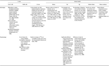
The broiler chicken was used as a model to study iron bioavailability with a unique duodenal loop technique for direct measurement of intestinal iron absorption. One-week-old chicks were allocated into iron-deficient v. iron-adequate treatment groups for 6 weeks. At week 7, birds were anesthetized and their duodenal loops were exposed. The loop was isolated and a nonocclusive catheter was inserted into the duodenal vein for blood sampling. A stable isotope solution containing 58Fe was injected into the loop. Blood samples were collected every 5 min for 120 min postinjection for measurement of 58Fe concentrations. Tako et al. (Reference Tako, Rutzke and Glahn2010) evaluated expression of proteins involved in Fe uptake and transfer as DMT1, FPN1 and Dcytb as an indicator of iron bioavailability. Table 1 summarizes the main advantages and disadvantages of the models studied.
Main advantages and disadvantages of the models

Conclusions
Many experimental models have been used since the past century in studies related to iron, most of which are still currently in use. In vitro models are usually simple, easy to handle and the results are obtained quickly with lower cost. These are some advantages in relation to studies in animal or humans. On the other hand, we have to consider the ethical aspect and only use animal and human studies when it is necessary.
Isotopic techniques are highly recommended in studies related to iron, but these are difficult in many routines worldwide because the experimental design requires a lot of special logistic conditions which are currently not feasible in many developing countries. Knowing about the main advantages and disadvantages of the in vitro and animal models used in studies related to iron is crucial to the researcher in the field of nutritional studies.