Introduction
The osteoglossiform fish Arapaima gigas (Schinz) (Arapaimidae), endemic to the Amazon Basin, has an important economic value due to the high quality of the flesh and valuable by-products in terms of scales and skin. It is actually considered an endangered species and attempts to culture it have been increasing to preserve the species and provide sustainable development. Studies on the parasites of arapaimas are therefore important for the successful culture of this fish, and the identification of disease-causing parasites is essential to plan a specific form of control.
Our research, carried within the auspices of the Pirarucu Management Project on Mexiana Island in the Amazon River Delta, Brazil, indicates that arapaimas are currently parasitized by two species of blood-feeding, tissue-dwelling philometrid nematodes, Nilonema senticosum (Baylis, 1922) and Rumai rumai Travassos, Reference Travassos1960 (Moravec, Reference Moravec1998; Thatcher, Reference Thatcher2006). In previous publications, we have studied N. senticosum obtained from arapaimas in Peru (Moravec et al., Reference Moravec, Scholz, Kuchta, Dyková and Posel2006) and Brazil (Santos & Gibson, Reference Santos and Gibson2007). In this study, we present a redescription of R. rumai, with additional data on N. senticosum, and comments on their relationship with the aquaculture system of Mexiana Island.
Materials and methods
Investigations into the parasites of 33 specimens of arapaimas, local name pirarucu, were carried out on fingerlings, elvers and adults (6.4–175 cm long) cultured in breeding tanks and channels at Fazenda Santo Ambrósio, Mexiana Island, Amazon River Delta (00°05′30″S, 49°34′50″W) (Pirarucu Management Project – License IBAMA 005-2007).
The nematodes were mechanically removed from capsules embedded in the tissues of the operculum, mouth cavity, tongue and head, using needles. They were washed in physiological saline and then fixed in hot 4% formaldehyde solution. For light microscopy, the nematodes were cleared with glycerine. Drawings were made with the aid of a Zeiss drawing attachment. After examination, the specimens were stored in 70% ethanol. All measurements are in micrometres unless otherwise stated. Fish names follow FishBase (Froese & Pauly, Reference Froese and Pauly2008).
For scanning electron microscopy (SEM), fixed specimens were postfixed in 1% osmium tetroxide in phosphate buffer, dehydrated through a graded acetone series, critical-point dried and sputter-coated with gold. The samples were examined using a JEOL ISM-7401F scanning electron microscope at an accelerating voltage of 4 or 8 kV.
For confocal laser scanning microscopy (CLSM) studies, the specimens fixed in 4% paraformaldehyde were incubated overnight with faloidin conjugated to fluorescein isothiocyanate (FITC) 1:700 in phosphate-buffered saline (PBS) (Sigma-Aldrich, Sāo Paulo, Brazil) and mounted on semi-permanent slides in PBS with 2.5% 1,4-diazabicyclo-(2,2,2)-octane-triethylenediamine and 50% glycerine at pH 7.2. Studies were performed using an Olympus BX51 with Fluoview version 3.2.
Specimens are deposited in the Helminthological Collection of the Instituto Oswaldo Cruz (CHIOC), Rio de Janeiro, Brazil (N. senticosum No. 35622 and R. rumai No. 35609) and the Helminthological Collection of the Institute of Parasitology, BC ASCR, in České Budějovice, Czech Republic (N. senticosum Cat. No. N-852 and R. rumai Cat. No. N-907).
Results
Rumai rumai Travassos, Reference Travassos1960
It is worth noting that, although we had examined all sizes of fish (6.5–179 cm long) and had carefully inspected every small slice of tissue throughout the body, not a single male was found, only females. Therefore, the male of R. rumai remains unknown.
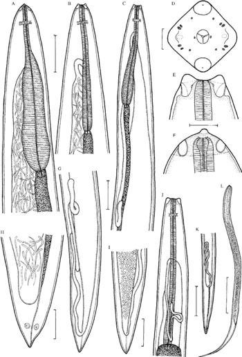
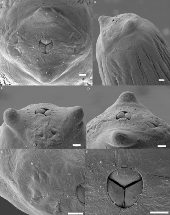
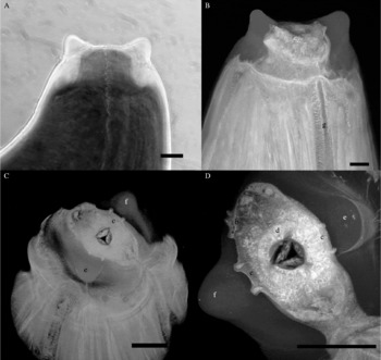
Redescription ( figs 1–3). Female (five gravid specimens; measurements of two non-gravid females in parentheses). Body of live gravid specimens red-coloured (yellowish after fixation), that of non-gravid females whitish, with smooth cuticle; anterior end blunt, posterior end conical; anterior part of body somewhat broader than posterior part. Lateral fields of thinner cuticle extend along body (see fig. 3B). Body length 52.22–106.94 (6.01–11.00) mm; maximum width at oesophageal region 816–952 (258–299); maximum width/length ratio of body 1:62–216 (1:23–37). Anterior end of body truncated in lateral view, rounded in dorsoventral view and almost square-shaped in apical view, bearing two conspicuously large (one dorsal and one ventral), horn-like, anterior projections 18–41 (15–18) high (figs 1D–F, 2A–D and 3A and B). Oral aperture large, oval to almost circular, surrounded by 14 small cephalic papillae arranged in two circles and pair of lateral amphids; eight elongate papillae of the external circle form two dorsolateral and two ventrolateral pairs; the internal circle consists of four small, submedian single papillae and two small, lateral papillae situated near the oral aperture (figs 1D and 2A–F). Peribuccal tissue, including both horn-like projections, appears to be sclerotized, forming a kind of cephalic shield connected to a broad, sclerotized ring encircling the anterior end of the oesophagus with contiguous circum-oesophageal tissue (figs. 1E and F and 3A–D). This whitish, hyaline sclerotized formation is broadest dorsally and ventrally, and narrowest laterally, resulting in a laterally elongate, oval shape of circum-oesophageal tissue in apical view (fig. 3C and D); length of cephalic sclerotized formation (including projections) 82–109 (48–54). Three oesophageal lobes not protruding from the mouth form flat surfaces, each bearing a slightly outlined narrow, transverse keel at the top (figs 1D and 2A–D and F). The oesophagus is relatively short, 2.00–2.69 (1.16–1.67) mm long (representing 2.2–3.8 (15.2–19.2)% of body length), without anterior inflation, consisting of a shorter and narrower anterior muscular portion and a much longer and wider posterior muscular–glandular portion which appears to be darker; muscular oesophagus 612–923 (381–476) long, with maximum width 54–68 (54); muscular–glandular oesophagus 1.36–1.77 (0.78–1.20) mm long, with maximum width 150–408 (68–82) (fig. 1A–C and J). Nerve ring encircles the muscular oesophagus 286–408 (222–258) from anterior extremity. Ventriculus absent. Oesophagus opens into intestine through a valve. Intestine light-coloured, relatively narrow anteriorly in gravid specimens and broad in non-gravid specimens; posterior end of intestine atrophied, forming narrow ligament attached to body wall near posterior extremity (fig. 1G–I and K). Poorly developed rectal glands present (fig. 1H). Posterior end of body conical, with rounded tip, lacking caudal projections (fig. 1G–I and K). Phasmids observed only in smallest, non-gravid specimen (fig. 1K). Ovaries narrow, long, reflexed, situated near anterior and posterior ends of body, with anterior ovary reaching anteriorly nearly to level of nerve ring, posterior ovary extending posteriorly to short distance from posterior end of body (fig. 1C, G, J and K). Uterus amphidelphic, in gravid specimens occupying major part of body, being filled with first-stage larvae, developing embryos and eggs; uterus of non-gravid specimens narrow, empty. Vagina and vulva absent. Anterior end of larvae blunt, with small dorsal larval tooth; posterior end sharply pointed (fig. 1L); larvae 528–546 long (n = 5), with maximum width 15–18; length of oesophagus 99–126 (18–23% of body length), of tail 114–126 (21–23%).
Rumai rumai Travassos, Reference Travassos1960, female. A, B, anterior end of gravid female (different specimens), dorsoventral and lateral views; C, anterior part of body of gravid female, lateral view; D, E, F, cephalic end of gravid female, apical, lateral and dorsoventral views, respectively; G, posterior part of body of gravid female, lateral view; H, I, caudal end of gravid female, lateral and dorsoventral views; J, K, anterior and posterior ends of smallest non-gravid female, lateral and dorsoventral views; L, larva from uterus. Scale bars: 500 μm (A–C, G, I), 30 μm (D), 100 μm (E, F, L), 200 μm (H, J, K).

Rumai rumai Travassos, Reference Travassos1960, scanning electron micrographs of cephalic end of gravid female. A, B, apical and subapical views; C, D, lateral and dorsoventral views; E, detail of lateral part of cephalic end, apical view; F, region of mouth opening, apical view. Abbreviations: a, amphid; b, submedian pair of cephalic papillae of external circle; c, submedian cephalic papilla of internal circle; d, lateral cephalic papilla of internal circle; e, dorsal horn-like cephalic protrusion; f, ventral horn-like cephalic protrusion; g, mouth with surfaces of three oesophageal lobes. Scale bars: 10 μm (A–F).

Rumai rumai Travassos, Reference Travassos1960, cephalic end of gravid female. A, light microscopy of cephalic end, lateral view; B–D, confocal laser scanning microscopy of cephalic end, lateral, subapical and apical views, respectively. Abbreviations: b, pair of submedian cephalic papillae of external circle; c, pair of submedian cephalic papillae of internal circle; d, lateral cephalic papilla of internal circle; e, dorsal horn-like cephalic protrusion; f, ventral horn-like cephalic protrusion; g, lateral field. Scale bars: 10 μm (A); 50 μm (B–D).

Male: unknown.
Host. Arapaima, Arapaima gigas (Arapaimidae, Osteoglossiformes).
Sites of infection. Encapsulated in tissues of mouth, tongue, operculum and head.
Locality. Fazenda Santo Ambrosio (00°05′30″S, 49°34′50″W), Mexiana Island (Amazon River Delta), Pará State, Brazil.
Prevalence and intensity. 21% (7 fish infected/33 fish examined); 2–44 nematodes per infected fish.
Nilonema senticosum (Baylis, 1922)
Host. Arapaima, Arapaima gigas (Arapaimidae, Osteoglossiformes).
Site of infection. Swimbladder.
Locality. Fazenda Santo Ambrosio (00°05′30″S, 49°34′50″W), Mexiana Island (Amazon River Delta), Pará State, Brazil.
Prevalence and intensity. 24% (8 fish infected/33 fish examined); 3–53 nematodes per infected fish.
Aquaculture data
Parasites negatively influence the health condition of their fish hosts, and the most pathogenic for the tank-reared arapaimas at Mexiana Island are apparently the gill-parasitic monogeneans Dawestrema spp., the anisakid nematode Goezia spinulosa (Diesing, 1939) and the two philometrid tissue-dwelling nematodes R. rumai and N. senticosum. Young cultured fish of the size-group c. 20 cm long are the most vulnerable to these parasites in terms of the high mortality observed.
Neither R. rumai nor N. senticosum were observed in arapaimas 6.4–15 cm long (n = 17). Infections by R. rumai were visible in fish 20–120 cm long (n = 7) and those of N. senticosum occurred in fish 20–175 cm long (n = 8). Simultaneous infections of both species occurred in four fish 70–88 cm long. The highest intensity of 44 R. rumai occurred in a fish 20 cm long and 53 N. senticosum were collected from a fish 71 cm long. The largest fish harbouring R. rumai was 120 cm long which had just four young nematode females; and 11 N. senticosum were collected from the largest infected arapaima of 175 cm in length.
Discussion
Travassos (Reference Travassos1960) described a new dracunculoid genus and species, Rumai rumai Travassos, Reference Travassos1960, based on a single female specimen recovered from the body cavity of Arapaima gigas in the Amazon River near Maicuru, Pará State, Brazil. This species has not been recorded since. This author assigned Rumai to the Philometridae, distinguishing it from other genera of this family by its remarkable cephalic structure. Although the original description of R. rumai is inadequate, subsequent authors (e.g. Rasheed, Reference Rasheed1963; Ivashkin et al., Reference Ivashkin, Sobolev and Khromova1971; Chabaud, Reference Chabaud, Anderson, Chabaud and Willmott1975; Moravec, Reference Moravec2006) recognized Rumai as a valid genus, highlighting the unusual structure of the cephalic end and a conical caudal extremity in the female.
A detailed examination of the numerous specimens comprising the present material, as well as of the holotype of R. rumai (CHIOC. No. 26636), confirmed their conspecificity and made it possible to redescribe in detail the morphology of this nematode species. Travassos (Reference Travassos1960) reported the body length of his specimen to be about 40 mm (our re-examination revealed that this is a stained gravid female permanently mounted on a slide in Canada balsam), but the body length of gravid females of this species can be more than double this, as indicated by the present results. Travassos (Reference Travassos1960) did not study the cephalic end in apical view, so his description of the cephalic structures is incomplete and somewhat misleading. He reported the cephalic end with a cuticular formation appearing pyramidal in lateral view and as slightly medially depressed in dorsoventral view; in fact, it is quite the opposite (see figs 1D–F and 2A–D), because the horn-like protrusions are dorsal and ventral. He reported only four cephalic papillae, but our SEM examination confirmed the presence of a total of 14 papillae arranged in two circles, typical of many philometrid species.
Females of some philometrids (mainly those parasitizing fish subcutaneous tissues), Philometra rischta Skryabin, 1923, for example, have at the cephalic end four large, strongly elevated, rounded, subapical, submedian projections regularly arranged around the mouth, each with two small papillae (Moravec, Reference Moravec2006; Sokolov & Kazakov, Reference Sokolov and Kazakov2007). In contrast, the two cephalic projections of R. rumai are located either dorsally or ventrally and seem to be fully sclerotized and deprived of any papillae. Similar dorsal and ventral cephalic projections are found in species of Dracunculus Reichard, 1759 (Dracunculidae) and Kamegainema Hasegawa, Doi, Araki & Miyata, 2000 (Micropleuridae), but they still retain double nerve endings; similarly to Rumai, representatives of the former genus may also have a sclerotized cephalic shield and a sclerotized support around the anterior end of the oesophagus (Moravec, Reference Moravec2006). The orientation of the square-shaped anterior end of R. rumai, with the corners situated dorsally, ventrally and laterally, is quite unique among the dracunculoids; in species of Philometra Costa, 1845, which also have a square-shaped anterior end, the corners are always submedian.
Travassos (Reference Travassos1960) illustrated the posterior portion of the oesophagus of R. rumai as markedly expanded; the present study shows that the width of the oesophagus is associated with the body size of the worms. In gravid females, the posterior part of the oesophagus varies from slightly expanded to very expanded (see fig. 1A and B). By the shape and structure, the oesophagus of R. rumai is more similar to species of Philonema Kuitunen-Ekbaum, 1933 and Micropleura Linstow, 1906 (both Micropleuridae) than to philometrids. The conical shape of the female caudal end in R. rumai is also a unique feature within the Philometridae (sensu Moravec, Reference Moravec2006).
The present study confirms that R. rumai, the type species of a monotypic genus, exhibits some taxonomically important features distinguishing it markedly from other philometrids and justifying the recognition of Rumai as a valid genus. Although, for the time being, Rumai is retained within the Philometridae, some morphological characters, such as the sclerotized formation of the female cephalic end, the presence of dorsal and ventral cephalic projections, the shape and structure of the oesophagus and the conical caudal extremity, indicate certain affinities with some representatives of the dracunculoid families Dracunculidae (Dracunculus) and Micropleuridae (Micropleura, Philonema). The actual phylogenetic position of Rumai can be only established when the hitherto unknown male of R. rumai is discovered and described, and the species is examined by molecular methods.
Both R. rumai and N. senticosum are blood-feeders, and thus likely weaken their fish hosts. Furthermore, the capsules containing R. rumai, located inside the mouth and under the tongue, appear to affect fish feeding. Elvers parasitized likely lose weight and, due to their poor health condition, they tend to die.
Among dracunculoid nematodes, the females are known to have a great number of first-stage larvae which are liberated into the aquatic environment when the female extrudes part of her body into the water, where it bursts. In the philometrid two-host life cycle, the released first-stage larvae are quickly eaten by copepods and moult twice in their haemocoel to become infective third-stage larvae (Moravec, Reference Moravec2006). In preliminary experiments, local unidentified cyclopid copepods were successfully infected with first-stage larvae of N. senticosum and R. rumai. This indicates that these crustaceans may play the role of intermediate hosts of both these nematode species.
In order to control parasitism by R. rumai and N. senticosum in the culture tanks of Mexiana Island, the live food supply for arapaima fingerlings and elvers, which is mainly composed of plankton, is now cooked. This simple technique, used to reduce the number of viable larvae in copepods, has visibly reduced the mortality of these fish in culture.
Acknowledgements
We would like to thank Dr Luiz Rabelo Neto from Grupo Reicon and Dr Rossana Venturieri for the facilities and support provided during the fieldwork. Dr Dely Noronha and Dr Luis Muniz, from the Helminthological Collection of the Instituto Oswaldo Cruz (IOC), Rio de Janeiro, kindly provided the holotype of Rumai rumai for study. We are grateful to Dr Marcelo Pelajo, Pedro Paulo de Abreu Manso and Bernardo Pascarelli, from Instituto Oswaldo Cruz, for providing the Laser Scanning Microscopic facilities. We also acknowledge the staff of the Laboratory of Electron Microscopy, Biology Centre, ASCR, České Budějovice, for technical help. This study was partly supported by the grant 524/06/0170 from the Grant Agency of the Czech Republic and by research projects of the Institute of Parasitology, BC ASCR (Z60220518 and LC522), the Fundação de Amparo à Pesquisa do Estado do Rio de Janeiro and PAPESV/FIOCRUZ/CNPq. We also thank Dr David Gibson, Natural History Museum, London for revising the English.