Introduction
In vitro production techniques have proven valuable in basic research and provide an inexpensive and readily available source of preimplantation bovine embryos (Sagirkaya et al., 2006). The production of bovine embryos in the laboratory includes three major steps: oocyte maturation, fertilization and embryo culture. While the culture environment after fertilization seems to be the most important factor in determining blastocyst quality, maturation of oocytes with a high degree of competence is also essential to achieve a high yield of blastocyst formation (Russell et al., Reference Russell, Baqir, Bordignon and Betts2006). Oocyte maturation is not only related to the resumption of meiosis (nuclear maturation), but also to the acquisition of developmental competence (cytoplasmic maturation) for postfertilization embryo development (Ikeda et al., Reference Ikeda, Kitagawa, Imai and Yamada2005). However, despite worldwide efforts by embryo producers to improve embryo culture systems, oocytes that have been matured in vitro are still less developmentally competent than those matured in vivo (Bordignon et al., Reference Bordignon, Morin, Durocher, Bousquet and Smith1997; van de Leemput et al., Reference van de Leemput, Vos, Zeinstra, Bevers, van der Weijden and Dieleman1999; Rizos et al., Reference Rizos, Ward, Duffy, Boland and Lonergan2002).
Neurotrophins and their receptors play an essential role in the differentiation and survival of neurons in the central and peripheral nervous systems (Jones et al., Reference Jones, Farinas, Backus and Reichardt1994). Their actions, however, do not appear to be limited to the nervous system (Feng et al., Reference Feng, Chen, Shelden and Seifer2003). There is increasing evidence for a role for neurotrophins in ovarian development and function, including oocyte maturation (Martins da Silva et al., Reference Martins da Silva, Gardner, Taylor, Springbett, De Sousa and Anderson2005). Neurotrophins include nerve growth factor (NGF), brain-derived neurotrophic factor (BDNF), and neurotrophins 3 and 4/5 (NT3 and NT4/5). They utilize two different classes of transmembrane receptor proteins, the tyrosine kinase (Trk) receptor and the neurotrophin receptor, p75. The p75 receptor is widely expressed and binds all neurotrophins. By contrast, the Trk receptors show selective affinity for different neurotrophins. NGF is the preferred ligand for TrkA, BDNF and NT4/5 are the preferred ligands for TrkB, and NT3 is the preferred ligand for TrkC (Barbacid, Reference Barbacid1994).
In many studies, it has been suggested that BDNF and its receptor TrkB have various ovarian functions including supporting ovarian follicular development (Paredes et al., Reference Paredes, Romero, Dissen, DeChiara, Reichardt, Cornea, Ojeda and Xu2004), initiation of primordial follicle growth (Spears et al., Reference Spears, Molinek, Robinson, Fulton, Cameron, Shimoda, Telfer, Anderson and Price2003; Harel et al., Reference Harel, Jin, Fisch, Feldberg, Krissi, Felz, Freimann, Tan, Ao and Abir2006), steroid metabolism (Chen et al., Reference Chen, Chen and Xing2004), oocyte maturation (Seifer et al., Reference Seifer, Feng, Shelden, Chen and Dreyfus2002; Kawamura et al., Reference Kawamura, Kawamura, Mulders, Gelpke and Hsueh2005; Martins da Silva et al., Reference Martins da Silva, Gardner, Taylor, Springbett, De Sousa and Anderson2005) and early embryo development (Kawamura et al., Reference Kawamura, Kawamura, Mulders, Gelpke and Hsueh2005; Martins da Silva et al., Reference Martins da Silva, Gardner, Taylor, Springbett, De Sousa and Anderson2005). Mice carrying a null mutation of the NGF gene are deficient in the development of primordial follicles (Dissen et al., Reference Dissen, Romero, Hirshfield and Ojeda2001). In another mouse study, treatment of cultured oocytes with BDNF enhanced first polar body extrusion and increased the competence of oocytes to complete preimplantation development (Kawamura et al., Reference Kawamura, Kawamura, Mulders, Gelpke and Hsueh2005). In cattle, however, BDNF alone was insufficient to enhance nuclear maturation of oocytes (Martins da Silva et al., Reference Martins da Silva, Gardner, Taylor, Springbett, De Sousa and Anderson2005).
Metformin (N,N′–dimethylbiguanide) has been used for many years in treating insulin resistance in type 2 diabetes and during the last decade it has also been used for ovulation induction in polycystic ovary syndrome (Harborne et al., Reference Harborne, Fleming, Lyall, Norman and Sattar2003; Lilja and Mathiesen, Reference Lilja and Mathiesen2006). Metformin stimulates insulin action by increasing insulin-stimulated glucose uptake in rat adipocytes (Matthaei et al., Reference Matthaei, Hamann, Klein, Benecke, Kreymann, Flier and Greten1991) and elevating receptor tyrosine kinase activity and inositol 1,4,5-trisphosphate production in Xenopus oocytes (Stith et al., Reference Stith, Goalstone, Espinoza, Mossel, Roberts and Wiernsperger1996). Other reports have demonstrated that metformin is able to regulate ovarian glutathione (GSH) content (Elia et al., Reference Elia, Sander, Luchetti, Solano, Di Girolamo, Gonzalez and Motta2006) and has a direct effect on ovarian steroidogenesis in humans (Mansfield et al., Reference Mansfield, Galea, Brincat, Hole and Mason2003). Recently, it was suggested that metformin enhanced the action of insulin in porcine oocytes and embryo by elevating the activity of the insulin receptor tyrosine kinase (Lee et al., Reference Lee, Kang, Lee and Hwang2005).
Accordingly, it can be hypothesized that the combination of BDNF and metformin has beneficial effects on the maturation of bovine oocytes. Thus, the aim of this study was to evaluate the effect of BDNF on the developmental potential of bovine in vitro fertilized (IVF) embryos when added during in vitro maturation (IVM) and/or in vitro culture (IVC). Furthermore, we investigated the possible collaborative effect of BDNF and metformin on nuclear and cytoplasmic maturation.
Materials and Methods
In vitro maturation of oocytes
Bovine ovaries were obtained from a local abattoir and transported to the laboratory within 2 h in 0.9% (v/v) NaCl solution at 30–35°C. Follicular contents from small antral follicles, 2–8 mm in diameter, were aspirated using an 18-gauge needle attached to a 10 ml disposable syringe. Cumulus–oocytes complexes (COCs) with evenly granulated cytoplasm and enclosed by more than three layers of compact cumulus cells were selected and washed three times in HEPES-buffered tissue culture medium-199 (TCM-199; Invitrogen) supplemented with 2 mM NaHCO3 (Sigma-Aldrich Corp.), 5 mg/ml BSA (Invitrogen), and a 1% (v/v) mixture of penicillin and streptomycin (Sigma-Aldrich Corp.). Groups of 30 to 40 COCs were cultured in 500 μl drops of bicarbonate-buffered TCM-199 supplemented with 10% (v/v) FBS, 0.005 IU/ml FSH (Antrin) and 1 μg/ml 17β–estradiol (Sigma-Aldrich Corp.) at 39°C, 5% CO2 in air.
Evaluation of the first polar body extrusion
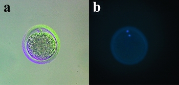
For evaluating the transition from metaphase I (MI) stage to metaphase II (MII) stage oocytes, COCs were stripped and processed for staining. After 22 h of maturation culture, cumulus cells were removed from oocytes by pipetting in 0.1% (v/v) hyaluronidase in HEPES-buffered TCM-199 and then the oocytes were stained with 5 μg/ml bisbenzimide (Hoechst 33342) for 5 min. The occurrence of first polar body extrusion in the oocyte was examined under an inverted microscope equipped with epifluorescence at ×200 magnification (Fig. 1).
Representative micrographs of in vitro matured oocytes. At 22 h after IVM, maturation was assessed by polar body extrusion (a, arrow) and nuclear staining (b), ×200.

Assessment of glutathione concentration
The GSH content of oocytes was measured by following the instructions of the Glutathione Assay Kit (Cat. No. 703002; Cayman). For sample preparation, cumulus cells were removed from the oocytes by pipetting in 0.1% (v/v) hyaluronidase. Then 30–40 oocytes were placed in a 1.5 ml Eppendorf tube and stored at −20°C until analysis. For deproteination of the sample, 5 μl of metaphosphoric acid (Sigma-Aldrich Corp.) was added. Then 50 μl of standards and samples were added onto the 96-well plate. The assay cocktail was prepared by mixing the following reagents in a 20 ml vial: MES buffer (11.25 ml), reconstituted cofactor mixture (0.45 ml), reconstituted enzyme mixture (2.1 ml), water (2.3 ml) and reconstituted DTNB (0.45 ml). A volume of 150 μl of freshly prepared assay cocktail was added to each well. The GSH level was evaluated using optical density levels measured with a microplate reader (Cat. No. A-5002; Tecan Systems Inc.).
In vitro fertilization and in vitro culture
After 22 h of IVM, bovine embryos were produced by IVF and cultured as previously described (Fukui, Reference Fukui1990; Fukui et al., Reference Fukui, Sonoyama, Mochizuki and Ono1990). Frozen–thawed spermatozoa were subjected to a swim-up technique and capacitated for 1 h, using Tyrode's albumin lactate pyruvate (TALP). The pooled supernatant medium containing motile spermatozoa was centrifuged three times at 500 g for 6 min and the sperm pellet was treated with 100 μg/ml heparin for 15 min at 39°C in a humidified atmosphere of 5% CO2 and 95% air. The COCs were washed three times in HEPES-buffered TALP washing buffer and washed once in IVF-TALP. Groups of six to seven oocytes (3 μl) were transferred into droplets (43 μl) of IVF-TALP overlaid with mineral oil (Sigma-Aldrich Co.) and inseminated at approximately 1 × 10⁶ spermatozoa/ml at 39°C in a humidified atmosphere of 5% CO2 in air. After 18–20 h, groups of six to seven presumptive zygotes were washed and cultured in 25 μl microdrops of the control modified synthetic oviductal fluid under mineral oil for 7 days at 39°C under humidified 5% CO2, 5% O2 and 90% N2 (Jang et al., Reference Jang, Bhuiyan, Jeon, Ko, Park, Kim, Kim, Kang, Lee and Hwang2006). The development of embryos was monitored and recorded at 2 and 7 days for cleavage and blastocyst formation, respectively.
Experimental design
In Experiment 1, to determine the effect of various concentrations of BDNF on the developmental potential of the oocytes and embryos in our previously established system (Cho et al., Reference Cho, Lee, Park, Lim, Shin, Kim, Lee and Hwang2002; Choi et al., Reference Choi, Lee, Lim, Kang and Hwang2002; Jang et al., Reference Jang, Bhuiyan, Jeon, Ko, Park, Kim, Kim, Kang, Lee and Hwang2006), we added 0, 10 or 100 ng/ml of recombinant human BDNF (Cat. No. B3795; Sigma-Aldrich Co.) to the IVM medium. After 22 h, the number of oocytes with a first polar body was recorded (Fig. 1). Moreover, in order to investigate the effective stage of BDNF supplementation, 10 ng/ml BDNF was added during the maturation and/or embryo culture period after in vitro fertilization.
Experiment 2 is an extension of Experiment 1 in that the effect of BDNF and metformin on IVM was further investigated. The control maturation medium was TCM-199 without serum and hormones, and experimental treatments were BDNF (10 ng/ml), metformin (10−5 M, Cat. No. D5035; Sigma-Aldrich Co.) (Lee et al., Reference Lee, Kang, Lee and Hwang2005) or a combination of BDNF and metformin. After 22 h of maturation culture, the rate of first polar body extrusion (Fig. 1) and the concentration of GSH were measured. Subsequently, the embryo developmental stage was monitored and recorded at 2 and 7 days.
Statistical analysis
Oocytes and IVF embryos were randomly distributed within each experimental group and the experiments were repeated at least three times. All data were subjected to analysis of variance (ANOVA) and to a protected least significant difference (LSD) test using general linear models (GLM) in a statistical analysis system (SAS) program to determine differences among experimental groups. When a significant treatment effect was found for an experimental parameter, data were compared by the least squares method. A p-value of less than 0.05 was considered statistically significant.
Results
Effect of BDNF on nuclear maturation of bovine oocytes
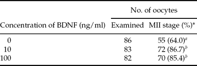
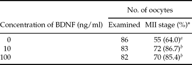
The effect of BDNF on nuclear maturation of bovine oocytes was examined. As shown in Table 1, supplementing the IVM medium with BDNF significantly increased the rate of MII oocytes at both 10 ng/ml (86.7%) and 100 ng/ml (85.4%) BDNF, when compared with the control (64.0%). No significant difference was observed between the two concentrations of BDNF.
Effect of supplementing brain-derived neurotrophic factor (BDNF) on maturation of bovine oocytes to metaphase II (MII) in a hormone- and serum-supplemented in vitro maturation medium.
*Percentage of oocytes examined.
a ,b Values for different superscripts are significantly different (p < 0.05).
Effect of BDNF supplement during IVM and/or IVC on developmental competence of bovine oocytes after in vitro fertilization
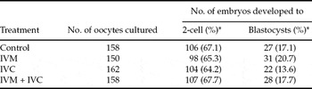
BDNF was added during IVM and/or IVC and the effects on developmental competence of bovine oocytes after in vitro fertilization were examined. Table 2 shows that supplementation with BDNF to our previously established IVM and/or IVC medium system did not show beneficial effect on the developmental competence of embryos. There were no significant differences in developmental competence among the experimental groups.
Effect of supplementing hormone- and serum-supplemented in vitro maturation (IVM) medium, in vitro culture (IVC) medium or both media with brain-derived neurotrophic factor (BDNF) on developmental potential of bovine oocytes after in vitro fertilization.

*Percentage of oocytes cultured.
The experimental concentration of BDNF was 10 ng/ml.
There were no statistically significant differences between the groups (p > 0.05).
Effects of adding BDNF and metformin per se during IVM
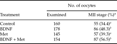
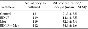
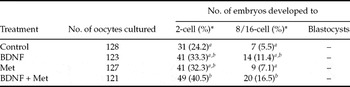
The effects of BDNF and metformin on nuclear maturation of bovine oocytes were examined. Both BDNF alone and the combination of BDNF and metformin significantly increased the rate of maturation of bovine oocytes to MII (48.3% and 56.5%). There was no significant effect from metformin alone on nuclear maturation in comparison with the control group (39.3% vs. 34.4%, Table 3). With regard to cytoplasmic maturation, the experimental groups (BDNF 14.6 ± 7.5, Met 32.0 ± 5.4, and BDNF + Met 34.9 ± 4.6 pmol, respectively) showed similar concentration of GSH when compared with the control group (21.3 ± 3.5 pmol, Table 4). The combination of BDNF and metformin significantly increased the rate of early development to 8–16-cell embryos (16.5%). There was no significant effect of BDNF (11.4%) or metformin alone (7.1%) on embryo development when compared with the control (5.5%, Table 5).
Effect of supplementing brain-derived neurotrophic factor (BDNF) and/or metformin (Met) on maturation of bovine oocytes to metaphase II (MII) in a chemically defined medium.
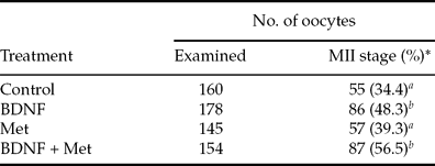
The experimental concentration of BDNF was 10 ng/ml and Met was 10−5 M.
*Percentage of oocytes examined.
a ,b Values for different superscripts are significantly different (p < 0.05).
Effect of supplementing brain-derived neurotrophic factor (BDNF) and/or metformin (Met) on glutathione (GSH) concentration of bovine oocytes in a chemically defined medium.

*The value was measured by Y = 2E−0.05X + 0.119, pmol.
The experimental concentration of BDNF was 10 ng/ml and Met was 10−5 M.
There were no statistically significant differences between the groups (p > 0.05).
Effect of supplementing a chemically defined in vitro maturation medium with brain-derived neurotrophic factor (BDNF) and/or metformin (Met) on developmental competence of bovine oocytes after in vitro fertilization.

The experimental concentration of BDNF was 10 ng/ml and Met was 10−5 M.
*Percentage of oocytes cultured.
a ,b Values for different superscripts in the same column are significantly different (p < 0.05).
Discussion
In the present study, we examined the effects of BDNF on different IVM media and the co-effects of BDNF and metformin. We demonstrated that BDNF at 10 or 100 ng/ml had a significant effect on first polar body extrusion, without a significant difference between the two concentrations (Table 1). In addition, BDNF promoted nuclear maturation of oocytes compared with our previously established IVM system (Cho et al., Reference Cho, Lee, Park, Lim, Shin, Kim, Lee and Hwang2002; Choi et al., Reference Choi, Lee, Lim, Kang and Hwang2002; Jang et al., Reference Jang, Bhuiyan, Jeon, Ko, Park, Kim, Kim, Kang, Lee and Hwang2006). Our results are consistent with previous studies, in which BDNF was shown to be an essential factor for first polar body extrusion as well as for cytoplasmic maturation (Seifer et al., Reference Seifer, Feng, Shelden, Chen and Dreyfus2002; Kawamura et al., Reference Kawamura, Kawamura, Mulders, Gelpke and Hsueh2005). In contrast, other researchers demonstrated that BDNF signalling is not associated with nuclear maturation in bovine oocytes (Martins da Silva et al., Reference Martins da Silva, Gardner, Taylor, Springbett, De Sousa and Anderson2005). This discrepancy in the effects of BDNF on oocyte maturation can be explained by species differences or by different maturation media.
In order to confirm the effect of BDNF on the developmental competence of bovine in vitro produced embryos, BDNF was added during IVM and/or IVC. In our study, there was no significant difference on developmental competence among the groups, suggesting BDNF treatment during IVM and IVC does not show any beneficial effects (Table 2). The insignificant effect of BDNF on the developmental potential of oocytes in this study may be due to the process of oocyte activation. In the present study, matured oocytes were inseminated with frozen–thawed semen, while in the previous study, which reported that BDNF-supplemented IVM medium significantly increased bovine blastocyst formation (Martins da Silva et al., Reference Martins da Silva, Gardner, Taylor, Springbett, De Sousa and Anderson2005), parthenogenetic activation was used.
TrkB, the specific receptor for BDNF, is a subgroup of receptor tyrosine kinase and has a tyrosine kinase domain (which phosphorylates proteins on tyrosine residues), a hormone binding domain, and a carboxyl terminal segment with multiple tyrosines for autophosphorylation. Because BDNF alone was insufficient to increase the developmental competence of oocytes compared with the control group (Table 2), metformin, the tyrosine kinase activator, was selected to enhance the action of BDNF. In Experiment 2, in order to investigate the effect of BDNF and/or metformin per se, we used chemically defined, serum-free media (TCM-199) without hormones and growth factors. The effective dose of BDNF was determined in Experiment 1 and the experimental concentration of metformin was selected according to a previous report (Lee et al., Reference Lee, Kang, Lee and Hwang2005). Overall, the use of chemically defined TCM-199 resulted in a lower competence in IVM and IVC in Experiment 2, compared with Experiment 1. This difference could be due to the lack of serum, hormones and growth factors that are necessary for oocyte maturation and development in chemically defined TCM. As a result, both BDNF alone and the combination of BDNF and metformin significantly increased the rate of oocyte maturation to MII (48.3 and 56.5%) compared with the control (34.4%, Table 3). Then, in order to further investigate the role of metformin, we analysed GSH levels, which play a critical role in the cytoplasmic maturation of oocytes. Unfortunately, however, the GSH concentration of oocytes did not increase when BDNF and metformin were supplemented during IVM (Table 4). Although there were no significant differences in GSH concentration, these results suggest that the combination of BDNF and metformin has a beneficial effect on the nuclear maturation of oocytes, which is one of the important factors for early embryo development. Indeed, in vitro matured oocytes cultured in medium supplemented with BDNF and metformin showed a significantly higher enhancement of the early embryonic developmental competence (8–16 cell) compared with the control group (16.5 vs. 5.5%; Table 5). Due to lack of effect, metformin alone did not improve oocyte maturation.
Taken together, the present study provides the first evidence that the combination of BDNF and metformin has a collaborative effect on bovine IVM. These results could contribute to the development of a more efficient bovine in vitro production system.
Acknowledgements
We acknowledge Dr Barry D. Bavister for his valuable assistance in the editing of this manuscript. This study was financially supported by the Korean MEST, through KOSEF (grant # M10625030005–08N250300510) and the BK21 program for Veterinary Science.