I. INTRODUCTION
Tissue engineering is one of the most important approaches to tissue replacement and organ repair including bone repair, Reference Praemer, Furner and Rice1,Reference Einhorn2 eye transplantation, Reference Keivyon and Tseng3 and heart transplantation. Reference Trulock, Christie, Edwards, Boucek, Aurora, Taylor, Dobbels, Rahmel, Keck and Hertz4 In tissue engineering, bioreactors can play a key role is the expansion of stem cell populations required for repair and also cell populations required for cell therapies.
The hematopoietic stem cells (HSCs) have been unquestionably important to therapies that involve blood and immune system replacement, including myeloma Reference Bladé, Samson, Reece, Apperley, Björkstrand, Gahrton, Gertz, Giralt, Jagannath and Vesole5 and leukemia. Reference Pavletic, Khouri, Haagenson, King, Bierman, Bishop, Carston, Giralt, Molina, Copelan, Ringdén, Roy, Ballen, Adkins, McCarthy, Weisdorf, Montserrat and Anasetti6 HSCs are of central importance to hematopoiesis and the formation of hematopoietic tissue in red bone marrow. They have the ability to replenish all cellular components of blood and to self-renew. Reference Thaler, Klausner and Cohen7 In bone marrow, HSCs reside in the endosteal niche, a microenvironment close to the surface of the highly porous trabecular bone located in the interior of large bones such as the femur. Once released from the endosteal niche, HSCs begin to differentiate. The HSCs in the endosteal niche live in association with a range of stromal cells known to effect HSC immobilization and control the HSC fate. Of these stromal cells, perhaps the most important are the osteoblast populations. Reference Oostendorp, Harvey, Kusadasi, de Bruijn, Saris, Ploemacher, Medvinsky and Dzierzak8,Reference Taichman and Emerson9 While there has been much work in this area, there is still much to learn. Some researchers have demonstrated that osteoblasts are necessary and rate-limiting for the HSC niche function. Reference Visnjic, Kalajzic, Rowe, Katavic, Lorenzo and Aguila10,Reference Visnjic, Kalajzic, Gronowicz, Aguila, Clark, Lichtler and Rowe11 If the endosteal niche is to be simulated in a bioreactor core during cell culture, scaffolds that mimic conditions in the niche will also play an important role in HSC maintenance and proliferation. Reference Wilson and Trumpp12 The three-dimensional architecture and the chemistry of the material must simulate the physiological conditions found in vivo. Reference Fuchs, Tumbar and Guasch13,Reference Jing, Fonseca, Alakel, Fierro, Muller, Bornhauser, Ehninger, Corbeil and Ordemann14 With the excellent bioactivity, biocompatibility, and osteoconduction, Reference Habibovic and de Groot15 hydroxyapatite (HA) has been applied in a wide range of studies on bone regeneration. Reference Gauthiera, Müllerb, von Stechowb, Lamy, Weiss, Bouler, Aguado and Daculsi16–Reference Daculsi18 In addition, a smaller number of studies have examined the effect of HA scaffolds on bone marrow cell culture. Reference Schmelzer, Finoli, Nettleship and Gerlach19 Indeed, studies Reference Shang, Wang, Liu, Shi, Cui and Cao20–Reference Marra, Szem, Kumta, DiMilla and Weiss23 have reported successful application of HA scaffolds in the bone marrow cell niche, the specific anatomic location that regulates bone marrow cell participation in tissue generation, maintenance, and repair. Reference Scadden24 Several reports have also highlighted the effect of calcium concentration on the HSC proliferation and the differentiation. Scadden reported that the high ionic calcium concentrations that exist near active osteoclasts might influence stem-cell function. Reference Scadden24 A high Ca2+ concentration has also been reported near the endosteal niche by Adams et al. Reference Adams, Chabner, Alley, Olson, Szczepiorkowski, Poznansky, Kos, Pollak, Brown and Scadden25 in a calcium-receptor deficient mice model. In these conditions, fewer HSCs were found in the bone marrow and more HSCs were found in the circulatory system. Nakamura et al. Reference Nakamura, Matsumoto, Sasaki, Egusa, Lee, Nakano, Sohmura and Nakahira26 also reported that calcium ions affect osteogenic differentiation, in which preosteoblasts would differentiate to osteoblasts. In Nakamura’s research, the osteoblasts with Ang-1, which is the surface marker for cell proliferation, have the ability to localize the HSCs to their cell surfaces. This will be very important to the immobilization, proliferation, and maintenance of HSCs in the endosteal niche and in bioreactor cultures.
This study is focused on the development of scaffold materials for bone marrow HSC culture. In particular, biphasic calcium phosphate scaffold materials are being designed to mimic the open architecture of trabecular bone in the endosteal niche, which is capable of releasing calcium ions into the stem cell microenvironment close to the bone surface and control HSC fate. Coating the scaffold surfaces with highly soluble calcium rich phases is expected to provide an initial burst of calcium ions into culture, which would quickly exhaust. Therefore, the calcium rich phases should be incorporated into the ceramic in such a way so as to maintain calcium release in a long term culture. Two alternative ways of realizing biphasic calcium phosphate ceramics are compared, the first involves infiltration of partially sintered HA scaffolds and the second involved a more conventional mixed powder processing route. The infiltration method has recently been applied to culturing HSCs and Human Umbilical Vein Endothelial Cells (HUVECs). The steady release of calcium ions from the infiltrated HA scaffold could maintain the stem cell population over 42 days of culture and stimulate the HSCs differentiate down the hematopoietic linage toward red blood cells and lymphocyte cells for the immune system. In this paper, the degradation of this new biphasic “blood ceramic” is examined with emphasis on the effect of ceramic processing on the release of calcium ions into saline over a period of a month.
II. MATERIALS AND METHOD
A. Materials
HA powder (3Ca3(PO4)2Ca(OH)2, Sigma-Aldrich, St. Louis, Missouri) was calcined at 900 °C for 1 h to ensure phase purity and no second phases were detected by X-ray diffraction (XRD). After calcination, the powder was milled in water for 24 h and was dried and granulated again by pestle and mortar.
B. Ceramic processing
Two different processing methods were used to process the biphasic ceramics in this study. The first produced dense materials by pressing pellets to allow for flat polished surfaces that were used to quantify the change in phase distribution and damage on surfaces exposed to saline and the second involved processing highly porous scaffolds more suited to bioreactor applications.
1. Pellets
In the pellet processing, there were three pellet conditions, (i) the single phase HA pellet, (ii) HA pellet infiltrated with a 5 M Ca2+ ion solution (5 M HA), and (iii) HA-CaCO3 pellet (10 wt% HA) processed by mixed powder processing. The HA powder was pressed into 13 mm diameter pellets at a pressure of 30 MPa with a resulting green density of 49.5%, based on the mass and the volume of the pellet determined from its measured dimensions. After pressing, the pellets were sintered in air at 1100 °C for 1 h, to give a relative bulk density of 67% measured by the Archimedes method. Importantly the comparison of the measured apparent density and the bulk density showed that all the porosity was open as would be expected in the initial stage and early intermediate stage sintering. The samples were heated at a rate of 5 °C/min and cooled at a rate of 10 °C/min during sintering.
The infiltrant was prepared from calcium nitrate tetrahydrate (Ca(NO3)2·4H2O, Alfa Aesar, Ward Hill, Massachusetts) dissolved in deionized-water. The concentration studied was 5 mol/L. The partially sintered pellets were immersed in the calcium nitrate solution and evacuated to remove all the air from the pores. After 24 h, the pellets were removed from the calcium nitrate solution. While still saturated with the calcium nitrate solution, drops of ammonia hydroxide solution (NH4(OH), J.T. Baker, Center Valley, Pennsylvania) at pH = 12.5 were applied onto the surface of the pellets and then the pellets were immersed in the same ammonium hydroxide solution for 30 min. Finally, the infiltrated pellets were allowed to dry in room air for 24 h. The pellets were then heat treated at 900 °C to achieve crystallization of CaO Reference Chakraborty, Bag, Pal and Mukherjee27 and then sintered to high density, 97.8(±1)%, at 1300 °C for 1 h. The remnant porosity was closed. In addition, HA control pellets were prepared with the same processing steps except infiltration.
For the HA-CaCO3 made using the more conventional mixed powder processing (10 wt% HA), it was important that the pellets be processed to give, under ambient conditions, a similar fraction of CaCO3 on the surface of the infiltrated HA pellets. The sintered surface of the 5 M HA pellets was scanned and analyzed by the XRD (Empyrean, The Analytical X-ray Company, Boulder, CO) with a cobalt target. The measured CaCO3 percentage on the 5 M HA pellet surface was 10(±0.5)%. Therefore, the samples made using the mixed powder method were processed with a fraction of calcium carbonate that would result in 10 wt% CaCO3 percentage in the final product if all the calcium derived from the added CaCO3 was converted back to calcium carbonate on exposure to ambient conditions. The powder mixing was accomplished by adding enough calcium nitrate to HA powder and converting it to fine CaCO3 during calcination. The ground HA powder was slowly added into ethanol containing dissolve calcium nitrate, the concentration of which was determined based on the weight percentage of CaCO3 that would exist in a pellet after calcination at 900 °C. Before the HA powder was adding, the solution was magnetically stirred for better blending. The stirring process continued for 0.5 h and then the suspension was dried under a heat lamp. The mixed powder then was grounded again and calcined at 900 °C before pressing into pellets at 30 MPa. The resulting pellets were then partially sintered in air at 1100 °C for 1 h with a rate of 5 °C/min in heating and 10 °C/min to simulate the partial sintering applied to the infiltrated samples. Finally, the mixed pellets were fully sintered to high density, at 1300 °C for 1 h using the same sintering treatment applied to the infiltrated samples.
In addition to sintering for 1 h at 1300 °C, samples of the infiltrated HA and the mixed HA pellets were also sintered at 1300 °C for 0.5, 2, and 5 h during the final sintering treatment to compare the effects of the processing method on densification. After sintering the bulk density of the samples was determined using the weight of the samples and their dimensions. Both the infiltrated and the mixed pellets were then mounted in epoxy and the sintered surfaces were lightly ground with a 45 μm diamond-grinding wheel. Then the surfaces were re-polished in oil with 15 μm diamond and finally polished in oil with 6 μm diamond and 1 μm diamond. The polished surfaces, which were close to the initial sintered surface of the pellets, were then kept under vacuum until they were immersed in saline.
2. Scaffold processing
To create infiltrated foams, HA ceramic foams were created by an emulsion method and then infiltrated with a 5 mol/L calcium nitrate solution. First, ammonium polymethacrylate polyelectrolyte dispersant (Darvan C, RT Vanderbilt, Co., Norwalk, Connecticut) was added to deionized (DI) water and adjust pH to 5.5 with HCl solution. At this pH condition, a cationic surfactant (benzethonium chloride, Sigma-Aldrich) is known to be active and could stabilize alkane droplets in the consequent preparation of the emulsion. Reference Barg, Soltmann, Andrade, Koch and Grathwohl28 After adding the HA powder to the pH = 5 solution, the resulting suspension was mixed at 2500 rpm for 20 min, to get proper dispersion. After the high speed mixing, the cationic surfactant (benzethonium chloride, Sigma-Aldrich) was added to the suspension and mixed at 2500 revolutions per minute (rpm) speed for 2 min. The final step was the addition of 10% volume of heptane to create the emulsion after a final 2 min of high speed mixing. To make the foam expand and form connected pores, the emulsion was poured into a sacrificial paper mold and placed in an incubator with 60% humidity for 1 h and then the humidity was decreased to 40% until the foam had fully dried. The foam and the paper mold were then fired in furnace to 900 °C using 2 °C/min heating rate and a 10 °C/min cooling rate. The foams were then partially sintered in air at 1000 °C for 1 h, using a heating rate 5 °C/min with cooling rate at 10 °C/min.
The calcium-rich biphasic HA foams were further processed by infiltrating the partially sintered HA foams with a 5 mol/L solution of calcium nitrate for 24 h in an evacuated container to make sure all the air was removed from the partially sintered foam. The foams were then immersed in ammonia hydroxide solution at pH 12–13 for 1 h to precipitate calcium hydroxide and calcium carbonate within the struts of the foam and on the surface of the struts. After drying, the foams were fired at 900 °C to achieve in situ crystallization of CaO in the pores, and finally sintered to high density at 1300 °C for 1 h. In this final sintering, the pores between the HA particles are closed leaving the CaO inside the foam walls. The heat rate for sintering was 5 °C/min while the cooling rate was 10 °C/min. After fully sintering, the HA foams and 5 M HA foams were kept in the laboratory air with 40–60% humidity. At this condition, CaO on the surface of the foam is expected to transform into CaCO3. Reference Chatterji29,Reference López-Arce, Gómez-Villalba, Martínez-Ramírez, Álvarez de Buergo and Forta30
C. Characterizations and the solubility test
1. Scanning electrode microscopy and XRD
The polished surfaces of the pellets and the surfaces of the foams were examined using scanning electron microscope (SEM; XL30 SEM with energy-dispersive X-ray spectroscopy [EDS], Philips/FEI Company, Hillsboro, Oregon, and JSM 6610LV, JEOL USA, Peabody, Massachusetts) after immersion in saline. In addition, some control samples were observed without immersion in saline. All the samples were coated with palladium prior to observation. Phase contrast was used to identify phases and qualitative chemical compositions were determined by energy-dispersive spectroscopy. XRD (Empyrean, the Analytical X-ray Company, Boulder, Colorado) was used on the pellet surfaces and crushed foams for phase identification using a cobalt target.
2. Three-dimensional imaging of the foam samples by micro-CT
Before soaked into Tris-buffered saline (TBS), the scaffold samples were scanned by micro-CT (SKYSCAN 1272, Micro Photonics, Inc., Allentown, Pennsylvania). After the solubility experiment, the same scaffolds were dried and scanned again at the similar position in the sample. The resolution of the micro-CT for the scans is 1.5 µm. The scaffold samples were 4–5 mm in diameter.
3. The solubility test on the pellets with polished surfaces
After polishing the pellets were removed from the mounting resin and each pellet was placed in 25 mL TBS (Thermo-Fisher Scientific, Waltham, Massachusetts) at pH = 7.4. These solutions were sealed and held at 20 °C for 1, 24, 72, 168, 336, and 672 h. At the end of each time period, samples were taken out, dried, and stored in a desiccator.
The sintered surfaces of the pellets corresponding to each time point were examined by XRD for phase identification, and the microstructures on the polished surfaces were observed by SEM. The Ca2+ concentration in the TBS surrounding each sample was measured at times ranging from 1 h to 672 h using a calibrated Ca probe (Accumet™ Polymer Membrane Combination ISEs, Fisher Scientific™, Waltham, MA) during the degradation test. The resolution of the probe is 0.04–39,000 ppm. Three sample repeats were used for each measurement.
4. The solubility test in the scaffold model
The HA and the 5 M Ca(NO3)2 solution infiltrated HA (5 M HA) were cut into 0.04(±0.005) g pieces and stored in a dry container. In the solubility test, the samples were soaked in TBS at room temperature (20 °C). There were 6 time points (1, 24, 72, 168, 336, and 672 h) and 3 samples for each different infiltration concentration and each time point. During immersion, each sample was placed in one sealed tube, which contained 2 mL TBS. The samples were removed from the saline after the appropriate immersion time. They were dried at the room temperature and prepared for SEM analysis.
Before the samples were soaked in TBS, each sample was scanned by micro-CT (SKYSCAN 1272, Micro Photonics, Inc.) and rescanned after the solubility test as described in Sec. II. C. 2.
Again, the Ca2+ concentration in TBS surrounding each sample was measured from 1 h to 672 h using a calibrated Ca probe (Accumet™ Polymer Membrane Combination ISEs, Fisher Scientific™, Waltham, MA) during the degradation test. The resolution of the probe is 0.04–39,000 ppm.
III. RESULT AND DISCUSSION
A. The effect of processing on sintered density
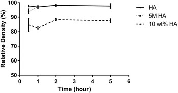
The relative densities of the biphasic materials, sintered for different times at 1300 °C, are shown in Fig 1. Interestingly, there is no significant difference between the densities of the phase pure HA and the biphasic 5 M HA infiltrated sample. Both of these materials achieved the same limiting density, about 97%, after only 1 h at 1300 °C. In contrast, the sample made by mixing showed a much lower density after 1300 °C for 0.5 h. There was no further densification of the mixed samples as the sintering time increased to 5 h. This implies that the mixed sample has also reached a limiting density after 0.5 h suggesting that the pore structure created by the mixing method was coarser than those created in HA and the infiltrated samples prior to presintering.
The relative density measurement of HA, 5 M HA, and 10 wt% HA pellets sintered at 1300 °C.

It is clear that the infiltration method has provided a clear advantage when one wishes to achieve a high sintered density at 1300 °C.
B. Phase distribution and microstructure of the biphasic materials
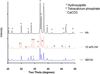
Figure 2 shows the XRD results for the pellet surfaces of the HA, 5 M HA, and 10 wt% HA after sintering at 1300 °C. The HA main peaks (37.1°, 37.5°, and 38.4° for a cobalt target) are shown in all three conditions. The CaCO3 peaks (34.6° and 41.7°) are also identified on the 5 M HA and 10 wt% HA pellets surfaces. Tetracalcium phosphate (TTCP) peaks (35.2° and 36.4°) were also detected on the 10 wt% HA pellet surface but not on the infiltrated pellet surface. TTCP is expected to form during the sintering of calcium phosphate ceramics for which the Ca/P ratio is greater than 1.67. However TTCP is not stable at room temperature and is expected to decompose to HA and CaO on cooling from the sintering temperature. Reference Moseke and Gbureck31 Clearly TTCP on the surface of the 10 wt% HA sample was retained at room temperature.
The XRD results for the HA, 5 M HA, and the 10 wt% HA pellet surfaces after sintering.

The XRD result showed that the fraction of the CaCO3 phase on the surface of 5 M HA was 10 wt%. This is a consequence of the in situ chemical reactions that take place when CaO on the ceramic surface is exposed to moisture. The finely divided CaO resulting from infiltration transforms to Ca(OH)2 and then CaCO3. Reference López-Arce, Gómez-Villalba, Martínez-Ramírez, Álvarez de Buergo and Forta30 The microstructure of these materials has been described in detail in our previous study of HA infiltrated with 2 mol/L Ca(NO3). Reference Zhang, Schmelzer, Gerlach and Nettleship32 The measured Ca/P ratio for clusters of Ca-rich particles and the adjacent matrix in the 5 M HA was estimated as 1.97, by EDS. This ratio is much higher than that expected for stoichiometric HA (1.67), and also higher than the average Ca/P ratio in locations away from the Ca-rich, which was 1.60. The average Ca/P ratio away from the second phase suggests that the phase pure HA is calcium deficient relative to the EDS analysis of a standard sample of stoichiometric HA powder.
The surface of the 10 wt% HA pellet surface showed 57% of TTCP and 43% of HA. The synthesis of TTCP has been studied previously. The TTCP synthesize temperature depends on the powder processing conditions. Reference Moseke and Gbureck31,Reference Liao, Duan, Li, Zheng, Yang, Zhou and Zou33 Carayon and Lacout studied calcium phosphates’ heat transformation behavior. Reference Carayon and Lacout34 TTCP was obtained successfully at 1300 °C in the presence of HA-seeds. Reference Gbureck, Barralet, Hofmann and Thull35 Therefore, it is possible that TTCP could form on the surface of the pellets made in this study. The EDS results on the 10 wt% HA pellet surface showed that the Ca/P at a Ca-rich location was 2.33. The average Ca/P on a relative large area of the surface was 1.73, which agrees with the theoretical Ca/P ratio for the TTCP/HA mixture with this phase distribution. In a previous study, Liao et al. Reference Liao, Duan, Li, Zheng, Yang, Zhou and Zou33 reported that a mixture of TTCP, HA, and CaO could be obtained after heating at temperatures above 1200 °C. A mixing method similar to that used in this study resulted in little TTCP formation at 1300 °C. Higher calcination temperatures caused more extensive TTCP formation suggesting that the kinetics of TTCP formation are sluggish. Another study chemically synthesized finer TTCP crystallites and achieved 95% purity TTCP at 1230 °C. Reference Jalota, Tas and Bhaduri36 This suggests that the size of the HA-seed and the CaO particles affects the kinetics of TTCP formation. The benefit of the mechanochemical activation is also demonstrated for TTCP formation by Romeo et al. Reference Romeo and Fanovich37 and Moseke. Reference Moseke and Gbureck31 It is not clear why TTCP did not form on the sintered surface of the infiltrated samples in this study. However, it is reasonable to speculate that infiltration may have resulted in coarse CaO particles during in situ formation, which resulted in slow TTCP formation at 1300 °C and therefore no TTCP could be detected. In contrast, the mixing method is thought to have produced finer and well-dispersed CaO particles that lead to extensive TTCP formation on the sintered surface during sintering. In summary, it is quite clear that the powder processing method strongly effects the phase distribution on the sintered surface. The effect of these phase distribution differences on calcium ion release and surface morphology on exposure to TBS will now be described.
C. Immersion of biphasic pellets in TBS
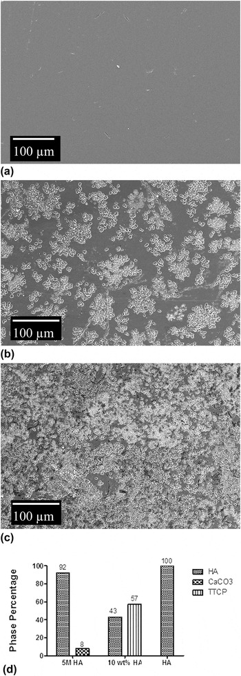
The pellets with the polished surfaces were soaked in TBS for different time periods. After the solubility test, the samples were examined to compare the evolution of the surface pitting and the Ca2+ release over 672 h. The Ca2+ concentrations in the liquid surrounding the pellets were measured using a calcium probe. As the TTCP phase appeared on the sintered surface of the mixed samples, attention was also paid to the decomposition of this phase and its effect on the surface morphology. Figure 3 shows the HA, 5 M HA, and 10 wt% HA pellet surfaces after 1 h in TBS.
The SEM images for (a) HA, (b) 5 M HA, and (c) 10 wt% HA pellet after 1 h in TBS, and (d) the phase fractions on the pellet surfaces.

Extensive pitting appeared on the surfaces of the 5 M HA pellets and the 10 wt% HA pellet after only 1 h. No such defects were observed on HA pellet. The absence of defects on the surfaces of the HA supports the hypothesis that the transformation of soluble Ca-rich phases [the 8% CaCO3 on the 5 M HA surface and 57% on the 10 wt% HA surface in Fig. 3(d)] is the main reason for the surface pitting. In the 5 M HA pellet, the infiltration appears to produce clusters of the second phase particles that are thought to be CaO at the sintering temperature. The volume expansion Reference Deng, Hong, Lan and Tang38 associated with the consequent hydration of closely spaced CaO particles on the surfaces when exposed to moisture is thought to cause cracking along the HA grain boundaries and removal of HA grains to give the observed large surface defects. Reference Zhang, Schmelzer, Gerlach and Nettleship32 The polished surface of the 10 wt% HA pellet has much more extensive damage than 5 M HA. This is thought to be due to rapid dissolution of the large amounts (57%) of TTCP and pit formation. TTCP is known to have a much higher solubility than HA, amongst the highest solubility of the crystalline calcium phosphates. Thus, the defects occupy a larger area fraction, as shown in Fig. 3(c).
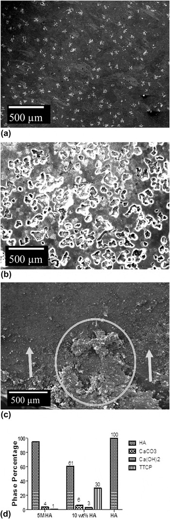
Figure 4 shows pits on the polished surfaces after 672 h. Figure 4(d) shows that the fraction of calcium rich phase on the surface of the 5 M HA sample is reduced and Fig. 4(b) shows more pitting and the pits are deeper than after 1 h. Interestingly, Fig. 4(c) shows that the polished surface of the 10 wt% HA has been completely removed after 672 h [circled in Fig. 4(c)] and partially covered by the surface deposit [arrowed in Fig. 4(c)]. Also, Fig. 4(d) suggests that the amount of TTCP on the surface of this sample has decreased significantly to 30% while the amount of HA has increased to 61%. In addition, minor amounts of CaCO3 and Ca(OH)2 are also present. The SEM observations and XRD results for the mixed sample (10 wt% HA) are consistent with the hydration of TTCP due to the following equation Reference Moseke and Gbureck31,Reference Gbureck, Hofmann and Barralet39 :
 $$3{\rm{C}}{{\rm{a}}_4}{\left( {{\rm{P}}{{\rm{O}}_4}} \right)_2}{\rm{O}} + 3{{\rm{H}}_2}{\rm{O}} \to {\rm{C}}{{\rm{a}}_{10}}{\left( {{\rm{P}}{{\rm{O}}_4}} \right)_6}{\left( {{\rm{OH}}} \right)_2} + 2{\rm{Ca}}{\left( {{\rm{OH}}} \right)_2}\quad .$$
The SEM images for (a) HA, (b) 5 M HA, and (c) 10 wt% HA pellet at 672 h, and (d) the percentages of phases on the pellet surface. The deposit on the surface was circled while the removed polish surface is arrowed in (c).

Instead of simply dissolving, TTCP appears to react with water to form HA which is much less soluble and results in the surface deposit. The EDS analysis of the surface deposit gave a Ca/P ratio of 1.63 consistent with HA formation. Calcium oxide and calcium hydroxide can result from the reaction with water. However, CaO will quickly hydrate to form calcium hydroxide, which will then form calcium carbonate in humid ambient conditions. After 672 h, there is also some pitting on the surface of the HA control sample but is very small and cover much less of the surface compared to the infiltrated and mixed samples.
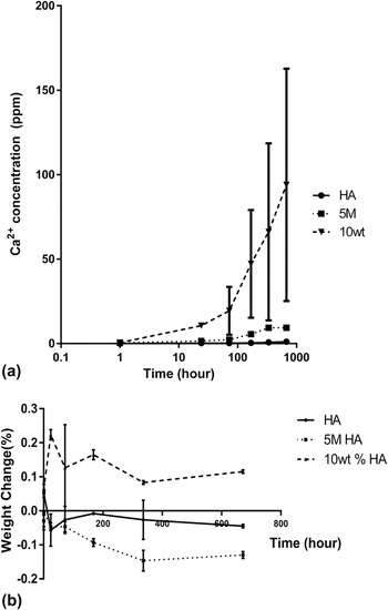
During the 672 h immersion, the Ca2+ concentrations in TBS were measured for all three ceramics. Figure 5(a) shows that the differences between the three conditions are significant. 5 M HA resulted in a significantly higher Ca2+ concentration in the liquid compared to the HA condition. In turn, the difference between the 10 wt% HA and the 5 M HA is considerable, especially at the longer time points. This is consistent with the fraction of soluble phases detected on the surfaces of the sample prior to immersion in saline. The 5 M HA sample surface contained 8 wt% CaCO3, which is responsible for the higher calcium release compared with HA. The much higher calcium content of the saline surrounding the 10 wt% HA sample is consistent with the large proportion of TTCP on the sintered surface (57 wt%) and its decomposition to HA in saline. This results in the surface deposit of low solubility HA and the highly soluble calcium rich second phases (Ca(OH)2 and CaCO3 which subsequently dissolve to increase the calcium content of TBS).
(a) Ca2+ concentration and (b) the weight change of the HA, 5 M HA, and the 10 wt% HA pellets over 672 h in TBS.

The weight changes of the pellets were recorded over 672 h. Three samples were removed from TBS for each time point and each processing condition, and then dried and weighed again. Figure 5(b) shows that the weight changes were small, leading to larger error bars. However, there were significant differences between the samples at the longer time points. The HA samples all showed some weight loss probably due to the small amount of pitting observed on the surfaces of the HA after 672 h. There is much more weight loss for the 5 M HA samples at the longer time points which is consistent with the dissolution of the CaCO3 second phase detected on the sintered surface and the higher calcium content of the saline at the longer soak times. In contrast, the 10 wt% quickly gains weight. Again, this is quite consistent with the phase distribution on the sintered surface and the results of immersion in TBS. In this case, the high fraction of TTCP on the sintered surface quickly caused pitting within 1 h but then the decomposition to give HA in the surface deposit caused the weight of the sample to increase as hydroxyl groups are incorporated in the crystal structure.
D. Immersion of biphasic foams in TBS
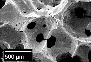
In bioreactors, the biphasic materials must combine the release of calcium with a highly porous architecture that simulates trabecular bone and the endosteal niche. The processing of HA foams used in this study involves an aqueous emulsion. Unfortunately, the presence of the highly soluble phases such as CaCO3 in the processing of the foams dramatically decreased the stability of the ceramic particle suspension used to create the emulsion and so direct mixing was abandoned in favor of the infiltration method to make a biphasic 5 M HA foam. Figure 6 shows the architecture of the foams, which contained approximately 90% open porosity and very little closed porosity measured by micro-CT. The degradation of the infiltrated foam in TBS was thus compared with that of a HA foam made using the same emulsion method without the consequent infiltration step.
The highly porous structure of the 5 M HA and HA scaffolds produced to simulate trabecular bone in the endosteal niche.

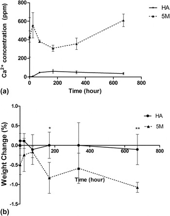
In the scaffold immersion experiments, focus was placed on the Ca2+ release into TBS and the consequent weight change in the scaffolds. Three repeats were measured for each time point and each scaffold condition. Figure 7(a) shows a considerable difference between the calcium concentration of the saline surrounding the 5 M HA and the HA condition over 672 h. The Ca2+ concentration increased significantly from 24 h to 72 h in the HA condition. From 372 h to 672 h, the Ca2+ concentration stayed constant. In contrast, for the 5 M HA condition, the Ca2+ concentration increases rapidly at the initial time points. The Ca2+ concentrations are several hundred ppm above that of the HA scaffold. From 336 h to 672 h, the Ca2+ concentration increased significantly for the 5 M HA foam. Figure 7(b) shows the associated weight change of the scaffolds over 672 h in TBS. The variability between the samples is too high to attribute any weight change for the HA samples which is consistent with the low concentration of calcium in TBS even after 672 h. In contrast, the 5 M HA sample showed increasing weight loss, which is consistent with the presence of the highly soluble calcium rich phase in the sintered scaffold, the transformation to CaCO3 on the surface and consequent dissolution to give high calcium concentration in TBS surrounding the samples. The differences in weight between the HA and the 5 M HA sample were significant at 168 and 672 h.
(a) The Ca2+ concentration in the TBS. (b) Weight change of the sample after immersion in TBS. Data are given as mean ± standard deviation from n = 3 repeats, asterisks indicate statistical significant differences (***P < 0.001, **P < 0.01, *P < 0.05).

Due to the fact that the scaffolds were highly porous, it was not possible to examine the effect of TBS on the porosity of the samples by density measurement. Therefore, micro-CT was used to determine the total pore fraction in the scaffolds after immersion for different periods of time. In each case, the individual foams were scanned before immersion and then scanned once again after immersion. Three repeats were used for each time point.
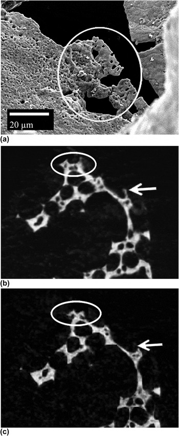
After being removed from TBS, the scaffolds were dried, scanned by micro-CT, and observed in SEM. The observations are then compared with similar observations on scaffolds that had not been exposed to TBS. Figure 8 shows SEM images to illustrate the effect of TBS exposure on the scaffold surface. Before the solubility test, there were no pits on the surfaces of the scaffolds. The short-term solubility behaviors of the scaffold conditions are similar to that of the pellet conditions. At 1 h time point, there was no pit on the HA scaffold surface, while pits appeared on the 5 M HA scaffold surface. After longer immersion times, the difference between the surfaces of the two scaffold conditions was more obvious. Pits did appear on the HA scaffold surface after 33 h immersion in TBS, which were very similar to those on the pellet surface in Fig. 4(a). In contrast, Fig. 8(a) shows that the pits on the surface of the 5 M HA scaffold appear to cause the fragmentation and removal of several grains (circled) on the thin edges of the struts created by the emulsion foaming process after 336 h of immersion. This fragmentation of the surface of the infiltrated foam will contribute to the weight loss of the scaffold but not to a corresponding increment of Ca2+ in the media. Large-scale fragmentation of the scaffold surface by this mechanism would not necessarily be desirable in bioreactors since this would cause at least some of the attached cell populations to become detached from the scaffold and float freely in the bioreactor core. This could affect cell fate and might even interfere with the flow of media through the bioreactor.
(a) 5 M HA are scaffold surface after 336 h in TBS, the circled region shows effects of dissolution of calcium rich phase, (b) micro-CT of a 5 M HA scaffold before immersion in TBS, and (c) micro-CT photo for the same 5 M HA sample at the same cross section after immersion in TBS for 672 h. The circles and the arrows in (b) and (c) show the evidence of material loss by fragmentation.

After immersion in saline for 672 h, the HA scaffold structure did not show any visible change under micro-CT scanning. This is to be expected given the low solubility of HA and the relatively low Ca2+ concentration in TBS, even at long soak times. In contrast, the comparison of micro-CT images of the same section of 5 M HA scaffold before and after immersion provided evidence of surface fragmentation. Compared to the section in Fig. 8(b), there are some small pieces of scaffold removed at the top of the 5 M HA section in the Fig. 8(c). Circles and arrows are drawn on the sections to help locate regions were fragments have been removed. Therefore, the weight decrease and porosity increase observed for the 5 M HA scaffold in Fig. 7(b) is not entirely due to simple dissolution of the calcium rich phase and the attendant pitting. Fragmentation also contributes to the weight loss and the porosity increase of the foam when immersed in TBS for long period.
In summary, biphasic calcium phosphate ceramics have been processed to incorporate highly soluble calcium rich phases. These materials are capable of continuously releasing calcium ions into saline over a period of 28 days. The powder processing method has been shown to control surface phase distribution, surface fragmentation, and calcium release. Calcium is known to control HSC fate in culture; therefore, the ceramic processing method could be used to influence the outcome of HSC culture in bioreactors.
IV. CONCLUSION
Biphasic calcium phosphate ceramics containing highly soluble calcium rich phases have been developed for culturing HSCs. HA has been combined with highly soluble calcium rich phases by both infiltration and by mixing. The phase distribution on the sintered surface of these ceramics under ambient conditions was sensitive to the processing method used. Mixing of the phases during powder processing resulted in a large fraction of TTCP on the sintered surface. When placed in TBS, TTCP reacted with water to remove the surface and give an insoluble HA deposit, which lead to weight gain in the sample. The hydration of TTCP also formed Ca(OH)2 and CaCO3, which results in relatively large amounts of Ca2+ released into TBS. Infiltration of partially sintered HA with calcium nitrate resulted in HA with a calcium carbonate minor phase. The dissolution of CaCO3 in the TBS resulted in surface pitting, the release of Ca2+, and weight loss.
The processing of highly porous biphasic scaffolds by impregnation was compatible with the emulsion method used for direct foaming. The scaffold was able to release Ca2+ into saline by dissolution of CaCO3 on the surface of the scaffold. The dissolution correlated with weight loss of the scaffold and surface pitting. Minor amounts of surface fragmentation were caused by the pitting, which will contribute to the weight loss of the scaffold when immersed in TBS. However, the surface fragmentation was not widespread and is not expected to significantly affect the immobilization of attached cells on the scaffold during culture.
ACKNOWLEDGMENTS
The authors would like to thank the National Institutes of Health for funding this work through award #1R01HL108631-01.