Milk proteins contain many peptide segments, including the 64 C-terminal amino acids of kappa-casein, known as caseinomacropeptide (CMP). CMP is the third most abundant protein/peptide in whey proteins produced from cheese. It is a heterogeneous group of polypeptides formed through the chymosin cleavage of kappa-casein (Delfour et al., Reference Delfour, Jolles, Alais and Jolles1965), with completely conserved post-translational modifications (phosphorylation and glycation) of the parental protein (Villumsen et al., Reference Villumsen, Jensen, Thu Le, Moller, Nordvang and Nielsen2015). CMP has two structural forms: glycosylated (gCMP) with sugar residues and the non-glycosylated form (aCMP) (Brody, Reference Brody2000). These two forms are present at the same ratio in native CMP derived from bovine milk (Thoma-Worringer et al., Reference Thoma-worringer, Sorensen and López Fandiño2006). aCMP exists in two forms, A and B, both of which have a molar mass of around 6.8 kDa. The molar mass of gCMP has been reported as 9–11 kDa, depending on the type of sugar and the level of glycosylation (Kreuβ et al., Reference Kreuβ, Strixner and Kulozik2009). CMP is a random-coil peptide with high conformational flexibility (Ono et al., Reference Ono, Yada, Yutani and Nakai1987; Kreuβ et al., Reference Kreuβ, Strixner and Kulozik2009); it is a peptide acid with pL between 4 and 5 (Thoma-Worringer et al., Reference Thoma-worringer, Sorensen and López Fandiño2006). CMP has received considerable attention due to its putative health-promoting effects, such as body weight reducing activity, inhibition of bacterial and viral adhesion, regulation of blood circulation (Thoma-Worringer et al., Reference Thoma-worringer, Sorensen and López Fandiño2006) and applications in diets for phenylketonuric patients due to the lack of phenylalanine in its sequence (Mollé and Léonil, Reference Mollé and Léonil2005).
CMP also has thermal stability, high solubility, and solubility in acidic conditions. It has been suggested that CMP could be used in the development of innovative food products with enhanced bioactivity and technological attributes, especially for those with special dietary requirements (Abd El-Salam et al., Reference Abd El-Salam, El-Shibiny and Salem2009). Studies have indicated that by varying the pH, concentration and temperature, CMP solutions can form gel. Furthermore, the addition of CMP in food matrices results in a more orderly and structured gel (Thoma-Worringer et al., Reference Thoma-worringer, Sorensen and López Fandiño2006). Whey protein forms a gel at a moderate concentration (about 12% w/w). A higher whey protein content in a product might increase the firmness of the product, which could result in undesirable textural changes. Whey protein can be modified in various ways to suppress its ability to increase a gel firmness (Purwanti et al., Reference Purwanti, Peters and Goot2013). One option is to add CMP in the preparation of the gel with whey protein isolate (WPI). However, there is little existing information regarding the functional properties of the gelation of CMP and its incorporation into food matrices (Thoma-Worringer et al., Reference Thoma-worringer, Sorensen and López Fandiño2006).
This study attempted to prepare high-protein-gel networks using mixtures of CMP and WPI at different compositions, and to analyse the effects on gel properties. The strength of the hydrogel network, and the ultrastructural characteristics based on CMP/WPI, were examined regarding incorporation into food matrices for the specific purposes of protein supplementation.
Materials and methods
Materials
The WPI used to make the gel was donated by Kraki & Kratschmer (Santo André, São Paulo, Brazil) and contained 92.31% (w/w) protein (Kjeldahl analysis, nitrogen to protein conversation factor of 6.38). The moisture content of the WPI powder was 3.45% (w/w), the ash 2.15% (w/w) and fat 1.31% (w/w), of which 0.92% (w/w) was lactose (Folin and Wu, Reference Folin and Wu1920; AOAC, 2010). The CMP used in this study came from two different systems of ultrafiltration (UF) of sweet whey; it was purchased from SOORO (Marechal Cândido Rondon, Paraná, Brazil). Membrane cut-offs of 50 kDa, 30 kDa and 5 kDa were used. The CMP1 sample originated from the 30 kDa retentate derived from the 50 kDa permeate, and was dried by lyophilisation. The CMP2 sample originated from the 5 kDa retentate derived from the 30 kDa retentate, and was dried by lyophilisation. The CMP3 sample originated from the 5 kDa retentate derived from the 30 kDa retentate, and was spray-dried. The CMP samples came from UF with two different drying forms, lyophilisation and spray-drying. The difference in drying, as well as the denaturation of proteins during heat treatment, is crucial for the gelling properties of dispersions (Svanborg et al., Reference Svanborg, Johansen and Abrahamsen2016).
The CMP1, CMP2 and CMP3 powder contained 71.94%, 72.81%, 73.74% (w/w) protein, 11.85%, 11.92%, 12.40% (w/w) lactose and 7.83%, 5.44%, 5.33% (w/w) ash (Folin and Wu, Reference Folin and Wu1920; AOAC, 2010). Of the CMP samples, CMP1 had the highest ash content, as well as the lowest protein and lactose contents.
Preparation of protein gels
The experimental design consisted of two variables (CMP and WPI), and three different concentrations of total protein (30%, 45%, 50% (w/v)) which were used for gel preparation. The CMP1, CMP2 and CMP3 samples were weighed and used to achieve concentrations of 15–30% (w/v), as well as done for the WPI samples. Samples of mixtures of CMP with WPI were dissolved in deionised water at room temperature, under stirring for 15 min. The volume was completed to 25 mL in a volumetric flask which was placed in an ultrasonic bath for 10 min. The samples with concentrations of WPI and CMP were as follows: control (30% WPI, w/v); A, B and C (30% CMP, w/v); 1 (15% WPI and 15% CMP, w/v); 2, 4 and 6 (30% WPI and 15% CMP, w/v); 3, 5 and 7 (30% WPI and 20% CMP, w/v), as shown in Table 1. All the analyses were performed in duplicate at all design points.
Thirty-minute treatment variables related to protein dispersion, comprising CMP1, CMP2, CMP3 and WPI samples

Note: (A), (B), (C) samples comprising only fraction of CMP isolate, which remained liquid with the treatment; (Control) sample without blending with CMP. (WPI) whey protein isolate sample. (CMP2) sample originated from the 5 kDa retentate derived from the 30 kDa retentate. (CMP3) sample originated from R5R30 with spray-drying. (CMP1) sample originated from the 30 kDa retentate. The protein concentration in the second column: 30 (15 : 15) means 30% protein with 15% WPI and 15% CMP; 45 (30 : 15) means 45% protein, of which 30% WPI and 15% CMP; 50 (30 : 20) means 50% protein, of which 30% WPI and 20% CMP. The CMP composition may be CMP1, CMP2 or CMP3.
a Samples analysed by SEM.
Preparation of heat-induced gels and rheological behaviour
The samples were transferred into plastic tubes (adapted syringes) with a diameter of 2.5 cm and length of 10 cm; they were then immersed in a water bath at 70°C for 30 min. However, in sample 1(15% WPI, 15% CMP2) a temperature of 80°C was used, and in samples 4 (30% WPI, 15% CMP3) and 5 (30% WPI, 20% CMP3) the temperature was 75°C. The intention was to firstly ascertain the temperature at which a sample would form a gel and, at the end of the experiment, to compare the samples with the same temperature parameters. Thereafter, the gels were immediately cooled in an ice bath and stored at 10 ± 1°C for 24 h.
Rheometry was conducted in a RheoStress RS75 HAAKE cone and plate rheometer (Karlsruhe, Germany), using a HAAKE thermal circulating bath (Karlsruhe, Germany) for optimal temperature control, and a PP35/s rotor model to perform the readings. The data were processed using RHEOWIN 3 JOB MANAGER (Karlsruhe, Germany) software. All the gel samples were kept at 25 ± 1°C and were analysed within 24 h of gelatinisation. Liquid paraffin was added around the plate edges to prevent dehydration of the samples. Dynamic oscillation measurements were performed under controlled temperature (25 ± 1°C), and strain ranging from 1 to 50 Pa. As the linear viscoelastic region (LVR) is frequency dependent, the test was performed at frequencies of 0.05 Hz and 10 Hz; and the result was the measurement of the complex modulus G*, and the phase angle in the established stress range. The LVR was established so that the deformation would not destroy the temporary internal connections between molecules or aggregates, which could lead to loss of viscosity and irreversible dissipation of energy as heat (Schramm, Reference Schramm2006).
The frequency sweeps from 0.05 Hz to 10 Hz were performed at constant stress within the LVR. The results were the values of elastic modulus G′, viscous modulus G″ and complex viscosity η* as a function of frequency. This information enabled the comparison of the characteristic viscoelastic profile of the systems formed. The data reported are the means of at least two individual samples, with an experimental error lower than 10%.
Scanning electron microscopy
Samples were processed as follows: washed four times in 0.1 m cacodylate buffer, pH 7.4 at 37°C; fixed in Karnovski solution (2.0% glutaraldehyde, 4.0% paraformaldehyde, 1 mm CaCl2 in cacodylate buffer 0.1 m, pH 7.2–7.4); washed again in cacodylate buffer 0.1 m pH 7.2–7.4; post-fixed with 1% osmium tetroxide (diluted in 0.1 m sodium cacodylate pH 7.4), washed in 0.1 m cacodylate buffer pH 7.2–7.4; and dehydrated by increasing concentrations of ethanol (30%, 50%, 70%, 90% and 100% (twice) for 10 min at each concentration). Samples were treated in a Balzers critical point dryer and then coated with gold using Balzers Union, SCD 030 (Balzers Union Ltd, Balzers, Lichtenstein) equipment. The samples of hydrogel subjected to the cryofracture technique were gently washed with gradients of glycerol (15, 30 and 50) in 0.8 m cacodylate buffer at pH 5.7, for 15 min each, and then rinsed. These samples were transferred into a container filled with liquid nitrogen and fractured by mechanical means (Fu et al., 2007); the fragments were then washed again with glycerol, as described above. They were subsequently dehydrated through a series of ethanol baths at increasing concentrations (20–99%). The fragments were then critical-point dried in BAL-TEC CPD 030 (Critical Point Dryer, Liechtenstein, Germany) equipment. The specimens were mounted on copper nails and sputter-coated with gold-palladium (SCD 030 – Balzers Union FL 9496). Specimens were observed in a JEOL JSM – 6360LV SEM (Jeol, Tokyo, Japan) field-emission scanning electron microscope operating at 15 kV.
Statistical analysis
The experiments and measurements were repeated at least three times. One-way analysis of variance (ANOVA) was applied to evaluate effect of treatments on the measured variables. The differences between the means were assessed at a significance level of P < 0.05 using the Tukey´s post hoc test (JAMOVI, version 1.8, computer software).
Results and discussion
Protein gel
Initially, a protein dispersion was prepared to serve as control sample (WPI). Subsequently, the objective was to establish a high-protein concentration during a 30 min treatment at 70°C for gel formation at the point of sol−gel phase change. Control sample (WPI) formed strong gel. Samples A (30% CMP2), B (30% CMP3) and C (30% CMP1) (Table 1) did not produce gels during 30 min treatment at 70°C, because CMP is highly hydrophilic and heat stable, due to its random spiral structure and negative charges carried by glutamate, aspartic acid, carboxyl groups, phosphorylation sites, and the carbohydrate chain at neutral pH (Smith et al., Reference Smith, Edwards, Palmano and Creamer2002). Despite this, several studies have shown that CMP can still interact with WPI through non-covalent interactions and affect their heat-induced gelation (Gaspard et al., Reference Gaspard S, Sharma and Fitzgerald2021). The remaining gel samples were composed of WPI mixed with CMP isolate (Table 1).
All the gels were yellowish, with the exception of sample 1, which formed a weak, whitish opaque gel with syneresis. As water holding capacity of gels in its tridimensional network demonstrates the strength of interactions, weak gels are formed by weak interactions and tend to lose more water (Burgardt et al., Reference Burgardt, Zuge and de Bonna Sartor2015). The increase in the proportion of CMP in relation to WPI caused an increase in the opacity of the gels (sample 1). This fact is due to the reduction of distance between the CMP chains in the solar state, which increased the possibility of aggregation and formation of junction zones (Ikeda and Foegeding, Reference Ikeda and Foegeding1999). The presence of CMP prevented the formation of a solid whey protein gel after the temperature necessary for gel formation was reached, as shown in a previous study (Gaspard et al., Reference Gaspard S, Sharma and Fitzgerald2021). In addition, the samples with CMP3 (4 and 5), which were spray-dried, required an additional heating of 5°C to obtain gel formation. This was also reported in a previous study, where in heat-induced gelation, CMP increased the denaturation temperature of β-lactoglobulin (β-LG) by up to 3°C and increased the gelation temperature by up to 7°C (Gaspard et al., Reference Gaspard S, Sharma and Fitzgerald2021).
Studies of β-LG-CMP mixes at neutral pH showed that the formations of β-LG-CMP aggregates were driven by electrostatic interactions and/or by hydrogen bonding. At neutral pH, both β-LG and CMP are negatively charged, which may cause a weaker interaction between the CMP and unfolded whey protein (Svanborg et al., Reference Svanborg, Johansen and Abrahamsen2016).
Rheological behaviour of gels
Rheological analyses were conducted to study the structure and viscoelasticity of a complex system, as well as the effect of different concentrations of CMP and WPI on gel formation, and the interaction between them. Oscillatory analysis is sensitive to chemical composition and the physical structure of samples. Initially, stress scanning was performed to determine the LVR of the samples. Tension selection was based on stress scanning at 0.05 and 10 Hz. According to Kutschmann (Reference Kutschmann2003), greater stability is expected for samples that have a wider LVR range and higher elastic modulus G′ values, as found in the Control sample, with an LVR range of 200–1000 Pa and G′ of 35 940 for frequency of 0.05 Hz, and 100–1000 Pa and G′ of 43 140 for frequency of 10 Hz.
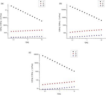
Depending on the particular sample, the tension was set at values ranging from 1 to 50 Pa for frequency scanning. At these tension ranges the gel structure was preserved because it remained within the LVR. Figure 1 shows the mechanical spectrum of the gel samples, in which all samples had elastic modulus values (G′) significantly higher than the viscous modulus (G″) across the frequency range (Brummer, Reference Brummer2006). They had a predominantly solid aspect, behaving as viscoelastic gel-like systems, which reflected the existence of a three-dimensional network. The G′ and G″ moduli were practically independent (Steffe, Reference Steffe1996) and both modules tended to be parallel, with a slight increase at high frequencies, meaning that this feature was slightly frequency-dependent. Such a spectrum characterises gel networks and emulsions with high internal structural strength (Brummer, Reference Brummer2006). Moreover, it is noteworthy that the addition of CMP isolate decreased the elastic (G′) and viscous (G″) modulus values of the samples (Table 2). The control sample had a lower tan δ value, which means greater difference between G′ and G″, corresponding to a stronger gel. The complex dynamic viscosity (ŋ*) of the control sample was much higher than the other samples, reaffirming its stronger gel structure. There were significant differences (P < 0.05) between the control sample and the samples enriched with CMP1, CMP2 and CMP3 in terms of G′, G″ and ŋ*.
Frequency vs. G′, G″ and complex dynamic viscosity (η*). (1) control: 30% WPI; (2) sample 3: 30% WPI and 20% CMP2; (3) sample 7: 30% WPI and 20% CMP1.

Values of shear or elastic storage modulus (G′), shear loss or viscosity modulus (G″), complex dynamic viscosity (η*), and tan δ (ratio G″/G′) at a frequency of 1 Hz

Data represent mean of three replicates ± standard deviation. Superscript* indicates significant differences (P < 0.05).
The samples with higher G′ values were control, 1 and 4 (Table 2). Samples 1 and 4 refer respectively to the compositions of 30% (15/15) of WPI/CMP2, and 45% (30/15) WPI/CMP3, treated for 30 min at 80°C and 75°C. Sample 1 had tan δ = 0.168, and sample 4 had tan δ = 0.163. These values were not significantly different in terms of the rigidity of the gel formed (P > 0.05). The complex dynamic viscosity values were approximately equal for both systems.
In comparison to the control, sample 1 was treated at a higher temperature (80°C) and had the same protein concentration (30%) but a different composition (WPI/CMP2). This showed a somewhat lower G′ value and a weaker gel. Even at a temperature that was 10°C higher, the sample with a mixture of CMP (sample 1) produced a significantly weaker gel. Comparing the control sample with samples 2, 4 and 6 (which were enriched with over 15% of the fractions CMP2, CMP3, and CMP1) more structural strength was observed in the control gel.
Although it was to be expected that by increasing the protein concentration there would be greater cross-linking, leading to a higher rate of gelation and, hence, greater gel strength (Otte et al., Reference Otte, Schumacher, Ipsen, Ju and Qvist1999), the addition of CMP, unlike the behaviour of polysaccharides and other proteins, led to a reduction in the strength of the gel network. Gaspard et al. (Reference Gaspard S, Sharma and Fitzgerald2021) confirmed the potential of CMP to control heat-induced denaturation and gelation of whey protein. Thus, although the tested variables (composition, concentration and temperature) interfered with each other in the resistance of the gel network, this study demonstrated that there was no significant difference between CMP1, CMP2, CMP3 regarding gel strength (P > 0.05).
Scanning electron microscopy analysis of gels formed by the polymerisation of proteins (WPI/CMP) in different concentrations
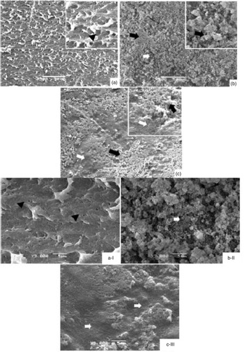
The compact pattern of polymerisation on the outer surface of the different gels was viewed by SEM (Fig. 2, micrographs A, B and C). The panoramic photo A (Fig. 2) shows that the gel containing 30% WPI (control) had a regular morphological pattern, with microstructures evenly distributed along its surface. Lamellar structures were distributed all over the gel surface. Under higher magnification, photo A showed evidence of homogenous porosity. Rheological analysis demonstrated that the control gel had the strongest structure of the three gels shown, and also the highest elastic modulus (G′) value. In contrast, the gels shown in photos B and C had coarse structures with clumps of aggregates.
SEM micrographs (500× magnification) of hydrogel enriched with different concentrations of proteins: (a) 30% WPI; (b) 50% (30% WPI and 20% CMP2); (c) 50% (30% WPI and 20% CMP1), after 30 min polymerisation at 70°C. (WPI) whey protein isolate sample. (CMP1) sample originated from the 30 kDa retentate. (CMP2) sample originated from the 5 kDa retentate derived from the 30 kDa retentate. The insertions in the zoomed micrographs (a, b, c) show the surface of the hydrogel at 2000× magnification (bar = 5 μm). Corresponding indications are: ( ) compact without the presence of pores, (
 ) particulate materials, (
 ) porosity and (
 ) aspect labile and looser, composed of the aggregation of a fibrillar pattern containing vesicular aggregates of smaller diameter. Micrographs A-I, B-II and C-III correspond to samples (1), (2) and (3) respectively. External surface morphology of hydrogels with increased magnification, 3000X. Corresponding indications are: (
 ) compact without the presence of pores, (
 ) particulate materials, (
 ) porosity.

The gel shown in photo B presented a precipitation of material on the network; these particles were clumped all over the surface. Of all the analysed gels, the gel shown in micrograph C (sample 7) showed a denser aspect on the surface. There was a large amount of particulate matter deposited on large areas in the form of plaques. This gel, which also corresponded to rheological sample number 7, had the lowest elastic modulus (G′) value and the weakest gel structure of the three gels analysed by SEM.
A previous study (Loria et al., Reference Loria, Pilosof and Farías2018) found that, in the absence of salts, CMP mainly presented a monomeric form at pH 7.0. However, if salt is added, the hydrophobic associations of CMP are promoted due to screening of electric charges. The presence of sodium and calcium ions can strongly influence the flow properties of protein suspensions (Thomar et al., Reference Thomar, Benyahia and Durand2014), and can, therefore, be a vital parameter to control its assembly properties (Loveday et al., Reference Loveday, Wang, Rao, Anema, Creamer and Singh2010; Wang et al., Reference Wang, Liu, Cao, Yongsawatdigul, Xiong and Liu2018). This may explain the microscopic differences between gel 7 (micrograph C), which had a higher salt content, from the image of gel 3 (micrograph B).
Figure 2 (b-II) shows the outer surface of the gel enriched with 50% protein, 30% WPI and 20% CMP2 (sample 3). This micrograph shows a massive deposition of solid particulate material clumped over the entire surface, making it highly irregular in topography. The outer surface was formed exclusively by the deposition and stacking of aggregate particles (zoomed Fig. 2b). This gel (rheological sample number 3) had intermediate structural strength compared to the others. The results agreed with previous findings that the size of gel-forming particles and the protein concentration can affect gel structure. Furthermore, the mixtures of several different proteins showed dense microstructures and coarse structures with clumps of aggregates (Donato et al., Reference Donato, Schmitt, Rouvet and Bovetto2009). According to Svanborg et al. (Reference Svanborg, Johansen and Abrahamsen2016) it is suggested that the aggregates in the β-LG-CMP-systems consisted of β-LG surrounded by CMP monomers and have a similar chaperone-like behaviour as some caseins, thus stabilising the β-LG at neutral conditions.
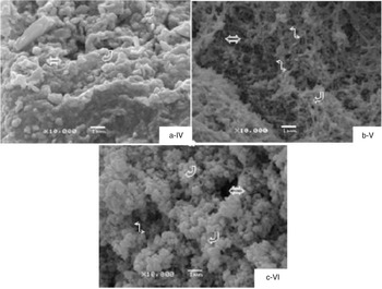
The ultrastructural analysis of the internal architecture of the respective gels, after fracture by freezing and breakage, as shown in Figure 3 (a-IV, b-V and c-VI), makes clear that the pattern of polymerisation consisted of globular structures. These latter were either isolated or fused to each other in the different tested gels, forming vesicles of variable sizes, as shown in micrographs A-IV and C-VI. Micrographs B-V and C-VI also showed a certain discontinuity and porosity, reflecting low elasticity and a relatively weaker gel network, as was confirmed by rheological analysis. Svanborg et al. (Reference Svanborg, Johansen and Abrahamsen2016) found that the microstructures of gels became porous and coarse as the CMP concentration increased. Gels without CMP were more resistant to stress and strain, with a 3% decrease in the linear viscoelastic region of G′ than gels containing 9–33% CMP (Svanborg et al., Reference Svanborg, Johansen and Abrahamsen2016). These findings confirm the experimental responses of the present study. Kuhn et al. (Reference Kuhn, Cavallieri and Cunha2011) concluded that some micrographs of a gel composed of WPI with added salt showed greater discontinuity and porosity, which was related to the low rigidity and elasticity of these gels. The increased porosity of a structure can be attributed to an obstruction in the interaction between proteins by some other element, as well as increased electrostatic repulsion (Kuhn et al., Reference Kuhn, Cavallieri and Cunha2011). Micrograph B-V depicts a unique internal pattern in the gel formed, compared to the other samples. This mesh appeared less dense and more labile; it was characterised by the aggregation of a fibrillar pattern, as well as vesicular aggregates of smaller diameter than the other gels. The gel network also presented a looser aspect in SEM, collaborating with results from Gaspard et al. (Reference Gaspard S, Sharma and Fitzgerald2021), who reported that the addition of CMP strongly affected the structure of heat-induced whey protein, resulting in a thinner chain structure.
Micrographs (10 000× magnification) of cryofracture (bar = 1 μm) of hydrogel. (a) 30% WPI; (b) 50% (30% WPI and 20% CMP2); (c) 50% (30% WPI and 20% CMP1). (WPI) whey protein isolate sample. (CMP1) sample originated from the 30 kDa retentate. (CMP2) sample originated from the 5 kDa retentate derived from the 30 kDa retentate. Micrographs A-IV, B-V, and C-VI correspond to samples (1), (2) and (3) respectively. The corresponding indications are: ( ) indicates matrix formed by globular structures; (⇔) polymerisation of the internal architecture pattern of the hydrogel; (
 ) less dense pattern and looser aspect.

Veith and Reynolds (Reference Veith and Reynolds2004) demonstrated that the presence of CMP in WPI was injurious to gel strength and water-holding properties. Compared to globular proteins, such as β-LG, which is present in the largest amount in whey proteins, CMP is a peptide that has different properties involved in the formation of gel. The injurious effect of CMP on the strength of the gel and the water holding capacity, suggests that CMP does not incorporate itself into the protein gel network and competes for connection to water.
In conclusion, we prepared a gel containing a high percentage of protein by mixing WPI and CMP, thereby providing a network of decreased strength. The best overall result was achieved by gel 7 (micrograph C), which contained 50% protein, 30% WPI and 20% CMP1, and which was heat-treated at 70°C for 30 min. SEM showed a structure with coarse aggregates and clumps at the surfaces, with a relatively denser aspect. In contrast to what is typically observed for polysaccharides and proteins, the addition of CMP to the gel resulted in a weak gel network. In addition, because it underwent an ultrafiltration process without the 5 kDa retentate, the CMP1 sample had a higher salt content, which contributed to reducing the strength of the gel network. However, the higher salt content seemed to have more influence on the microstructure than on the rheological parameters.