Introduction
The twenty-first century is witnessing its worst healthcare crisis with the coronavirus disease 2019 (Covid-19) pandemic. Frontline caregivers, who are mostly medical and nursing residents, have needed to adapt to rapidly updated regulations, extreme work environments and cumbersome protective gear within a short time-span.Reference Vindrola-Padros, Andrews, Dowrick, Djellouli, Fillmore and Gonzalez1,Reference Dutta and Azgaonkar2
Several complications have been documented with personal protective equipment (PPE). These brought about necessary changes in PPE design and the re-scheduling of optimum duty hours, and helped re-orient the attitude and perception of caregivers regarding the proper use of such kit.
Here, the authors describe their own experience of non-traumatic fracture of the laryngeal framework that resulted from voluntary cough-holding whilst wearing a PPE suit which was too small with a flexed, stiff neck. A rare event by itself, such a posture-related complication has not been hitherto reported with PPE use. This paper highlights the importance of donning appropriately sized PPE suits to prevent potentially serious posture-related complications. The paper also discusses the pressure dynamics around the vocal folds during the event of voluntary cough-holding in a flexed, stiff neck, and offers plausible mechanisms of the laryngeal injury that followed.
Case report
A 28-year-old male otolaryngology resident presented to his faculty with pain and difficulty in swallowing, and swelling in front of his neck with crepitus. During his rotation in the coronavirus disease ward the previous night, he remembered having suppressed a paroxysm of cough whilst being seated in his PPE suit with a flexed neck in order to prevent his goggles from fogging. He felt a sudden release of pressure, which was immediately followed by intense pain and development of the presenting symptoms. There was no respiratory difficulty or change in voice. He disclosed further that he was wearing a PPE suit that was too small for his stature as it was the largest size available at that time. This made him flex his neck to about 30 degrees whilst sitting, with restricted neck movements.
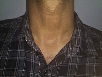
On examination, there was subcutaneous emphysema in the anterior neck (Figure 1). Tenderness, but no fracture line or bony crepitus could be detected on gentle, careful palpation. Flexible fibre-optic laryngoscopy findings were unremarkable.
The otolaryngology resident presented with subcutaneous emphysema in the anterior neck (visible to the observer as suprasternal fullness) following voluntary cough-holding whilst negotiating within a personal protective equipment suit that was too small with a flexed, stiff neck. He could feel the crepitus from the level of the lower part of thyroid cartilage up to the root of the neck.

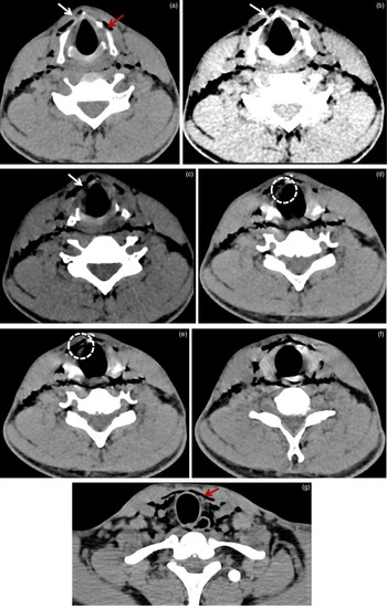
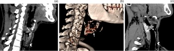
A non-contrast computed tomography (CT) scan (Figures 2 and 3) revealed a non-displaced fracture in the lower end of the partially ossified right thyroid lamina, with an evident tear in the cricothyroid membrane. The fracture could also be visualised in the three-dimensional virtual reality reconstruction of the laryngeal framework. The subcutaneous emphysema could be seen extending from the level of the fracture to the root of the neck.
Non-contrast computed tomography scan of the neck (serial axial cuts) show a non-displaced fracture (white arrows) in the lower end of the partially ossified right thyroid ala ((a & c) bone windows; (b) soft tissue window corresponding to part (a)), extending inferomedially to involve the cricothyroid membrane (dotted circles in (d & e) represent bone windows). Subcutaneous emphysema (red arrows) can be seen throughout, from the level of the fracture up to the root of the neck (a–g).

Non-contrast computed tomography scan of the neck (parasagittal sections; soft tissue windows) show the non-displaced fracture in the lower end of the partially ossified right thyroid ala ((a); solid arrow) along with the subcutaneous emphysema ((a & c); arrowheads). A three-dimensional virtual reality reconstruction of the laryngeal framework also reveals the fracture ((b); solid arrow). The parasagittal section with the head and neck partly rotated to the right shows the tear in the cricothyroid membrane ((c); dotted arrow).

The resident was a non-smoker and non-drinker, who regularly exercised and was otherwise healthy. He was reassured and advised to rest for a week. He was kept under observation, and given analgesics, oral prednisolone (at 1 mg/kg body weight per day for 7 days) and a proton pump inhibitor. His symptoms gradually disappeared over the next 4 days, and he recuperated uneventfully.
Discussion
India, along with USA and Brazil, is one of the worst hit nations by the Covid-19 pandemic.3 Its second wave left India's health infrastructure overwhelmed. The challenges momentarily seemed insurmountable, with a dearth of healthcare providers and medical consumables. Residents across clinical disciplines were given roster duties in the busy coronavirus disease wards or intensive care units.Reference Dutta and Azgaonkar2
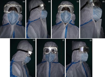
The patient described herein was a resident on the coronavirus disease ward on the night of the incident described. He was taller than the average adult Indian man, and a PPE suit appropriate for his stature was not available in the store on that shift. He had to wear a smaller-sized PPE coverall whose upper-half had to be negotiated lest the front zipper stretched and released. In order to maintain the integrity of the suit, the resident patient kept his head flexed (Figure 4). Besides causing claustrophobia, this also limited his neck movements. With this compromised posture, an attempt at stifling a paroxysm of cough to prevent fogging of the goggles led to rupture of his laryngeal framework.
Recreation of postures in a smaller-sized (a–e) and appropriate-sized personal protective equipment (PPE) (f & g). When the resident patient donned the smaller-sized PPE suit and sat erect, the front zipper and anterior wall of the coverall became stretched ((a) front view; (b) lateral view) and tended to release ((c) front view; (d) lateral view). He had to flex his neck by about 30 degrees to keep the zipper relaxed, intact and in place (e). These problems were eliminated when he was in a PPE coverall of appropriate size ((f) front view; (g) lateral-oblique view). Note that the Velcro was kept unfastened in all photographs to show the position of the zipper within.

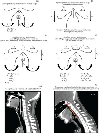
The natural aerodynamics of cough involves a forceful release of air through highly co-ordinated, complex, sequential neuromuscular events comprising inspiratory, compressive and expulsive phases.Reference Brooks4 The momentary glottic closure during the compressive phase allows considerable subglottic pressure to develop, with sustained isometric contraction of the expiratory (chest) muscles effectively acting as a piston (Figure 5a). Subsequent forceful expulsion of air mimics a positive displacement compressor. The process is aided by contraction of the extrinsic muscles of the laryngohyoid complex that lowers the hyoid and helps eject the gust of air, adding to the recoil property for the compressor system (the expulsion–recoil couplet) (Figure 5b).
Parts (a–d) show the airflow and pressure dynamics around the vocal folds. During the compressive phase of cough (a), the effective subglottic pressure (eP1) – a function of the expired air pressure (P1) accentuated by isometric chest compression (f1) – produces turbulence (T1) of proportional intensity, acting as a piston against the closed glottis. During the expulsive phase (b), the effective subglottic pressure and turbulence decreases following Bernoulli's principle. The expulsion is propelled by the recoil brought about by contraction of the infrahyoid muscles (R1) (the expulsion–recoil couplet). The associated back-and-forth neck thrust (Th1) accommodates the turbulence during forceful air release. Thus, the subglottic turbulence (T) is directly proportional to the effective subglottic pressure (eP) and inversely related with the recoil (R) and neck thrust (Th). Sustained voluntary glottic closure during cough-holding prolongs the compressive phase (c). With continued isometric chest compression (f2), the effective subglottic pressure (eP2) increases with a proportionately high turbulence (T2). This is in spite of potential leakage of air volume (p) that tends to be minimal as the expulsive phase is consciously suppressed. There is minimal neck thrust (Th2) and much reduced recoil (R2) because of attenuated infrahyoid muscle contraction, helping to prevent expulsion of air, thereby increasing the turbulence. When the neck is flexed and stiff (as when wearing a personal protective equipment (PPE) suit that is too small) (d), the neck thrust (Th3) decreases further, so does the recoil (R3), because the power of the infrahyoid muscles are grossly diminished following lowering of the hyoid. The effective subglottic pressure (eP3) increases as a result of complete sealing off of the glottis by a back thrust (p'). All these increase the turbulence (T3). The back thrust is produced by the pressurised leaked air (p) facing obstructions in its trajectory because of the reduced angle of the supralaryngeal tract and its vertical limb assuming a bend. Parts (e) and (f) are the mid-sagittal computed tomography cuts of the resident patient archived from follow-up imaging. The supralaryngeal tract could be seen with its vertical and horizontal limbs at right angles (a) when the head and neck were erect (e). However, when the neck was flexed (as when wearing a PPE suit that is too small) (f), the vertical limb assumed a bend (yellow line), and the angle (a', formed by intersection of the extended horizontal limb and a tangent drawn in red through the point of maximum convexity in the curved vertical limb) reduced. These kinks in the expiratory airflow tract caused the back thrust (p'). It sealed off the glottis, increasing the effective subglottic air pressure (eP3) and the resultant turbulence (T3).

A voluntarily closed glottis during the suppression of cough aims to abut the expulsive phase, but in effect accentuates the compressive phase. The resultant air release is brief, following momentary and partial vocal fold abduction during the continued effort at glottic closure. The intraluminal pressure consequently increases manifold, as does the turbulence (Figure 5c).
This effect is greatly accentuated by neck flexion in at least three plausible ways (Figure 5d). First, the expelled air-jet trajectory follows the supralaryngeal tract, which, depending upon the degrees of neck flexion, reduces from the normal right-angle to variable acute angles, with its vertical component assuming a bend (Figure 5e and f). These kinks in the pressurised air-jet trajectory tend to produce a back thrust that prevents further air escape through the glottis. This effectively seals the glottic chink, adding to the subglottic pressure and turbulence.
Second, flexion of the neck lowers the hyoid. Contraction of infrahyoid muscles during the expulsive phase therefore becomes largely ineffective. This attenuates the recoil handle of the expulsion–recoil couplet, preventing or reducing the release of air from the voluntarily sustained closed glottis.
Third, the back-and-forth neck thrust that would have otherwise negotiated the turbulence on the laryngotracheal and pharyngeal framework during the expulsive phase would be minimised because of restricted movement of the flexed neck in a PPE suit that was too small. Thus, an attempt to stifle a cough would considerably increase the subglottic air pressure and turbulence as a result of a prolonged compressive and minimal expulsive phase, aggravated by the lack of neck thrust.
These factors synchronise to generate enough force to rupture the laryngeal framework. The problem is aggravated when the neck remains flexed with restricted movements, as when wearing a PPE suit that is too small.
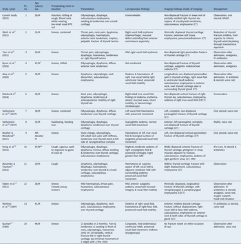
Non-traumatic laryngeal injuries or fracturing following an attempt to suppress a cough or sneeze is extremely rare, with only 13 patients on record in the PubMed/Medline database (Table 1).Reference Sbeih, Tierney, Bryson and Kominsky5–Reference Quinlan16 That such injuries have exclusively affected young to middle-aged men can be explained by their higher pulmonary capacity. Congenital anomalies and focal demineralisation could be the predisposing factors, but evidence is inconclusive in light of inadequate case strength.Reference Alexander and Toynton13
Cases of non-traumatic injury to laryngeal framework due to cough and sneeze*

*Identified in the PubMed/Medline database. †Chronic smoker, with a past history of obstructive sleep apnoea and hypertension. ‡Patients recovering from upper respiratory tract infection. Pt. no. = patient number; M = male; PPE = personal protective equipment; CT = computed tomography; NSAID = non-steroidal anti-inflammatory drug; IV = intravenous; CECT = contrast-enhanced computed tomography; MRI = magnetic resonance imaging; ICU = intensive care unit
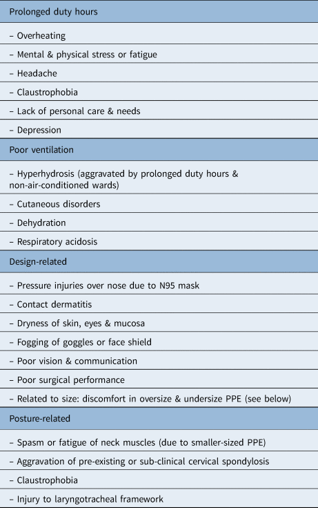
The PPE kits are indispensable for Covid-19 caregivers given their proven role in preventing virus transmission. However, wearing them under extraordinary circumstances has resulted in several complications (Table 2). Complications related to attitudes and perceptions associated with the proper use of PPE kits are potentially avoidable, and, interestingly, have seldom been emphasised and possibly go unreported. Significant complications include those that are posture-related, which can be obviated by choosing the right sized PPE.
Complications associated with PPE

PPE = personal protective equipment
Whilst PPE suits are unisex, an individual should consciously select the size appropriate for his or her stature. A larger suit may be uncomfortable, but a smaller one may cause serious injuries due to physical compromise, like neck spasm, aggravation of pre-existing or sub-clinical cervical spondylosis, and, rarely, rupture of the laryngeal framework. Maintaining an uninterrupted supply of PPE during this pandemic has been a real challenge for the already burdened public health infrastructure in most nations across income groups.Reference Vindrola-Padros, Andrews, Dowrick, Djellouli, Fillmore and Gonzalez1 The clinical experience reported here acknowledges the critical situation, and underlines the need for an adequate supply of properly sized PPE suits for Covid-19 caregivers.
This ongoing pandemic has threatened a nation's capability to surmount issues related to its infrastructure, economy and managerial resources in the face of an unforeseen global emergency. In addition, at a more physical and emotional level, it has put the human spirit to its ultimate test of survival through endurance, optimism and courage.
It can be recalled how the resident patient met his faculty colleagues in the early morning following that eventful night shift – anxious, perplexed and trembling – with a sense of impending doom written over his face. Scheduled duties in the coronavirus disease ward, long hours within a PPE suit, limited time to eat, drink and conduct toilet necessities, restricted communication, and too-close encounters with death, suffering and helplessness made the young residents emotionally charged.Reference Dutta and Azgaonkar2 In such a situation, an unexpected, accidental physical injury during coronavirus disease duty will accentuate their stress manifold.
• Posture-related complications associated with wearing personal protective equipment (PPE) kits are seldom emphasised and documented
• Wearing a smaller sized PPE suit compromises posture, and necessitates a flexed and restricted neck to prevent stretch and release of the coverall zipper
• Non-traumatic laryngeal framework injury following a stifled cough is a rare but a potential posture-related airway complication associated with smaller sized PPE
• This paper explores relevant pressure dynamics that explain this unusual airway injury, accounting for the compromised (flexed and restricted) neck position
• Coronavirus disease 2019 caregivers need to use appropriately sized PPE, to minimise posture-related complications
Periodic and flexible leave, reassurance, proper medical care, and psychological support need to be arranged by the authorities on the merit of individual incidences. Taking due care of the emotional health of coronavirus disease caregivers is therefore of utmost importance from a humanitarian perspective, and for the care delivery system to run smoothly. This clinical record documents the crisis at a personal and professional level, between a teacher and his student, and underlines the need to preserve the humane element in this global health emergency.
Conclusion
The Covid-19 caregivers need to be aware of potentially avoidable posture-related complications that result from donning inappropriately sized PPE suits. This paper illustrates one of the rarest of such injuries from an apparently trivial incidence of voluntary cough-holding, and provides theoretical explanations using pressure dynamics. It serves as an important caveat for this group of PPE-related complications that are seldom highlighted and documented.
Acknowledgement
The authors wish to thank Anup Kumar Sadhu (consultant at EKO CT & MRI Scan Centre, Medical College and Hospital, Kolkata), for his valuable help in interpreting the radiological images.
Competing interests
None declared