INTRODUCTION
Primary progressive aphasia (PPA) is a clinical syndrome characterized by progressive decline in speech or language abilities over time and occurs due to neurodegenerative disease (Mesulam & Weintraub, Reference Mesulam and Weintraub1992). Three main variants of PPA are recognized: a semantic variant (svPPA), a nonfluent/agrammatic variant (nfvPPA), and a logopenic (“word poverty”) variant (lvPPA) (Gorno-Tempini et al., Reference Gorno-Tempini, Dronkers, Rankin, Ogar, Phengrasamy, Rosen and Miller2004, Reference Gorno-Tempini, Hillis, Weintraub, Kertesz, Mendez, Cappa and Grossman2011). Each of the variants has specifically defined clinical features, distinct atrophy patterns, and a likelihood of pathological subtype (Gorno-Tempini et al., Reference Gorno-Tempini, Hillis, Weintraub, Kertesz, Mendez, Cappa and Grossman2011).
The semantic variant is characterized by fluent speech and a loss of semantic knowledge, including but not limited to word meaning, is associated with predominantly left anterior temporal lobe atrophy, and pathologically with frontotemporal lobar degeneration (FTLD), most often caused by FTLD TAR DNA Binding Protein, Type C (TDP-C). NfvPPA is recognized by apraxia of speech, impairments in articulation and grammar, left fronto-insular atrophy, and is most often associated with FTLD Tau and/or TDP pathology. LvPPA is unique in that it is most often associated with Alzheimer’s disease (AD) pathology and biomarkers (Leyton, Britton, Hodges, Halliday, & Kril, Reference Leyton, Britton, Hodges, Halliday and Kril2016; Spinelli et al., Reference Spinelli, Mandelli, Miller, Santos‐Santos, Wilson, Agosta and Meyer2017) and is now considered a variant of early-age-of-onset AD (Dubois et al., Reference Dubois, Feldman, Jacova, Hampel, Molinuevo, Blennow and Bateman2014). LvPPA is identified by word finding pauses and impairments in phonological short-term memory (Henry et al., Reference Henry, Wilson, Ogar, Sidhu, Rankin, Cattaruzza and Seeley2014) and atrophy that extends along the posterior portion of the left superior/middle temporal gyri into the inferior parietal lobule (supramarginal and angular gyri).
Since language difficulties can confound both task performance and comprehension of instructions on many neuropsychological measures, broader examination of cognitive domains other than language in PPA could allow researchers and clinicians to better understand the full spectrum of cognitive impairment in PPA and how it changes with disease progression, and potentially improve differential diagnosis. Even though exclusion criteria for PPA include initial and functionally significant impairments in visuospatial processing and visual memory, that is, by definition patients with PPA do not complain of difficulties with visuospatial functioning at presentation, many patients with PPA present with low scores on formal testing on tasks that are largely thought to be visuospatial in nature and can develop functional difficulties in this cognitive domain as the disease progresses.
There is limited research examining cognitive abilities other than language in PPA (Butts et al., Reference Butts, Machulda, Duffy, Strand, Whitwell and Josephs2015; Foxe et al., Reference Foxe, Leyton, Hodges, Burrell, Irish and Piguet2016; Foxe, Irish, Hodges, & Piguet, Reference Foxe, Irish, Hodges and Piguet2013; Ramanan et al., Reference Ramanan, Flanagan, Leyton, Villemagne, Rowe, Hodges and Hornberger2016), which confines interpretation of performance discrepancies in cognitive domains to subjective analysis. Our goal in this study was to explicitly study an understudied area of cognition in PPA to better describe and understand the cognitive profile of the different PPA variants as well as provide insights into the types of non-language tests that can help with differential diagnosis.
Among the three PPA variants, the most difficult differential diagnosis based solely on speech and language tasks is between nfvPPA and lvPPA, as both variants have difficulties in speech output and relatively intact semantic knowledge. Additionally, the short-term phonological memory deficits associated with the lvPPA variant can complicate assessment of grammar. Therefore, non-language domains, such as visuospatial tasks, may help to reveal other important differences between the lvPPA and nfvPPA variants that might aid in differential diagnosis. Because the lvPPA variant is known to have an atrophy pattern that begins in the temporal-parietal junction, includes medial temporal atrophy and longitudinally progresses in the parietal lobes bilaterally (Rohrer et al., Reference Rohrer, Caso, Mahoney, Henry, Rosen, Rabinovici and Gorno-Tempini2013), whereas the parietal lobes are spared in nfvPPA, tasks that associate with parietal lobe functioning, such as visuospatial tasks, may be particularly helpful in understanding longitudinal clinical presentations in the PPA variants, especially when the aphasia is severe and difficult to assess.
We sought to investigate visuospatial abilities of all three PPA variants, using tasks distributed across visuospatial cognitive domains. Specifically, we report on an exploratory factor analysis of all our visuospatial data, four separate a priori composites of visuospatial functioning (construction, immediate recall, delayed recall, and executive functioning), specific performance on 11 individual neuropsychological measures, and change over time. We hypothesized that the lvPPA variant would have the most difficulty with visuospatial tasks compared to the other PPA variants and controls given that the lvPPA variant has more atrophy in regions typically associated with visuospatial functioning (in particular, the parietal lobes) than the other variants. Over time, we expected that the lvPPA group would have a more significant decline in performance on visuospatial tasks.
METHOD
Participants/Recruitment
Individuals with PPA were recruited and diagnosed through UCSF’s Frontotemporal Dementia Program Project Grant and Alzheimer’s Disease Research Center. A diagnosis of PPA, determined using the consensus criteria established in 2011 (Gorno-Tempini et al., Reference Gorno-Tempini, Hillis, Weintraub, Kertesz, Mendez, Cappa and Grossman2011), and fluency in English were necessary for inclusion in the study. Baseline assessments from 156 participants were included (34 with logopenic variant, 74 with semantic variant, and 48 with nonfluent/agrammatic variant). Exclusion criteria for the PPA group consisted of a score below 10 on the Mini-Mental State Exam (MMSE) or Clinical Dementia Rating (CDR) greater than 2.
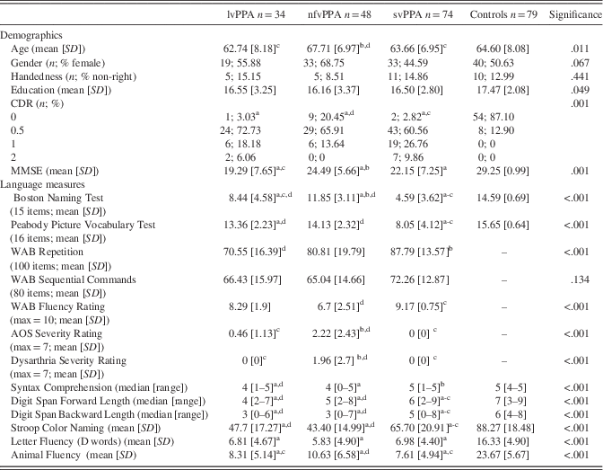
Seventy-nine control participants were recruited through the UCSF Hillblom Study on Healthy Aging and selected based on equivalent testing procedures to our PPA group. Exclusion criteria for controls were a history of major illness, including psychiatric illness, and a score below 27 on the MMSE or CDR greater than 0.5. Table 1 outlines the demographic characteristics of the participants. All participants provided informed written consent; the study was approved by the UCSF Committee on Human Research and conducted in accordance with the Helsinki Declaration.
Demographics and language scores

a vs controls.
b vs lvPPA.
c vs nfvPPA.
d vs svPPA.
WAB=Western Aphasia Battery; AOS=Apraxia of Speech.
Demographic variables were compared using analysis of variance with Bonferroni post hoc tests. There were significant group differences in age at baseline, education, and CDR and MMSE scores (Table 1). The nfvPPA group was significantly older than the other PPA groups, but not controls. Although there was a significant group difference in amount of education, follow-up testing did not reveal specific between group significant differences after controlling for multiple comparisons. Control participants had significantly higher MMSE and significantly lower CDR scores than the PPA sample. Regarding MMSE scores, participants with lvPPA had significantly lower MMSE scores than those with nfvPPA (p=.001) and participants with svPPA had similar MMSE scores to both the lvPPA and nfvPPA groups (p’s>.1). On the CDR, participants with nfvPPA had significantly lower totals than the svPPA group (p=.007) and marginally lower scores than the lvPPA group (p=.06); the lvPPA group had similar CDR scores to the svPPA groups (p>.6). Groups had similar distributions of gender and handedness.
Follow-up assessments were available on 83 participants (17 with logopenic variant, 43 with semantic variant, and 23 with nonfluent/agrammatic variant). The mean visit gap (14.64±6.4 months) did not differ by diagnosis, p>.20 (see Table 4). At follow-up, there was a significant group difference in age; the nfvPPA group was significantly older than the svPPA group. There were no significant group differences at follow-up in education, gender, or handedness.
Neuropsychological Assessment
Neuropsychological testing was administered by research staff or neuropsychology fellows who were trained and supervised by neuropsychologists. Nurses performed the CDR assessment. Neuropsychological testing covered screening of global cognition, processing speed, immediate recall, working memory, visual memory, visuospatial abilities, and executive functioning. In particular, we used an abbreviated Beery VMI to assess visuomotor integration (copying of geometric forms of increasing difficulty), Benson figure copy and recall to examine visuomotor figure construction and visual figure delayed recall (Kramer et al., Reference Kramer, Jurik, Sha, Rankin, Rosen, Johnson and Miller2003; Possin, Laluz, Alcantar, Miller, & Kramer, Reference Possin, Laluz, Alcantar, Miller and Kramer2011), an abbreviated VOSP Number Location as a measure of visuospatial localization (Warrington & James, Reference Warrington and James1991), WAIS Block Design for visuospatial construction and Spatial Span Forward and Backward for visual attention and working memory, WMS Visual Reproduction I and II for visuomotor figure construction and delayed recall (Wechsler, Reference Wechsler1997), a modified and abbreviated trails B type test (visuomotor sequencing that alternates between numbers and days of the week) to evaluate timed visuospatial sequencing and switching (Kramer et al., Reference Kramer, Jurik, Sha, Rankin, Rosen, Johnson and Miller2003), and DKEFS Design Fluency filled dots for figural fluency (Delis, Kaplan, & Kramer, Reference Delis, Kaplan and Kramer2001).
Language measures were also used to confirm PPA diagnoses. We used the fluency rating, sequential commands, and repetition from the Western Aphasia Battery (Kertesz, Reference Kertesz1982); an apraxia of speech and a dysarthria severity rating (Wertz & Rosenbek, Reference Wertz and Rosenbek1991); and abbreviated versions of the Boston Naming Test and Peabody Picture Vocabulary Test, syntax comprehension, digit span length forward and backward, rapid color naming, category fluency (animals), and phomenic fluency (“D” words) from our neuropsychological screen (Kramer et al., Reference Kramer, Jurik, Sha, Rankin, Rosen, Johnson and Miller2003).
Statistical Analyses
Factor and composite scores for a priori domains (total visuospatial, visuospatial construction, immediate recall, delayed recall, and visuospatial executive) were created based on a priori hypotheses about different cognitive domains within the visual-spatial province. Our a priori hypotheses for composite domains stemmed from previous literature that suggested distinct syndromic performances on visuospatial tasks, for example, studies that have reported that the lvPPA variant has difficulties with immediate and delayed recall of visual material (Foxe et al., Reference Foxe, Irish, Hodges and Piguet2013; Ramanan et al., Reference Ramanan, Flanagan, Leyton, Villemagne, Rowe, Hodges and Hornberger2016), and conversely, that the svPPA variant may have somewhat preserved visuospatial abilities particularly with regard to figure copying, attention, and speed (Butts, Machulda, Duffy, Strand, Whitwell, & Josephs, Reference Butts, Machulda, Duffy, Strand, Whitwell and Josephs2015; Viskontas, Reference Viskontas, Boxer, Fesenko, Matlin, Heuer, Mirsky and Miller2011) (see Table 3.).
Composite scores were created by averaging Z-scores from multiple tests that have similar neuropsychological features, for example, delayed recall, for each subject. Specifically, we divided the neuropsychological data into visuospatial subdomains of construction, immediate recall, delayed recall, and executive. The construction composite included performance on the abbreviated Beery VMI, Benson figure copy, and the WAIS Block Design. The immediate recall composite was based on scores on WMS Visual Reproduction I and WAIS Spatial Span Forward and Backward. The delayed recall composite was based on scores on WMS Visual Reproduction II and Benson recall. The visual executive composite included Spatial Span Backward, a ratio of modified trails number of correct lines divided by the completion time, and Design Fluency correct designs. Each subject’s score on each test was standardized based on the overall sample, and then mean Z-scores were computed for each subject for each composite set. We report the composite scores as group averages of these Z-scores.
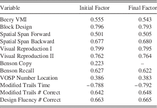
The visuospatial factor score was created using factor analysis on baseline data. Specifically, we used the principal-factor method, Kaiser cutoff of eigenvalues greater than 1, and promax ^ 3 oblique rotation because cognitive variables likely correlate. Regression-based score generation (Osborne & Costello, Reference Osborne and Costello2009) was used in the factor analysis. Initially, we analyzed a visual-spatial factor that included all visuospatial neuropsychological variables (Table 2). However, the initial Benson Figure copy factor loading was 0.22, which is less than a 0.3 cutoff (regression coefficient based on sample size), so that item was removed from the factor. The eigenvalue for the final visual-spatial factor was 4.88 and this factor explained 87.29 percent of the variance in the data. Subject to variable ratio was greater than 10:1 (only complete datasets were included in the factor generation (total N=117 based on controls=34, lvPPA=20, nfvPPA=25, svPPA=38). Participants with complete datasets did not differ from participants without complete datasets on age, handedness, gender, or education (all p’s > .59). However, there were significantly fewer participants with complete datasets who had CDR scores of 2 (n=1 compared to n=8; p=.034). Participants with complete datasets also had higher MMSE scores than participants without complete datasets (diff=3.89; p<.001).
Factor loadings for the first, and only retained, factor.

Note. Initial Factor loadings are given, as well as after removing unrelated variables (Final Factor).
Since the factor analysis included participants with lower disease severity, this likely makes our final factor scores more conservative, as we would expect that group differences by diagnostic group would be subtler among milder patients. Disease severity was included as a covariate in the statistical analyses. The one-factor model is the model that fit the data the best; the next highest factor had an eigenvalue of 0.76, which is below the Kaiser cutoff (for a scree plot see Supplementary Figure S1). To validate that this was in fact a visuospatial factor and not a general cognitive factor, we performed a sensitivity analysis. We ran the same factor analysis but added two language measures, abbreviated forms of confrontation naming and single word comprehension (Kramer et al., Reference Kramer, Jurik, Sha, Rankin, Rosen, Johnson and Miller2003). This analysis yielded a two-factor model, wherein the visuospatial data loaded onto the first factor with an eigenvalue of 5.37 and explained 68.43 percent of the variance while the two language measures loaded on a second factor that had an eigenvalue of 1.89 and explained 24.15 percent of the variance, which suggests that we identified a distinct visuospatial factor rather than a general cognitive factor.
In our analysis of change over time, there was not a significant difference among the PPA groups in time between visits, gender, handedness, or education. There were significant group differences in age at baseline.
Statistical analyses of demographics and neuropsychological scores between groups were performed using Stata version 14 or higher. Multivariate analysis of covariance was used to conduct omnibus significance testing; analysis of covariance and one-way analysis of variance were used to conduct follow-up tests. Non-parametric tests were conducted using Kruskal-Wallis and Dunn’s tests. Bonferroni correction was applied to all follow-up tests of group differences by number of group comparisons, such that findings were considered statistically significant at an alpha level < 0.008 (0.05 divided by six comparisons). Effect sizes were calculated based on Cohen’s d. Omnibus testing of group differences on all neuropsychological factors, composites, and specific tests included age at testing, education, and CDR total as covariates (Table 3). Follow-up tests were also adjusted for age, education, and CDR total differences because we were interested in differences due to visuospatial functioning, rather than absolute performance differences.
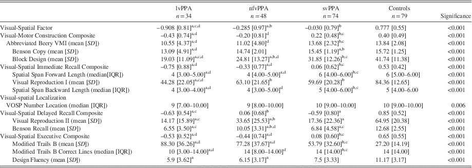
Neuropsychological test factors, composites, and scores

a vs controls.
b vs lvPPA.
c vs nfvPPA.
d : vs svPPA.
n.s.=not significant; IQR=interquartile range.
RESULTS
Cross-sectional Visuospatial Performance
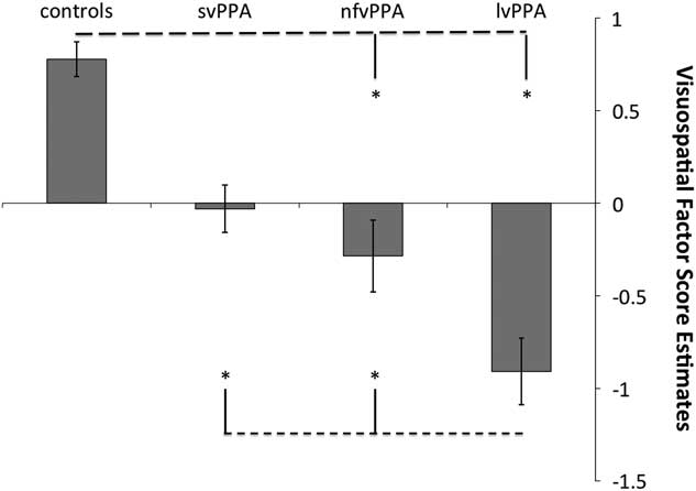
There was a significant group difference in performance on the visual-spatial factor after controlling for age, education, and CDR scores (Figure 1). Controls had significantly higher scores on the visual-spatial factor than the nfvPPA (p=.04; d=0.79) and lvPPA (p < .001; d=1.91) groups, but similar scores to the svPPA group (p>.9). The svPPA group performed better than the lvPPA group (p < .001; d=1.55) but did not differ from the nfvPPA group (p=.102). The nfvPPA group also had significantly better performance than the lvPPA group (p=.03; d=0.79).
Visuospatial Factor score estimates by group, unadjusted. *indicates a significant difference p<.05 after Bonferroni correction for multiple comparisons and adjusted for differences in age, education, and CDR totals.

Group differences on the visuospatial composite scores and individual tests are reported in Table 3 and summarized here.
Visuospatial construction
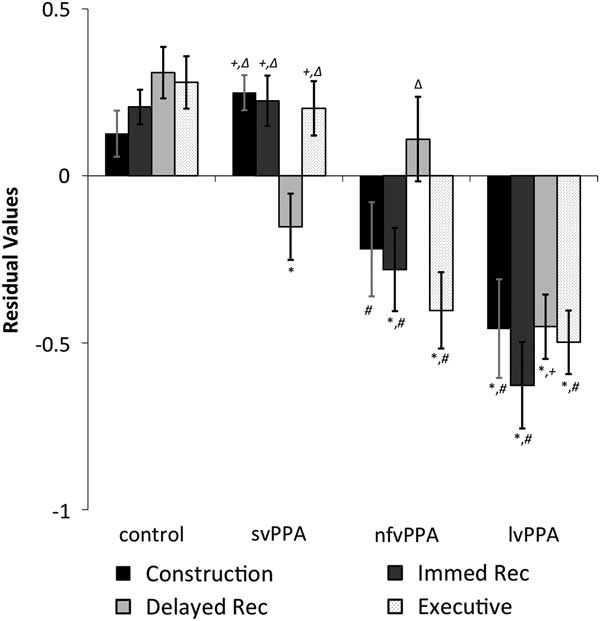
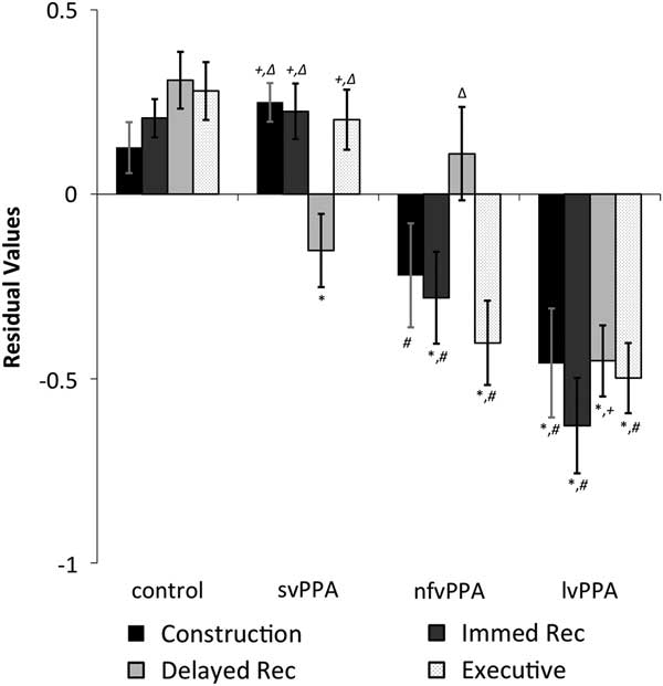
There was a significant main effect of group on the visuomotor construction composite. Post hoc analyses revealed that controls performed significantly better than the lvPPA group (p=.001; d=1.01) and marginally better than the nfvPPA group (p=.080; d=0.56). The svPPA group performed significantly better than the nfvPPA (p=.002; d=0.83) and lvPPA (p<.001; d=1.33) groups, but similarly to controls (p > .9). LvPPA and nfvPPA groups were not significantly different (p=.713).
Visuospatial immediate recall
There was a significant main effect of group on the visual-spatial immediate recall composite. Controls performed significantly better than the nfvPPA (p=.001; d=0.89) and lvPPA (p<.001; d=1.76) groups, but similarly to the svPPA group (p > .9). The svPPA group also performed significantly better than the nfvPPA (p<.001; d=0.79) and lvPPA (p<.001; d=1.43) groups. The nfvPPA group was not significantly different from the lvPPA group (p=.141). There was a significant group effect on both visual-spatial measures of immediate recall (Spatial Span and Visual Reproduction I) (p’s < .001).
Visuospatial localization
On a brief measure of visuospatial localization, there was a significant group difference.
Visuospatial delayed recall
A main effect of group was found on the visual-spatial delayed recall composite. Controls performed significantly better than the svPPA (p=.002; d=0.70) and lvPPA (p<.001; d=1.42) groups, but had a similar level of performance to the nfvPPA group (p > .9). The nfvPPA had significantly higher scores on the delayed recall composite than the lvPPA group (p=.008; d=0.88) but not the svPPA group (p=.407). This was the only composite that differentiated the nfvPPA from the lvPPA group. SvPPA and lvPPA variants had similar scores on the delayed recall composite (p=.346). On each of the delayed recall measures, there were significant group differences.
Visuospatial executive functions
On the visual executive composite, there was a main effect of group (Fig. 2). Follow-up analyses revealed that controls performed significantly better than the nfvPPA (p<.001; d=1.16) and lvPPA (p<.001; d=1.49) groups. The svPPA group also performed significantly better than the nfvPPA (p<.001; d=1.01) and lvPPA (p<.001; d=1.31) groups, but similarly to controls (p>.9). The nfvPPA and lvPPA groups performed similarly (p>.9).
Visuospatial composite residual values after adjusting for differences in age, education, and CDR totals. Symbols indicate a significant difference at p<.05 after Bonferroni correction for multiple comparisons. *vs. controls, #vs. svPPA, +vs. nfvPPA, ∆vs. lvPPA.
Visuospatial Performance Over Time
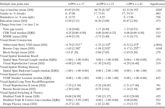
Declines in MMSE or CDR were not significant for any group, controlling for age at baseline differences. Furthermore, only two neuropsychological tests showed a significant group effect for change over time: Beery VMI (p<.002) and the Benson Figure Copy (p=.017) (Table 4.). Follow-up analyses demonstrated that the lvPPA group’s performance on the abbreviated Beery VMI declined by an average of 4.8 points in approximately a year and this amount of decline was significantly different from the other PPA groups (effect sizes: vs. nfvPPA=−1.16; and vs. svPPA=−1.47). There were no other significant differences between groups on change in performance on the abbreviated Beery VMI. On the Benson figure copy, the svPPA group showed a small improvement in performance of almost two tenths of a point and this small improvement was significantly different from the change seen in the other two PPA groups, both of which had a decline in performance by more than 1.5 points (effect sizes: vs. nfvPPA =0.94; vs. lvPPA=0.89).
Neuropsychological test scores change from time 1 to time 2

b vs lvPPA.
c vs nfvPPA.
d vs svPPA.
n.s.=not significant; IQR=interquartile range.
DISCUSSION
Our study aimed to elucidate the performance of patients with PPA on visuospatial neuropsychological tasks using a large sample of PPA participants and an extensive neuropsychological battery. We identified that one visuospatial factor could account for most of the variance in the data. Based on this factor, the lvPPA group had significantly worse overall performance. The svPPA group performed better than the lvPPA group on every visuospatial composite except the delayed recall composite where they performed similarly. The nfvPPA group had significantly higher scores than the lvPPA group on the delayed recall composite and this was the only composite that differentiated these two variants.
When performance over time was examined, only the lvPPA group had a significantly greater decline than other groups on the Beery VMI. The svPPA group showed a small improvement in performance over time on the Benson figure copy whereas the other PPA groups showed a slight decline, which was a significant group difference.
Difficulties in visuospatial tests were present in the PPA sample at baseline even after controlling for disease severity, which suggests that visuospatial functioning is affected in this population even if it is less severely affected than language deficits and does not result in a functional impairment in everyday life. Our study investigated visuospatial cognition across several tasks and sub-domains and showed that the different variants have distinct difficulties on visuospatial tasks that mirror their difficulties in language sub-domains and are consistent with patterns of anatomical damage in each variant. However, this is not the first time visuospatial difficulties have been reported in this population.
A few studies have expanded the non-linguistic cognitive literature base in PPA. Foxe et al. (Reference Foxe, Irish, Hodges and Piguet2013) found that patients with lvPPA performed as poorly as patients with classic Alzheimer’s disease (AD) on visuospatial short-term memory tasks. While the lvPPA patients performed worse on a verbal auditory (digit) span, they were similarly impaired on the spatial span. A subsequent analysis by Foxe et al. (Reference Foxe, Leyton, Hodges, Burrell, Irish and Piguet2016) demonstrated that this dissociation in span performance also related to distinct cortical thinning patterns. Both Foxe studies only investigated the lvPPA variant of PPA, which left a question remaining about the performance of the other two PPA variants on these tasks. Our study showed that both the lvPPA and nfvPPA groups were impaired on visuospatial immediate recall tasks but that the svPPA group was not, a pattern similar to their language profiles and differential patterns of dorsal versus ventral atrophy, respectively (Henry, Wilson, Babiak, Mandelli, Beeson, Miller, & Gorno-Tempini, Reference Henry, Wilson, Babiak, Mandelli, Beeson, Miller and Gorno-Tempini2016).
Butts et al. (Reference Butts, Machulda, Duffy, Strand, Whitwell and Josephs2015) studied all three PPA variants and found that there were significant group differences between the PPA variants on figure copying, some aspects of visual memory, and a visuospatial executive functioning task. In particular, the lvPPA variant had lower scores than the nfvPPA variant on visual memory, and lower scores than the svPPA variant on figure copying, visual immediate recall, and visual executive functioning. Many of our findings are similar to those reported by Butts et al. (Reference Butts, Machulda, Duffy, Strand, Whitwell and Josephs2015), with the exception of delayed recall in the svPPA group. Butts et al. (Reference Butts, Machulda, Duffy, Strand, Whitwell and Josephs2015) reported average visual recall in an svPPA sample; it is possible that sample size played a role in this difference and/or that this sample differed from ours in disease severity, length of disease, or degree of left versus right atrophy.
Classically, svPPA is associated with greatest atrophy in the left temporal lobe initially but volume loss in the right temporal lobe is nevertheless present, including the medial temporal lobe, and becomes more severe over time (Henry et al., Reference Henry, Wilson, Ogar, Sidhu, Rankin, Cattaruzza and Seeley2014; Kumfor et al., Reference Kumfor, Landin-Romero, Devenney, Hutchings, Grasso, Hodges and Piguet2016; Rohrer et al., Reference Rohrer, McNaught, Foster, Clegg, Barnes, Omar and Fox2008). Visuospatial memory impairments associated with right temporal lobe damage have been reported in the literature (Milner, Johnsrude, & Crane, Reference Milner, Johnsrude and Crane1997; Pigott & Milner, Reference Pigott and Milner1993). Therefore, there could be an anatomical basis for visuospatial delayed recall difficulties in svPPA. Further studies are needed to better understand when and how individuals with svPPA have difficulties with visuospatial delayed recall.
Ramanan et al. (Reference Ramanan, Flanagan, Leyton, Villemagne, Rowe, Hodges and Hornberger2016) analyzed non-verbal episodic memory in PPA and found that, of a few different episodic memory tests, performance on delayed recall of the Rey Complex Figure was the most powerful discriminator between lvPPA and nfvPPA patients, with the lvPPA patients more impaired. Our result of impairment in delayed visual recall in the lvPPA group supports the similar finding by Ramanan et al. (Reference Ramanan, Flanagan, Leyton, Villemagne, Rowe, Hodges and Hornberger2016) but extends the finding to include the domain of visual delayed recall; a finding that may relate to Alzheimer’s disease targeting medial temporal lobe structures (Ossenkoppele et al., Reference Ossenkoppele, Cohn-Sheehy, La Joie, Vogel, Moller, Lehmann and Rabinovici2015).
Overall, the lvPPA group had the lowest scores on the visuospatial factor, which was expected given that part of the clinical criteria for lvPPA includes parietal atrophy on structural MRI or hypometabolism on PET/SPECT. Future studies examining the neuroanatomical and neurometabolic correlates of visuospatial performance in the lvPPA group will help to clarify these associations.
One surprising outcome of this study is the degree of impairment the nfvPPA group evidenced on visuospatial tasks given that visuospatial processing is commonly thought of as a right parietal activity. One possible reason the nfvPPA displayed difficulty on these tasks is that several of the tasks rely on visuomotor abilities and nfvPPA has been associated with degradation of white matter pathways connecting the left inferior frontal gyrus (Broca’s area) to premotor and supplementary motor regions (Budisavljevic et al., Reference Budisavljevic, Dell’Acqua, Djordjilovic, Miotto, Motta and Castiello2017; Mandelli et al., Reference Mandelli, Caverzasi, Binney, Henry, Lobach, Block and Henry2014). In this sense, the deficits may relate more to motor planning and sequencing. However, further investigation is necessary to determine the underlying mechanism.
Of all the visuospatial variables we analyzed, decline in only two were found to have a main effect of group, raising the question of practice effects on several tests. Both tasks that showed decline were figure-copying tasks, and the lvPPA and nfvPPA groups showed the most decline. These tasks require integration of multiple cognitive abilities and poor performance can be due to damage within the dorsal frontoparietal network (Possin et al., Reference Possin, Laluz, Alcantar, Miller and Kramer2011).
Strengths of the study include a large PPA sample, investigation of visuospatial functioning overall and within different sub-domains, and a multiple time point perspective. Limitations include a relatively small follow-up sample. Additionally, although we tried to control for disease severity, the CDR may provide a biased perspective on disease severity for some PPA groups more than others because of a greater emphasis placed on memory impairments compared with other functions such as speech intelligibility. Future studies should include more time points with larger longitudinal samples and specifically investigate earlier patterns of cognitive dysfunction with the PPA population as well as more specific models and evidence for how visuospatial functioning is distributed across the brain.
In conclusion, performance on visuospatial cognition highlights differential patterns of performance in the PPA variants, likely in relation to the underlying cognitive and anatomical deficits. The results provide important information for differential diagnosis within the context of PPA and for understanding cognition in PPA more broadly.
ACKNOWLEDGMENTS
This work was funded by the National Institutes of Health (M.T., NINDS R01 NS050915), (M.T., NIDCD K24 DC015544), (B.M., NIA P50 AG03006), (B.M., NIA P50 AG023501), (B.M., NIA P01 AG019724); (M.T., State of California (DHS04-35516)); (B.M., Alzheimer’s Disease Center of California (03-75271 DHS/ADP/ARCC)); (J.K., Larry L. Hillblom Foundation); (B.M., John Douglas French Alzheimer’s Foundation); Koret Family Foundation; (B.M., Consortium for Frontotemporal Dementia Research); and (B.M., McBean Family Foundation). Disclosures; Dr. Possin receives research funding from Quest Diagnostics. Dr. Rabinovici receives research support from Avid Radiopharmaceuticals, GE Healthcare, and Piramal, and has received consulting fees or speaking honoraria from Roche, Eisai, Lundbeck, Piramal and Putnam. Dr. Kramer receives royalties from Pearson, Inc. for the California Verbal Learning Test. Dr. Bruce L. Miller receives grant support from the NIH/NIA and the Centers for Medicare & Medicaid Services (CMS) as grants for the Memory and Aging Center. As an additional disclosure, Dr. Miller serves as Medical Director for the John Douglas French Foundation; Scientific Director for the Tau Consortium; Director/Medical Advisory Board of the Larry L. Hillblom Foundation; Scientific Advisory Board Member for the National Institute for Health Research Cambridge Biomedical Research Centre and its subunit, the Biomedical Research Unit in Dementia (UK); and Board Member for the American Brain Foundation (ABF). Dr. Tempini has received personal compensation in an editorial capacity from Neuroimage Clinical. No other disclosures exist.
Supplementary material
To view supplementary material for this article, please visit https://doi.org/10.1017/S1355617717000984