Arrhythmias in tetralogy of Fallot
Since the first surgical repair of tetralogy of Fallot by Dr Lillihei in 1955,Reference Lillehei, Cohen and Warden1 the surgical approach to tetralogy of Fallot has undergone many advances, and although there is still no consensus to the timing and technique for optimal repair these patients are surviving well into adulthood. Unfortunately, there is still significant morbidity and mortality following surgical repair of tetralogy of Fallot, and a large proportion of the morbidity is related to arrhythmias, both atrial and ventricular. These arrhythmias can be the result of re-entrant tachycardias due to diseased myocardium around surgical scars, from myocardial fibrosis due to haemodynamic derangements due to residual cardiac lesions, or from primary myocardial disease. Therefore, arrhythmia surveillance is critical to the management of children and adults after surgical repair of tetralogy of Fallot.
The most significant haemodynamic derangement in these patients is right ventricular volume and pressure overload from pulmonary insufficiency and/or stenosis. Every patient will have some degree of right ventricular outflow tract dysfunction and these tend to worsen over time. Pulmonary insufficiency results in volume overload of the right ventricle, causing right ventricular chamber dilation. This in turn can contribute to the widening of the QRS duration, a finding that can be a predilection for ventricular arrhythmias and sudden cardiac death.Reference Abd El Rahman, Abdul-Khaliq and Vogel2, Reference Gatzoulis, Till, Somerville and Redington3 In addition, patients may be predisposed to decreased left ventricular systolic function, ventricular tachycardia, and sudden cardiac death.Reference Ghai, Silversides, Harris, Webb, Siu and Therrien4 The majority of research in this patient population has focused on ventricular arrhythmias that can lead to sudden death; however, these patients also are at risk for developing supraventricular re-entrant arrhythmias, such as atrial flutter and fibrillation, especially in older individuals. This article will review the mechanism of arrhythmias in patients with tetralogy of Fallot and describe arrhythmia management, in the acute and chronic setting.
Peri-operative arrhythmias
In the modern era, it is known that morbidity and mortality are improved when surgical repair of tetralogy of Fallot occurs at a young age.Reference Van Arsdell, Maharaj and Tom5 Some centres advocate for neonatal repair,Reference Hirsch, Mosca and Bove6 whereas other centres may provide palliative procedures in patients with insufficient pulmonary blood flow with a systemic-to-pulmonary arterial shunt and electively repair them at 3–6 months of age in order to minimise the surgical trauma to the right ventricle.Reference Morales, Zafar and Fraser7 Both approaches have low post-operative mortality and equal long-term outcomes.Reference Kanter, Kogon and Kishbom8 Arrhythmia substrates, such as ventricular pre-excitation, are uncommon in this patient population, although a baseline electrocardiogram should be obtained shortly after birth. In addition to confirming the diagnosis, an echocardiogram will evaluate the cardiac function and identify any lesions that may be haemodynamically significant and therefore precipitate arrhythmias, such as valvar insufficiency/stenosis and chamber dilation, although arrhythmias in this age group are uncommon. Continuous cardiorespiratory monitoring with a telemetry system should be routine during the hospitalisation. Tetralogy of Fallot is associated with genetic syndromes, such as DiGeorge, Velo-Cardio-Facial, and Alagille syndrome, which may have other comorbidities that can affect other organ systems and electrolyte regulation and therefore the predisposition for arrhythmias.
The operative period exposes the neonate with congenital heart disease to a vast array of physiological and non-physiological machinations that further compound the proclivity for arrhythmias. Diligent care to maintain a normal electrolyte and pH balance, ensuring no residual haemodynamic lesions that require intervention and maintenance of sinus rhythm is critical to minimise the risk of peri-operative arrhythmia development. Antiarrhythmic medications may be necessary when arrhythmias persist despite normalisation of the metabolic milieu, although these often improve in the following several months following surgery.
One post-operative arrhythmia following tetralogy of Fallot repair that is challenging to manage is junctional ectopic tachycardia. It occurs following ventricular septal defect repair or when there is traction applied close to the atrioventricular node and it generally leads to haemodynamic compromise. It is the result of irritation to the atrioventricular node and its associated conduction tissue. This irritation causes excitability of the atrioventricular node, resulting in a rapid narrow-complex tachycardia with atrioventricular dissociation – ventricular rate exceeds the atrial rate – as shown in Figure 1. Complex ventricular septal defect repair, especially those involving the perimembranous area, along with prolonged bypass times are at risk for junctional tachycardia.Reference Hoffman, Bush and Wernovsly9 Patients can have such profound haemodynamic compromise that might even require extreme mechanical support as rescue, such as extracorporeal membrane oxygenation. Junctional ectopic tachycardia generally occurs in the first 24–48 hours after surgery, and aggressive management is important. Weaning inotropes and cooling the patient are helpful. As a general approach, cooling below 35.5°C is avoided. Overdrive atrial pacing is often attempted to restore atrioventricular synchrony, which is successful as long as the junctional rate can be slowed with medications and other measures. Correcting acidosis and electrolytes are key strategies. Magnesium in particular should be given if tolerated. If these strategies do not work, then intravenous amiodarone should be promptly initiated. In a neonate with already compromised haemodynamics, care must be given when administering intravenous amiodarone. It should be given in doses of 5 mg/kg and run over 20–60 minutes. Pre-medicating infants with a bolus of calcium chloride often prevents the hypotension that can be seen with rapid amiodarone administration. Procainamide is a second agent that can be used to treat junctional tachycardia. It is less potent than amiodarone and therefore not used as commonly, but may be better tolerated.Reference Madapati, Byrum and Kavey10 There is increasing evidence that dexmedetomidine may be useful in post-operative junctional ectopic tachycardia.Reference Chrysstomou, Beerman and Shiderly11 Typically, once sinus rhythm is restored and post-surgical inflammation resolves, this arrhythmia subsides and does not require long-term treatment.
ECG depicting junctional ectopic tachycardia. Junctional ectopic tachycardia with a bundle branch block following surgical repair of tetralogy of Fallot in a 2-year-old boy. The arrows point to the atrial signals demonstrating the slow atrial rate after administration of amiodarone. ECG = electrocardiogram.

Atrioventricular block can also occur following surgery because of mechanical injury to the atrioventricular node, especially during closure of the ventricular septal defect. Atrioventricular demand pacing should be performed using temporary pacing wires and atrioventricular conduction should be monitored daily. If atrioventricular conduction does not return by post-operative day 10, a permanent epicardial pacing system should be placed.Reference Epstein, DiMarco and Ellenbogen12 Although late-onset atrioventricular node block was once thought to be a major cause of sudden cardiac death in these patients,Reference Quattlebaum, Varghese and Neill13 this is no longer thought to be true.
Treatment for peri-operative arrhythmias outside of the post-operative state will vary from institution to institution. Most arrhythmias seem to resolve within the first 6 months of surgery. Consideration should be made to wean off medication within 6–12 months of therapy and continue to monitor for arrhythmia recurrence. Immediate post-operative arrhythmias do not always predict recurrence later in life.
Late-onset arrhythmias
Adolescents and adults following repair of tetralogy of Fallot are prone to developing late-onset supraventricular and ventricular arrhythmias. They are relatively uncommon in the first two decades of life.Reference Khairy, Aboulhoson and Gurvitz14 However, as these patients are surviving late into adulthood, the incidence of arrhythmias becomes relatively high, and accounts for significant morbidity and mortality. The most common cause of death in patients after surgical repair of tetralogy of Fallot is sudden death, presumably from ventricular arrhythmias.Reference Gatzoulis, Balaji and Webber15, Reference Khairy, Dore and Talajic16 Fortunately, the incidence of sudden death is low, with an annual incidence of 0.15.Reference Silka, Hardy, Menashe and Morris17 Unfortunately, the arrhythmia burden in this patient population continues to be quite high. In one recent multi-centre study, 30% of adults with repaired tetralogy of Fallot had a sustained arrhythmia.Reference Khairy, Aboulhoson and Gurvitz14 The majority of these were atrial, although ventricular tachycardia was the most common single arrhythmia subtype. Intra-atrial re-entrant tachycardia and atrial fibrillation were the most common atrial arrhythmias, with atrial fibrillation being more common in those >55 years of age.
The surgical approach to the repair of tetralogy of Fallot has been shown to have a significant impact in the development of arrhythmias. Earlier repairs were performed using a transventricular approach. More contemporary approaches involve transatrial/transpulmonary approaches and valve sparring procedures when possible. Procedures to minimise a ventriculotomy seem to have a low arrhythmia burden in medium-term follow-up.Reference Dietl, Cazzanigna, Dubner, Perez-Balino, Torres and Favaloro18, Reference Nui, Morris and Morales19 Patients who are repaired at a later age or who have a longer cardiopulmonary bypass run may be factors for post-operative mortality, both early and late.Reference Hamada, Terai, Jibiki, Nakamura, Gatzoulis and Niwa20 The use of a transannular patch has been shown to be a risk factor for the development of late ventricular arrhythmias.Reference Gatzoulis, Balaji and Webber15
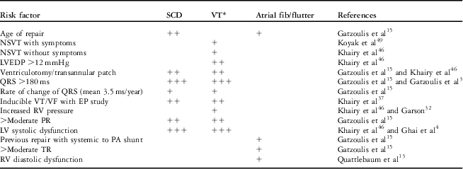
Nonetheless, early identification of patients at risk of arrhythmias, in particular ventricular arrhythmias and sudden death, remains challenging. There is no single identified risk factor, other than perhaps resuscitated sudden events, which predicts sudden death. Multiple studies have identified certain haemodynamic and electrophysiological properties that warrant further investigation and perhaps implantable cardioverter-defibrillator implantation for primary prevention. These risk factors include surgical correction at an older age,Reference Gatzoulis, Balaji and Webber15 the use of a transannular patch during the primary repair,Reference Gatzoulis, Balaji and Webber15 moderate to severe pulmonary regurgitation,Reference Gatzoulis, Balaji and Webber15 a history of ventricular tachycardia, a QRS duration >180 ms,Reference Lillehei, Cohen and Warden1 and left ventricular systolic and/or dysfunction.Reference Gatzoulis, Balaji and Webber15 In addition, patients with at least moderate tricuspid insufficiencyReference Gatzoulis, Balaji and Webber15 and the presence of right ventricular diastolic dysfunctionReference Quattlebaum, Varghese and Neill13 correlate with the development of atrial arrhythmias. Risk factors for the development of arrhythmias are summarised in Table 1.
Risk factors for arrhythmia development after tetralogy of Fallot repair.
EP = electrophysiology; ICD = implantable cardioverter-defibrillator; LV = left ventricular; LVEDP = left ventricular end-diastolic pressure; NSVT = non-sustained VT; PA = pulmonary artery; PR = pulmonary regurgitation; RV = right ventricular; SCD = sudden cardiac death; TR = tricuspid regurgitation; VF = ventricular fibrillation; VT = ventricular tachycardia
*Includes appropriate ICD discharge data, which do not necessarily predict SCD
The management of these patients, especially as they become adults, should therefore focus around identifying risk factors. These physiological effects are a continuum and progression is inevitable. Although a patient may not meet criteria at one point in time or be considered high risk at one time is not necessarily true in the following years. Careful diligence must be carried out when ascertaining symptomatology and routine diagnostic testing during follow-up visits. Non-invasive imaging is the mainstay for long-term follow-up of these patients, primarily with echocardiography, although cardiac magnetic resonance can perhaps more accurately measure right ventricular volumes and diastolic and systolic function, which are important variables in the development of ventricular arrhythmias.Reference Knauth, Gauvreau and Powell21 In addition, the degree of fibrosis can be readily characterised by cardiac magnetic resonance, a substrate for ventricular arrhythmias.Reference Babu-Narayan, Kilner and Li22 Both systolicReference Ghai, Silversides, Harris, Webb, Siu and Therrien4 and diastolic dysfunctionReference Aboulhoson, Lluri and Gurvitz23 have been found to be associated with arrhythmia development and should be assessed on every study performed in these patients.
As right ventricular dilation progresses, the QRS duration increases as a result of mechano-electrical interactions.Reference Abd El Rahman, Abdul-Khaliq and Vogel2, Reference Gatzoulis, Till, Somerville and Redington3 In addition to right ventricular dilation, regional wall motion abnormalities are commonly seen, which contributes to the electrical malady of the right ventricle following tetralogy of Fallot repair.Reference Vogel, Sponring and Cullen24 Ventricular mass and function has also been shown to cause heterogeneous depolarisation/repolarisation, resulting in a QRS prolongation.Reference Helbing, Roest and Niezen25 Multiple studies have demonstrated that a QRS duration >180 ms on a surface electrocardiogram has been associated with ventricular arrhythmias and sudden death, which can be considered a surrogate for the overall health of the right ventricle. Owing to the fact that the right ventricle is subject to more haemodynamic derangements and hence progressive hypertrophy, fibrosis, dilation, and dysfunction over time, the QRS can progressively become more prolonged. Several studies have shown that restoring pulmonary valve competency can slow the progression of haemodynamic derangements and the associated prolongation of QRS duration.
More advanced electrocardiographic parameters may prove to be useful in identifying patients at risk for the development of ventricular arrhythmias. Use of signal-averaged electrocardiogram was shown in one recent study to be more predictive of the development of ventricular arrhythmias than absolute QRS duration on a baseline surface electrocardiogram.Reference Russo, Folino and Mazzotti26 Microvolt T-wave alternans, a measure of repolarisation homogeneity – variation in beat-to-beat T-wave amplitude – and QT/JT dispersion, another marker for identifying ventricular repolarisation abnormalities, are other electrocardiographic modalities that have been studied in patients with tetralogy of Fallot, although they have not been shown to be superior to QRS duration in predicting risk of ventricular arrhythmias as of yet.Reference Chiu, Chiu and Wang27, Reference Berul, Hill and Geggel28 Unfortunately, these modalities are not readily available in many centres.
In addition to baseline electrocardiograms, ambulatory Holter monitoring can be a useful adjunct to detecting arrhythmias in these patients, especially in asymptomatic patients.Reference Czosek, Anderson and Khoury29 One recent study demonstrated that up to 40% of patients following repair of tetralogy of Fallot had arrhythmias on Holter monitoring, the majority of which were asymptomatic.Reference Rodriguez, Moodie and Neeland30 Therefore, these should be used routinely during follow-up in patients, especially in older patients in whom the arrhythmia risk is higher. Holters have not been shown to be very specific, however, in predicting which patients will go on to have sudden death.Reference Cullen, Celermajer and Franklin31
The presence of premature ventricular contractions was identified as a marker for sudden death in a study by Garson in 1979.Reference Garson, Nihill and McNamara32 It was noted that premature ventricular contraction were found on a resting electrocardiogram in 21 out of 217 adolescents and young adults after tetralogy of Fallot repair and seven of those patients died suddenly, whereas no sudden deaths were observed in the remaining 186 patients. These patients had more significant right ventricular systolic dysfunction and/or right ventricular hypertension, and it is likely that the premature ventricular contractions are a marker for worsening haemodynamics. Harrison et alReference Harrison, Harris and Siu33 demonstrated that the presence of frequent premature ventricular contractions predicted the development of sustained ventricular tachycardia. The appearance or increase in ectopy should therefore prompt an investigation to search for right ventricular abnormalities that may give one the propensity of ventricular arrhythmias and sudden death.
Exercise stress testing is a common outpatient test used in older children and adults after repaired tetralogy of Fallot. Arrhythmias are commonly seen during exercise testing, and can be as high as 73%.Reference Roos-Hesselink, Perlroth and McGhie34 Although ventricular tachycardia is seen in older patientsReference Wessel and Paul35 and in patients with higher right ventricular pressures,Reference Garson, Gillette and Gutgesell36 no studies have been able to demonstrate that exercise-induced arrhythmias predict sudden cardiac death.
Invasive electrophysiology studies are a useful tool to help identify those patients at high risk of sudden death. A multi-centre retrospective review by Khairy et al demonstrated the significance of programmed ventricular stimulation in repaired tetralogy of Fallot. Out of 252 patients, up to 35% had inducible ventricular arrhythmias, and in multivariate analysis inducible sustained ventricular tachycardia was an independent risk factor for the development of clinical ventricular tachycardia or sudden cardiac death.Reference Khairy, Landzberg and Gatzoulis37 In addition to risk stratification, both atrial and ventricular ablations can be performed using 3-D electroanatomical mapping to accurately define activation patterns and areas of scar with voltage mapping, critical for re-entrant arrhythmias commonly seen in these patients. Using this technology, up to 98% acute success has been demonstrated in post-operative atrial tachycardia ablations, although recurrence is common.Reference De Groot, Lukac and Schalij38 Successful ablation of ventricular tachyarrhythmias using voltage mapping to identify critical isthmuses for re-entrant arrhythmias can also be achieved.Reference Zeppenfeld, Schalij and Bartelings39Figure 2 demonstrates the four critical ventricular tachycardia isthmuses following surgical repair of tetralogy of Fallot, which are the targets for ablation of these ventricular arrhythmias. These occur between the right ventricular outflow tract patch and tricuspid annulus (isthmus 1), right ventricular scar from a ventriculotomy and the pulmonary valve (isthmus 2), scar from the ventricular septal defect repair to the pulmonary valve (isthmus 3), and the ventricular septal defect scar to the tricuspid annulus (isthmus 4).Reference Zeppenfeld, Schalij and Bartelings39
VT isthmus following tetralogy of Fallot repair. Schematic of the localisation of anatomic boundaries (blue lines) for VT after repair of CHD and the resulting anatomic isthmuses (red lines). CHD = congenital heart disease; RV = right ventricle; RVOT = right ventricular outflow tract; TA = tricuspid annulus; VSD = ventricular septal defect; VT = ventricular tachycardia. Modified from ZeppenfeldReference Zeppenfeld, Schalij and Bartelings39 with permission.
For patients at risk of ventricular arrhythmias and sudden death, the management strategy should focus on reducing the risk. Pulmonary valve replacement has clearly been shown to improve pulmonary regurgitation, reduce right ventricular chamber size, and stabilise the QRS duration. Multiple studies have shown that severe right ventricular dilation (>170 ml/m2) does not improve following pulmonary valve replacement and therefore may not prevent sudden death.Reference Knauth, Gauvreau and Powell21, Reference Therrien, Provost and Mechant40 Several guidelines have been published that help define timing of pulmonary valve replacements.Reference Warnes, Williams and Bashore41–Reference Baumgartner, Bonhoeffer and De Groot43 Whether this changes the risk of ventricular arrhythmias and sudden death still remains debatable. Gatzoulis et alReference Gatzoulis, Balaji and Webber15 showed that there was no sudden death in patients following pulmonary valve replacement late after surgical repair in a large cohort, concluding that pulmonary valve replacement may prevent sudden death. However, a more recent retrospective, single-institutional, match-controlled study did not demonstrate a decrease in ventricular tachycardia or survival in patients following late pulmonary valve replacement.Reference Harrild, Berul and Cecchin44 Nonetheless, preservation of right ventricular size and function in order to minimise fibrosis as an arrhythmia substrate and improve haemodynamics is imperative in determining the timing of pulmonary valve replacement. Intra-operative cryoablative techniques can be applied at the time of pulmonary valve replacement, which may decrease the future arrhythmia risk even further.Reference Therrien, Siu and Harris45
Implantable cardioverter-defibrillators have been shown to be successful in converting potentially life-threatening ventricular arrhythmias into sinus rhythm, potentially preventing sudden death.Reference Khairy, Harris and Landzberg46, Reference Berul, Van Hare and Kertesz47 Selecting those thought to be high enough risk to benefit from implantable cardioverter-defibrillator implantation still remains unclear. Experiences with implantable cardioverter-defibrillator therapy in tetralogy of Fallot are mixed. Many patients will receive an “appropriate” shock for ventricular tachycardia, although it is unclear whether all those shocks would have resulted in sudden death. In fact, tetralogy of Fallot itself is an independent risk factor for appropriate implantable cardioverter-defibrillator therapy in one multi-centre study looking at adults with congenital heart disease.Reference Yap, Roos-Hesselink and Hoendermis48 Unfortunately, these patients also receive a high number of inappropriate shocks, up to 42% in one study,Reference Koyak, de Groot and Bouma49 and high complication rates continue to be a major problem in this patient population. Therefore, careful consideration of risk factors using the tools mentioned above should be used to help guide which patients will benefit most from implantable cardioverter-defibrillators, as outlined in Figure 3.
Electrocardiographic risk factors for SCD. Known risk factors for sudden cardiac death following tetralogy of Fallot repair. (a) A prolonged QRS in a patient with tetralogy of Fallot, pulmonary atresia. A different patient with palpitations had a non-sustained episode of ventricular tachycardia on a Holter monitor (b) and subsequent inducible ventricular tachycardia/fibrillation on an EP study (c) and underwent ICD implantation. EP = electrophysiology; ICD = implantable cardioverter-defibrillator; SCD = sudden cardiac death; TOF = tetralogy of Fallot.
Conclusion
Although tremendous advances have been made to the surgical and medical management of patients with tetralogy of Fallot, the risk of arrhythmia development and sudden death remains significant. It is clear that pulmonary insufficiency leads to right ventricular dilatation, which results in scarring and electrical changes that predispose these patients to arrhythmias. Optimal timing for pulmonary valve replacement is still controversial. In addition, the ideal management strategy of patients with arrhythmias, in particular ventricular arrhythmias, can be debated. It does not seem that antiarrhythmics are particularly effective in preventing sudden death in themselves. However, not all patients with ventricular arrhythmias will have sudden death, and thus ongoing risk assessment is important. The technology of catheter ablation of arrhythmias, both in the catheterisation laboratory and intra-operatively, have improved and are necessary components to the treatment of arrhythmias. In addition, implantable cardioverter-defibrillator implantation remains the most effective way to prevent sudden death, although which patients require them is not always clear. Future multi-centre studies may help better answer some of these ongoing questions.