In developing countries such as India, many patients with conotruncal anomalies are palliated and many patients underwent primary repair of these anomalies at a later age, which predisposes these patients to dilation of the ascending aorta. Reference Chiostri, Piccinini and Vrancic1
Case report
A 5-year-old male child with a weight of 16 kg underwent modified Blalock–Taussig shunt through midline sternotomy (between innominate artery and right pulmonary artery) and left pulmonary artery plasty at 3 months of age presented to our institution with cyanosis for the last 2 months. On examination, patient had cyanosis with saturation on room air of 62%.
Echocardiography revealed a large single subaortic ventricular septal defect with long-segment pulmonary atresia with confluent pulmonary arteries, patent shunt with dilated ascending aorta and trivial aortic regurgitation.
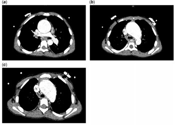
The computerised tomographic angiography (CTA) demonstrated confluent pulmonary artery with long-segment pulmonary atresia. Aneurysmally dilated ascending aorta with a size of 4.4 cm and proximal arch measuring 3.7 cm at the base of innominate artery, sinus, and annulus within normal range and no large aortopulmonary collateral (Fig 1a–c).
( a – c ) CTA showing confluent pulmonary artery with dilated ascending aorta and dilated arch.

Surgical technique
Redo median sternotomy was done and the heart along with right neck was then dissected and right carotid artery was looped with a vessel loop.
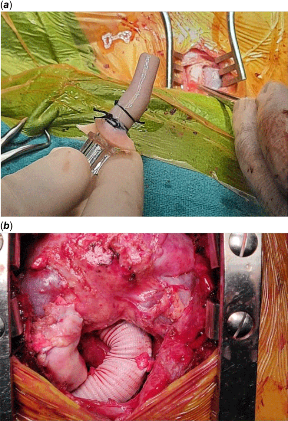
Heparin was given at a dose of 4 mg/kg and the carotid artery was anastomosed to innominate artery homograft (Fig 2a) and bi-caval cannulation was done and cardiopulmonary bypass was commenced. Shunt was clipped and taken down. Heart was vented through the right superior pulmonary vein. Aorta was cross-clamped and after cardioplegia supra-coronary ascending aorta was excised. Right ventricular outflow tract was opened and infundibular resection was done and ventricular septal defect was closed. Pulmonary artery was opened and the distal anastomosis was done end to side with aortic homograft.
( a and b ) Intraoperative image showing homograft innominate artery being anastomosed to carotid artery and final picture after the procedure.

#22 Dacron graft was taken and anastomosis was done at supra-coronary level. Since proximal arch was also dilated, hemi-arch repair was done using the open arch technique. Flows were reduced to one-third during that time., innominate artery and left carotid artery was clamped and near-infrared spectroscopy was observed which did not fall on the left side. Aortic cross-clamp was opened and the left subclavian artery was blocked using Fogarty catheter and the sucker was placed in descending thoracic aorta. Undersurface of the arch was excised and distal anastomosis of dacron graft was done with lower body circulatory arrest. After anastomosis clamped was released and full flows were resumed. Proximal anastomosis at the right ventricular outflow tract was reconstructed on beating heart using pericardial hood (Fig 2b).
Patient aortic cross-clamp time was 120 minutes and cardiopulmonary bypass time was 208 minutes with lower body circulatory arrest time of 17 minutes.
Post-operative transoesophageal echocardiography showed good biventricular function with no significant right ventricular outflow tract gradient and trivial aortic regurgitation.
Patient was extubated on post-operative day 1 and was discharged on post-operative day 10 in stable condition. Patient did not have any cognitive issues post-operatively. Patient was asymptomatic at 3 months of follow-up.
Biopsy of the excised aortic specimen showed elastic fragmentation, increased ground substance, medial necrosis, smooth muscle disarray, and fibrosis.
Discussion
Aortic dilatation is a known entity in corrected, uncorrected, and palliated conotruncal anomalies. Risk factors of aortic dilation in a patient with conotruncal anomalies are male sex, diagnosis of tetralogy of Fallot, pulmonary atresia seems to have aortic dilatation at an early age, Reference Francois2 right aortic arch, longer intervals between palliation and correction, older age at correction, a residual ventricular septal defect, and aortic regurgitation. Reference Vida, Triglia and Zanotto3
Mechanism of aortic dilation in these patients is either due to haemodynamic stress Reference Vida, Triglia and Zanotto3 and/or intrinsic pathology of aorta related to medionecrosis, elastic fragmentation, and elastic lamellae disruption. Reference Kim, Na, Baek and Yang4
Concomitant surgery of dilated aorta during primary repair is uncommon, however, few cases are reported. Kiziltan et al reported combined repair of tetralogy and aortic root replacement in 43-year old. Reference Kiziltan, Topcu, Ozbarlas and Kayaselcuk5 Benjamin Chiostri et al reported a combined Rastelli and Bentall’s procedure for a 32-year-old patient with good outcome. Reference Chiostri, Piccinini and Vrancic1
Although surgeries related to late complications with tetralogy group on the right side of heart have relatively good outcomes, however, patients which require surgery due to aortic pathology in these groups of patients have high mortality. Reference Vida, Triglia and Zanotto3
Although there no clear guidelines for intervening the aorta at primary surgeries in these repair but certain data can guide us for intervening on aorta for preventing the late surgeries on aorta. The upper limit of normal is defined as an indexed diameter of >21 mm/m2 and significant risk for complication from >27.5 mm/m2.Reference Francois 2
Another important implication we have to keep in mind is about the performance of prosthetic Dacron graft in aortic surgery, evidence shows the discrepancy between the elasto-mechanical properties of the grafts and of the native aorta as the responsible of worrisome sequelae both locally, at the anastomotic site, which may result in pseudoaneurysm formation and retrogradely, in term of valve dysfunction and ventricular workload. Hence, this patient who had been the replacement of ascending aorta graft at a young age showed to be followed up for long-term implications.Reference Spadaccio, Nappi and Al-Attar 6
The index case had significant factors for aortic dilatation with ascending aorta diameter of 4.4 cm, that is 44 mm/m2 indexed to body surface area, sinuses were normal, hence a decision was taken for supra-coronary replacement of ascending aorta, if sinus also had been dilated valve-sparing root replacement would have been the better option.
Conclusion
Hence, individualised approach to patient characteristics might help to plan the patient better who has dilated aorta. Expertise has to be there to conduct the surgery safely and give the benefit to patient.
Authors’ note
The authors declared that permission was granted by the patient parents to publish the case report.
Acknowledgements
None.
Financial support
This research received no specific grant from any funding agency, commercial, or not-for-profit sectors.
Conflicts of interest
None.