Introduction
Arachidonic acid is a 20-carbon polyunsaturated fatty acid derived from linoleic acid and linolenic acid. It is an essential precursor of prostaglandins, leukotrienes and the other eicosanoids. These lipid mediators regulate a wide range of physiological responses such as smooth muscle tone, vascular permeability and platelet aggregation. In addition, they play important roles in inflammation and immunopathological processes (Harizi et al., Reference Harizi, Corcuff and Gualde2008).
Most parasitic helminths lack the ability to synthesize polyunsaturated fatty acids de novo (Frayha & Smyth, Reference Frayha and Smyth1983). In microfilariae and adults of Brugia malayi, arachidonate is rapidly esterified and incorporated into phospholipids and neutral lipids (Longworth et al., Reference Longworth, Foster, Dvorak and Weller1985, Reference Longworth, King and Weller1987, Reference Longworth, Monahan-Earley, Dvorak and Weller1988; Liu & Weller, Reference Liu and Weller1990). In the presence of exogenous arachidonic acid, Taenia taeniaformis metacestodes may generate thromboxane (Leid & McConnell, Reference Leid and McConnell1983a), prostaglandins (Leid & McConnell, Reference Leid and McConnell1983b) and hydroxyeicosatetraenoic acid (HETE)-like compounds with chemotactic activities towards eosinophils (Potter & Leid, Reference Potter and Leid1986). With an active arachidonate cascade, cercariae of Schistosoma mansoni are able to produce eicosanoids for successful penetration and transformation (Fusco et al., Reference Fusco, Salafsky and Kevin1985, Reference Fusco, Salafsky and Delbrook1986). Moreover, adult worms of S. mansoni have a soluble, enzymatically active lipoxygenase (Lox) (Abdel Baset et al., Reference Abdel Baset, O'Neill and Ford-Hutchinson1995). The arachidonic-acid derived eicosanoids have been reported to play important roles in parasite physiology and host–parasite interactions (Daugschies & Joachim, Reference Daugschies and Joachim2000).
Angiostrongylus cantonensis is an aetiological agent of eosinophilic meningitis and eosinophilic meningoencephalitis in humans. This parasitic nematode usually inhabits the pulmonary arteries and right ventricle of rats. Its early larval stages develop in snails or slugs. Humans become infected after ingesting the infective (third-stage) larvae in contaminated intermediate or paratenic hosts or fresh vegetables. Infective larvae migrate from the bloodstream to the central nervous system (Alicata, Reference Alicata1965). At this site, they are exposed to a constant pool of free fatty acids in the cerebrospinal fluid (Pilitsis et al., Reference Pilitsis, Diaz, Wellwood, Oregan, Fairfax, Phillis and Coplin2001). They may use the essential fatty acids of the host as precursors of eicosanoids. In order to elucidate the potential importance of arachidonate metabolism in A. cantonensis, we determined the uptake and incorporation of arachidonic acid, as well as the release of eicosanoids in infective larvae.
Materials and methods
Maintenance of parasites
A life cycle of A. cantonensis (Taiwan strain) has been maintained in our laboratory by cycling through Biomphalaria glabrata snails and Sprague–Dawley rats (Wang et al., Reference Wang, Chao and Chen1989). Three weeks after infection with the first-stage larvae, the infective (third-stage) larvae were recovered from infected snails by digesting the tissues with 0.6% (w/v) pepsin–HCl (pH 2–3) for 1 h. They were washed with 0.85% sodium hypochlorite in normal saline and then suspended in a phosphate-buffered saline (pH 7.4) with 20 mm glucose (PBSG). These viable larvae were used in further experiments within 24 h.
Uptake and incorporation studies
To determine uptake, viable infective larvae suspended in PBSG at a density of 400 larvae/ml were incubated with 12 nm of [5,6,8,9,11,12,14,15-3H]arachidonic acid (Amersham, Arlington Heights, Illinois, USA) at 37°C. At different time intervals, aliquots (0.5 ml) of the culture were filtered through a piece of 10-μm nitrocellulose filter paper and washed three times with 15 ml ice-cold PBSG containing 120 nm of unlabelled arachidonic acid. The filter paper was dried under an infrared lamp and then soaked in 10 ml of Ultrafluor (National Diagnostics Inc., Somerville, New Jersey, USA). Radioactivity was determined by liquid scintillation spectrometry using the Beckman LS5000TA scintillation counter (Beckman Coulter, Fullerton, California, USA). Formalin-killed larvae were used as a control for the correction of non-specific fatty acid binding.
After the uptake experiment, the filtered infective larvae were disrupted ultrasonically. The supernatants were extracted using a modified method of Clancy & Hugli (Reference Clancy and Hugli1983) before adding isopropanol and 5 m citric acid at a ratio of 1:0.5:0.03 (v/v/v). The mixture was extracted again 5 min later with 1.5 volumes of diethyl ether. The organic phase was then concentrated in vacuum and resuspended in chloroform/methanol/water at a ratio of 2:1:0.01 (v/v/v). Radioactivity in the extracts was determined by liquid scintillation spectrometry.
Amounts of incorporated and residual fatty acids were determined by thin-layer chromatography (TLC). Polysilicic acid gel TLC sheets were developed with petroleum ether/diethyl ether/glacial acetic acid (90:15:1, v/v/v). Each sheet was cut transversely into 5-mm sections. These sections were eluted by methanol in scintillation vials and counted after adding scintillation fluid. Amounts of diglyceride, fatty acid, triglyceride and cholesterol ester, respectively, were compared with their corresponding reference values.
Classes of incorporated phospholipids were determined by high-performance liquid chromatography (HPLC) (Nissen & Kreysel, Reference Nissen and Kreysel1983). The Bio-Sil HO-10 column (Bio-Rad, Richmond, California, USA) was eluted with a programmed gradient from 97.5% to 85% acetonitrile at a flow rate of 1 ml/min. Absorbance at 214 nm and radioactivity of the elutents were detected synchronously by the Beckman 168 photodiode array detector (Beckman Coulter) and the Beckman 171 post-column in-line radioisotope detector (Beckman Coulter). Reference phospholipid standards included phosphatidylglycerol, phosphatidylinositol, phosphatidylserine, phosphatidylethanolamine phosphatidylcholine and lysophosphatidylcholine. In addition, the release of leukotriene B4 was quantified by radioimmunoassay according to the instructions of the manufacturer (Amersham Life Science, Buckinghamshire, UK).
Determination of fatty acid oxidation
As an index of fatty acid oxidation, radioactive carbon dioxide generated by infective larvae was determined. A suspension of 1000 larvae was incubated with 12 nm [U-14C]arachidonate in a flask with a central well containing 0.01 ml of 1.0 m hyamine hydroxide at 37°C for 30 min. The incubation was terminated by adding 1 ml of 4.2 m perchloric acid. Aliquots were removed from the central well. After shaking at room temperature for 60 min, the absorbed radioactive carbon dioxide was determined by liquid scintillation spectrometry.
Enzyme assays
After suspending 2000 infective larvae in a homogenate buffer (1.0 ml) containing Tris–HCl (50 mm, pH 7.0), sucrose (250 mm), KCl (20 mm), MgCl2 (6 mm) and dithioerythriotol (1 mm), the worms were homogenized using a ground-glass tissue homgenizer in an ice-bath. The homogenates were sonicated six times for 40 s each time with 1-min intervals and then centrifuged at 10,000 g and 4°C for 1 h. Supernatants were used for subsequent enzyme assays within 24 h.
Protein concentrations were determined by the method of Lowry et al. (Reference Lowry, Rosebrough, Farr and Randall1951) using bovine serum albumin as a standard. One enzyme activity unit was defined as the amount of enzyme catalysing the transformation of 1 nm of substrate/min or as otherwise specified under indicated conditions. Specific activities were expressed as units/mg protein. Enzyme activities were assayed at 37°C in 1.0 ml of assay mixture using a Beckman DU65 spectrophotometer (Beckman Coulter).
Assays of acyl-CoA dehydrogenases were carried out according to Fong & Schulz (Reference Fong and Schulz1978). Reaction mixtures contained potassium phosphate (66 mm), dichlorophenolindophenol (0.1 mm), phenazine methosulphate (1.0 mm), bovine serum albumin (0.03%, w/v), Triton X-100 (0.06%, w/v) and KCN (1 mm). The substrate for short-chain acyl-CoA dehydrogenases was butyryl-CoA and that for long-chain acyl-CoA dehydrogenase was palmitoyl-CoA. Reactions were started by adding crude extracts and enzyme activities were determined at 600 nm using a millimolar extinction coefficient of 20.
Crotonase was assayed according to Wakil & Mahler (Reference Wakil and Mahler1954). The reaction mixture contained Tris (50 nm), nicotinamide adenine dinucleotide (NAD; 1 nm), glutathione (GSH; 10 mm), crotonyl-CoA (1 mm) and reduced nicotinamide adenine dinucleotide (NADH; 0.2 units). Reactions were started by adding crude extracts and enzyme activities were measured at 340 nm using a millimolar extinction coefficient of 6.29.
Assays of 3-hydroxyacyl-CoA dehydrogenase (HADH) were performed according to Reichmann et al. (Reference Reichmann, Pette and Vrbová1983). Reaction mixtures contained triethanolamine (TEA)–HCl (100 mm), ethylenediaminetetraacetic acid (EDTA) (5 mm), NADH (0.28 mM) and acetoacetyl-CoA (0.25 mm). Reactions were started by adding acetoacetyl-CoA and enzyme activities were calculated from the changes in optical density at 340 nm.
Thiolases were assayed by a modified method of Barakat et al. (Reference Barakat, Brown and Henry1978) and Fong & Schulz (Reference Fong and Schulz1978). Reaction mixtures contained Tris (100 mm), MgCl2 (25 mm), KCl (50 mm) and coenzyme A (CoA; 0.18 mm). The substrate for acetyl-CoA acyltransferase was 3-ketodecanoyl-CoA (0.02 mm) and that for acetyl-CoA acetyltransferase was acetoacetyl-CoA (0.128 mm). Reactions were started by adding crude extracts.
Results
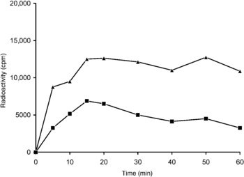
Infective larvae of A. cantonensis took up [3H]arachidonic acid in a time-dependent manner (fig. 1). The uptake rapidly increased from an initial rate of 778 cpm to a peak at 20 min. The level then had a decreasing trend up to 40 min and increased to another peak at 50 min before decreasing again up to 60 min. No appreciable radioactivity was detected in the aliquots from the culture of larvae fixed with formalin, suggesting the absence of non-specific fatty acid binding. After 60 min of incubation, more than 92% of the larvae remained viable.
Uptake and incorporation of arachidonic acid by infective larvae of Angiostrongylus cantonensis. Viable infective larvae were incubated with [3H]arachidonic acid (12 nm) at 37°C. Levels of uptake and incorporation at different time intervals were determined by detecting the radioactivity in each aliquot. Formalin-fixed larvae were used as a control. cpm, counts/min. The figure represents two independent experiments.

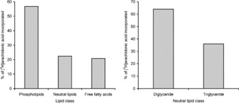
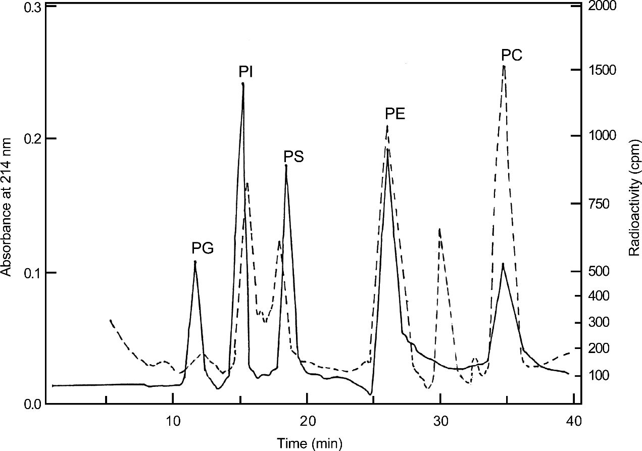
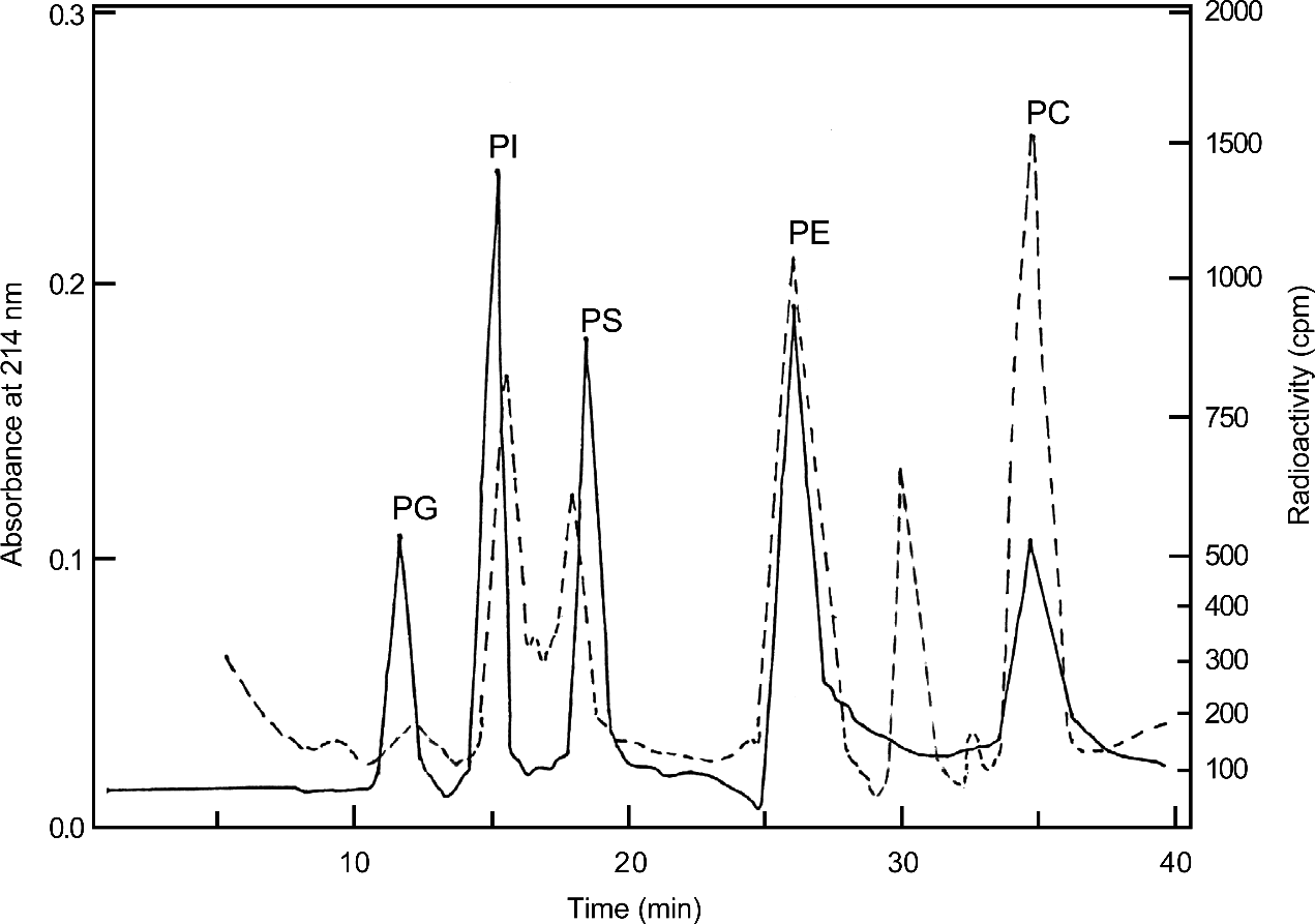
Fatty acid incorporation also exhibited a time-dependent pattern. The rate of incorporation also rapidly increased and reached a peak within 15 min. However, radioactivities in the lipid pools gradually decreased to 65.7% of the peak at 60 min. In extracts of the larvae incubated with [3H]arachidonic acid for 60 min, radioactivities were detected in the phospholipids (56.8%) and neutral lipids (22.4%). Moreover, the remaining 20.8% existed as free fatty acids. In neutral lipids, 64.0% of radioactivity was detected in diglycerides and 36.0% in triglycerides (fig. 2). In the phospholipid pool, the predominant fatty acids were phosphatidylcholine, phosphatidyethanolamine and phosphatidylinositol, with a small amount of phosphatidylserine (fig. 3). By radioimmunoassay, leukotriene B4 was detected in culture medium of viable infective larvae and the level increased with time. In the cultures of formalin-fixed larvae, only negative results were found.
Incorporation of arachidonic acid into lipid classes by infective larvae of Angiostrongylus cantonensis. Total lipid was extracted from the larvae incubated with [3H]arachidonic acid (12 nm) for 60 min at 37°C and separated by thin-layer chromatography using chloroform/methanol/water (2:1:0.01) as a solvent system. The values are means of two independent experiments.

Chromatogram of phospholipids incorporated by infective larvae of Angiostrongylus cantonensis incubated with [3H]arachidonic acid (12 nm) for 60 min at 37°C. The Bio-Sil HO-10 column was eluted with a programmed gradient from 97.5% to 85% acetonitrile at a flow rate of 1 ml/min. Absorbance at 214 nm (solid line) and radioactivity (dashed line) of the eluates were detected synchronously. PG, Phosphatidylglycerol; PI, phosphatidylinositol; PS, phosphatidylserine; PE, phosphatidylethanolamine; PC, phosphatidylcholine; cpm, counts/min. The values are means of two independent experiments.
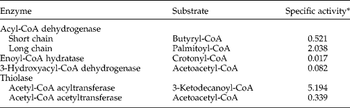
In the cultures of viable infective larvae with [U-14C]arachidonate, radioactive carbon dioxide was detected and the level increased with time. Moreover, substantial activities of short- and long-chain acyl-CoA dehydrogenase, acetyl-CoA acyltransferase and acetyl-CoA acetyltransferase and low levels of enoyl-CoA hydratase and 3-hydroxyacyl-CoA dehydrogenase were detected in crude extracts of the parasite (table 1).
Activities of enzymes related to fatty acid oxidation in crude extracts of infective larvae of Angiostrongylus cantonensis.
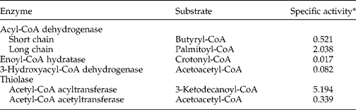
* Specific activity was expressed as nmol/min/mg protein at 28°C.
Discussion
Uptake is the intake of exogenous molecules through different mechanisms such as diffusion, active transport or endocytosis, whereas incorporation refers to the utilization of exogenous molecules via different metabolic pathways. However, the molecules taken up by an organism may or may not be incorporated. In the present study, we have demonstrated that infective larvae of A. cantonensis are able to take up and incorporate exogenous arachidonic acid into the lipid pools. Although the exact mechanism of uptake remained unclear, a surface protein expressed on schistosomula of S. mansoni after exposure to human serum has been demonstrated to be a receptor for low-density lipoproteins (Rumjanek et al., Reference Rumjanek, McLaren and Smithers1983). Moreover, by the action of monopalmitoylphosphatidylcholine, the schistosomula may lyse adherent human red blood cells and immobilize cell membrane components (Golan et al., Reference Golan, Brown, Cianci, Furlong and Caulfield1986). These findings indicate that parasitic helminths not only take up lipids from the serum via a receptor but also obtain lipids from the cell membrane of the host. Since the pattern of arachidonic acid uptake by infective larvae of A. cantonensis does not change in the observed time interval, it may not only occur via simple diffusion. However, the role of receptors or other mechanisms on the transport of lipids require further studies.
Since the cerebrospinal fluid is a constant pool of free fatty acids (Pilitsis et al., Reference Pilitsis, Diaz, Wellwood, Oregan, Fairfax, Phillis and Coplin2001), infective larvae of A. cantonensis may utilize these molecules as a source of energy. By TLC, we have shown that arachidonic acid was esterified into phospholipids and neutral lipids by infective larvae. In addition, phosphatidylcholine was found to be the major phospholipid by HPLC. It has been reported that the synthesis of phosphatidylcholine in mammals may occur by fatty acyl exchange, de novo synthesis from choline, methylation of phosphatidylethanolamine or base exchange between phospholipids (Pelech & Vance, Reference Pelech and Vance1984). In adult S. mansoni, phosphatidylcholine was also found to be the major phospholipid and the synthesis of phosphatidylcholine occurred by de novo synthesis from choline. Moreover, female Dirofilaria immitis may synthesize phosphatidylcholine by way of choline and cytidine 5’-diphosphocholine (Kennedy pathway) and also by S-adenosylmethionine-mediated sequential methylation of phosphatidylethanolamine (Bremer–Greenberg pathway) (Srivastava & Jaffe, Reference Srivastava and Jaffe1985). Further studies are required to determine the pathways of phosphatidylcholine synthesis in A. cantonensis.
The arachidonic acid-derived eicosanoids are well-known powerful immune modulators. Release of eicosanoids has been reported in S. mansoni (Fusco et al., Reference Fusco, Salafsky and Kevin1985, Reference Fusco, Salafsky and Delbrook1986), D. immitis (Weller et al., Reference Weller, Longworth and Jaffe1989) and T. taeniaformis (Leid & McConnell, Reference Leid and McConnell1983a, b; Potter & Leid, Reference Potter and Leid1986). In this study, we demonstrated the release of leukotriene B4 by infective larvae of A. cantonensis. Since leukotrienes have been demonstrated to be important mediators of immediate hypersensitivity reactions and inflammation (Samuelsson, Reference Samuelsson1983), this lipid mediator may play a role in the pathogenesis of angiostrongyliasis.
In mammals, fatty acid oxidation has been considered to occur via mitochondrial and peroxisomal β-oxidation. In mitochondrial β-oxidation, dehydrogenation of the β-carbon band of fatty acyl coenzyme A is rate limiting (Osmunden, Reference Osmunden1981). There are at least three mitochondrial fatty acyl coenzyme A dehydrogenases, distinguished by their preferred substrates (short-chain, medium-chain and long-chain fatty acyl coenzyme A) (Hall, Reference Hall1981; Thorpe, Reference Thorpe1981). Peroxisomal β-oxidation involves transfer of electrons, O2 and H2 to NAD such that H2O2 and NADH are formed (Lazarow & de Duve, Reference Lazarow and de Duve1976).
Adult or larval parasitic helminths have not been demonstrated to possess a functional β-oxidation pathway, since most of them do not utilize significant amounts of lipids even during starvation or under aerobic conditions. However, the presence of Krebs cycle has been suggested in A. cantonensis (Shih & Chen, Reference Shih and Chen1982). In this study, we demonstrated that infective larvae oxidize exogenous fatty acid into carbon dioxide. Moreover, activities of fatty acid oxidation enzymes were also detected in crude extracts. These findings indicate that a functional β-oxidation pathway exists in infective larvae of A. cantonensis and the parasite may oxidize exogenous free fatty acids as an additional source of energy in this developmental stage.
Acknowledgements
This work was supported in part by a grant from Chang Gung University (Grant no. CMRP 170052).