Introduction
Dogs are considered to be one of the most valuable models for studying human genetic diseases because of their genetic, anatomical and physiological similarities to humans (Ostrander et al., Reference Ostrander, Galibert and Patterson2000; Sutter and Ostrander, Reference Sutter and Ostrander2004; Mack, Reference Mack2005). However, because germline-transmissible embryonic stem cells, which are generally used for production of transgenic mice (Bradley et al., Reference Bradley, Evans, Kaufman and Robertson1984; Robertson, Reference Robertson1991; Pluck and Klasen, Reference Pluck and Klasen2009), have not been available in the canine species (Schneider et al., Reference Schneider, Wolf, Braun, Kolb and Adler2009), transgenesis in canines has been hampered. To overcome this problem, somatic cell nuclear transfer (SCNT) using transfected cells was employed and successfully produced the first transgenic dogs (Hong et al., Reference Hong, Kim, Jang, Oh, Park, Kang, Koo, Kim, Kwon, Koo, Ra, Kim, Ko and Lee2009b).
The combination of SCNT and transgenic technologies has been considered to be an effective and economical approach to producing disease-specific transgenic animals (Brunetti et al., Reference Brunetti, Perota, Lagutina, Colleoni, Duchi, Calabrese, Seveso, Cozzi, Lazzari, Lucchini and Galli2008). This approach also allows reliable prediction of the expression pattern of the foreign gene and embryonic developmental potential of the donor cells. Although many transgenic animals have been born as a result of SCNT and successfully expressed the transgene, mosaic expression or silencing of transgenes has also been reported in cloned embryos and animals (Park et al., Reference Park, Lai, Cheong, Cabot, Sun, Wu, Rucker, Durtschi, Bonk, Samuel, Rieke, Day, Murphy, Carter and Prather2002; Brunetti et al., Reference Brunetti, Perota, Lagutina, Colleoni, Duchi, Calabrese, Seveso, Cozzi, Lazzari, Lucchini and Galli2008). There have been conflicting results regarding the developmental capacity of embryos reconstructed with transfected versus non-transfected donor cells (Zakhartchenko et al., Reference Zakhartchenko, Mueller, Alberio, Schernthaner, Stojkovic, Wenigerkind, Wanke, Lassnig, Mueller, Wolf and Brem2001; Arat et al., Reference Arat, Gibbons, Rzucidlo, Respess, Tumlin and Stice2002; Forsberg et al., Reference Forsberg, Strelchenko, Augenstein, Betthauser, Childs, Eilertsen, Enos, Forsythe, Golueke, Koppang, Lange, Lesmeister, Mallon, Mell, Misica, Pace, Pfister-Genskow, Voelker, Watt and Bishop2002; Lee et al., Reference Lee, Ock, Yoo, Kumar, Choe and Rho2005b). Thus, in vitro studies of various transgenic donor cells may provide useful information prior to generating live animals (Illmensee et al., Reference Illmensee, Levanduski and Zavos2006; Sugawara et al., Reference Sugawara, Sugimura, Hoshino and Sato2009).
Interspecies somatic cell nuclear transfer (iSCNT) has the potential to become a useful tool for addressing basic questions about nuclear-cytoplasmic interactions between species (Song et al., Reference Song, Lee, Kim, Kim, Park, Kim, Chang, Han, Lee, Lee and Koo2009; Wang et al., Reference Wang, Beyhan, Rodriguez, Ross, Iager, Kaiser, Chen and Cibelli2009). It has also been proposed as an alternative for the therapy of infertility, the production of stem cells and the cloning of endangered species or animals whose oocytes are difficult to obtain, as in canines (Uhm et al., Reference Uhm, Gupta, Kim and Lee2007; Lee et al., Reference Lee, Kim, Park, Jeong, Lee, Park, Choi, Kim, Jeong, Kim, Hyun and Hwang2008; Oh et al., Reference Oh, Kim, Jang, Kim, Hong, Park, Park, Park, Sohn, Kim, Shin and Lee2008; Wang et al., Reference Wang, Beyhan, Rodriguez, Ross, Iager, Kaiser, Chen and Cibelli2009). Previous studies have shown the possibility of producing cloned dog embryos using bovine (Westhusin et al., Reference Westhusin, Burghardt, Ruglia, Willingham, Liu, Shin, Howe and Kraemer2001; Murakami et al., Reference Murakami, Otoi, Wongsrikeao, Agung, Sambuu and Suzuki2005) and porcine (Lee et al., Reference Lee, Kim, Park, Jeong, Lee, Park, Choi, Kim, Jeong, Kim, Hyun and Hwang2008) oocytes. However, transgenic canine cells have not previously been used as nuclear donor cells in iSCNT studies.
In the present study, cell viability and transgene expression of green fluorescent protein (GFP)-transfected canine fetal fibroblasts were determined. Then, in vitro embryonic development of cloned embryos derived from transgenic and non-transgenic cell lines with bovine in vitro matured oocytes was investigated.
Materials and methods
Transfection and cell culture
Canine fetal fibroblasts (designated as BF3), isolated as described in a previous report, were used for transfection (Hong et al., Reference Hong, Kim, Jang, Oh, Park, Kang, Koo, Kim, Kwon, Koo, Ra, Kim, Ko and Lee2009b). The advantages of gene transfection into target cells using non-viral vectors are safety and convenience (Niidome & Huang, Reference Niidome and Huang2002). In the present study, BF3 cells were transfected with constructs containing the GFP and puromycin resistance genes using FuGENE 6® (Roche Molecular Biochemicals). Stable transfectants were isolated with puromycin selection (2 μg/ml) for 2 weeks. GFP-transfected cells were frozen and stored. Cells from passage numbers four to six were used as nuclear donor cells for SCNT and analyses. Prior to SCNT, cells were thawed, cultured for 3 to 4 days until confluent and retrieved from the monolayer by trypsinization for 2 to 5 min.
Cell viability assay
Levels of viability were determined by the MTT [3-(4,5-dimethylthiazol-2-yl)-2,5-diphenyl tetrazolium bromide] assay. Briefly, cells were plated onto 96-well tissue culture plates, then MTT solution (10 μl) was added to each well. The dishes were mixed by shaking briefly on an orbital shaker and then incubated at 37 °C for 4 h. The MTT solution was removed and 200 μl dimethyl sulfoxide (Sigma-Aldrich Corp.) was added into each well to dissolve the formazan by pipetting up and down several times. The absorbance (optical density, OD) was measured on an ELISA plate reader with a test wavelength of 570 nm and a reference wavelength of 630 nm to obtain a sample signal (OD 570–630).
GFP expression in the canine fetal fibroblasts
For measurement of GFP expression of the cells, approximately 1 × 10⁶ cells were placed into a 12 × 15 mm test tube and spun down by centrifugation for 5 min at 300 g. The supernatant was removed and cells were resuspended in 500 ml of phosphate-buffered saline (PBS). Hoechst 33342 stock solution (5 μg/ml, 5 ml) was added and incubated at 37 °C for 45 min. Analyses were performed using a fluorescence activated cell sorter (FACS) Calibur (Becton–Dickinson).
In vitro maturation of bovine oocytes
Mature bovine oocytes were prepared as described in previous reports (Hong et al., Reference Hong, Jang, Oh, Koo, Park, Park, Kang and Lee2009a). Bovine ovaries were obtained from a local abattoir and transported to the laboratory within 2 h in 0.9% (v/v) NaCl solution at 30–35 °C. Follicular contents from small antral follicles 2–8 mm in diameter were aspirated using an 18-gauge needle attached to a 10 ml disposable syringe. Cumulus–oocyte complexes (COCs) with evenly granulated cytoplasm and enclosed by more than three layers of compact cumulus cells were selected and washed three times in HEPES-buffered tissue culture medium 199 (TCM-199; Invitrogen) supplemented with 2 mM NaHCO3, 5 mg/ml BSA (Invitrogen), and a 1% (v/v) mixture of penicillin and streptomycin (Sigma-Aldrich Corp.). Groups of 30 to 40 COCs were cultured at 39 °C, 5% CO2 in air in 500 μl drops of bicarbonate-buffered TCM-199 supplemented with 10% (v/v) fetal bovine serum (FBS), 0.005 IU/ml follicle stimulating hormone (FSH) (Antrin) and 1 μg/ml 17β-estradiol (Sigma-Aldrich Corp.).
Interspecies somatic cell nuclear transfer
After 22 h of maturation culture, cumulus cells were removed by repeated pipetting in 0.1% (v/v) hyaluronidase in HEPES-buffered TCM-199. Oocytes with a first polar body were selected and enucleated as described in previous reports (Jang et al., Reference Jang, Bhuiyan, Jeon, Ko, Park, Kim, Kim, Kang, Lee and Hwang2006). A canine donor cell that expressed the GFP gene was detected under an inverted microscope equipped with a GFP-specific filter (EX 450–490 nm, BA 520 nm) and deposited into the perivitelline space of each enucleated bovine oocyte. After microinjection, a couplet of donor cell-cytoplast complexes was induced to fuse using two pulses of direct current, 35–40 V/cm for 15 μs each using an Electro-Cell Fusion apparatus (NEPA GENE Co.). Fusion was subsequently confirmed by microscopic examination (×100). Chemical activation was induced by incubating the reconstructed embryos in modified synthetic oviductal fluid (mSOF) containing 5 μM ionomycin (Sigma-Aldrich Corp.) for 4 min at 39 °C (Cho et al., Reference Cho, Bhuiyan, Shin, Park, Jang, Kang, Lee and Hwang2004). Reconstructed iSCNT embryos were then washed and further incubated for 4 h in mSOF supplemented with 1.9 mM 6-dimethylaminopurine (Sigma-Aldrich Corp.). In order to monitor in vitro development, groups of six to seven transgenic and non-transgenic iSCNT embryos were cultured in 25 μl microdrops of mSOF under mineral oil for 7–10 days at 39 °C in a humidified atmosphere of 5% CO2, 5% O2 and 90% N2.
Statistical analysis
Embryos were randomly distributed within each experimental group. All data were subjected to analysis of variance (ANOVA) and a protected least significant difference (LSD) test using general linear models (GLM) in a statistical analysis system (SAS) program to determine differences among experimental groups. When a significant treatment effect was found for an experimental parameter, data were compared by the least squares method. A p-value of less than 0.05 was considered statistically significant.
Results and Discussion
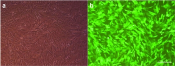
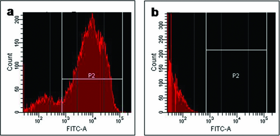
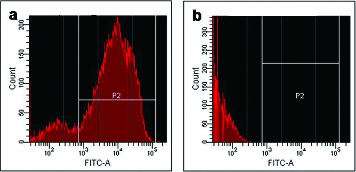
GFP is the most widely used reporter gene in transgenic research (McCreath et al., Reference McCreath, Howcroft, Campbell, Colman, Schnieke and Kind2000; Keefer et al., Reference Keefer, Baldassarre, Keyston, Wang, Bhatia, Bilodeau, Zhou, Leduc, Downey, Lazaris and Karatzas2001; Ono et al., Reference Ono, Shimozawa, Ito and Kono2001; Lee et al., Reference Lee, Kim, Hyun, Lee, Jeon, Nam, Jeong, Kim, Kim, Han, Ahn, Kang, Lee and Hwang2005a; Jang et al., Reference Jang, Bhuiyan, Jeon, Ko, Park, Kim, Kim, Kang, Lee and Hwang2006). In the present investigation, GFP gene-transfected canine fetal fibroblasts were isolated using a non-viral vector. The transfected cells expressed strong green fluorescence under an inverted microscope equipped with a GFP-specific filter (Fig. 1). FACS analysis also showed that a mean of 85.9% of transfected cells were positive for GFP expression (Fig. 2). Then, cell viability was measured by the MTT assay in which increased absorbance indicates increased viability. The viability of GFP-transfected fetal fibroblasts (mean OD = 0.25) was not significantly different from non-transfected fetal fibroblasts (mean OD = 0.35).
Morphology of GFP-transfected fibroblasts and expression of green fluorescence under UV light. (a) Visible light image. (b) Fluorescence image. Scale bars, 200 μm. (See online for a colour version of this figure.)

FACS analysis of GFP expression in (a) GFP transfected cells; and (b) non-transfected canine fetal fibroblasts. (See online for a colour version of this figure.)
Then, the embryonic developmental potential of canine GFP-transfected cells was examined by creating interspecies embryos via SCNT using bovine oocytes. There was no difference between the two groups in terms of fusion rates (transgenic iSCNT embryos, 73.1% vs. non-transgenic iSCNT embryos, 75.7%), cleavage rates (69.7% vs. 73.8%) and development to the 8–16-cell stage (40.1% vs. 42.7%). Our data indicate that the bovine oocyte is able to reprogram canine somatic cells and support their early embryonic development (Table 1). Interestingly, the percent of cleavage and subsequent embryo development to the 8–16-cell stage of canine iSCNT embryos was higher than that of a previous report in which canine intraspecies SCNT embryos were constructed (Hong et al., Reference Hong, Kim, Jang, Oh, Park, Kang, Koo, Kim, Kwon, Koo, Ra, Kim, Ko and Lee2009b).
Development and GFP expression of iSCNT embryos

a SCNT-BF3 means nuclear transfer with canine fetal fibroblasts and bovine oocytes.
b SCNT-BF3/GFP means nuclear transfer with canine fetal fibroblasts infected with the GFP gene.
There were no statistically significant differences between the groups (p > 0.05).
However, the iSCNT embryos derived from canine fibroblasts did not reach the preimplantation stage, consistent with a previous study in which porcine oocytes were used (Lee et al., Reference Lee, Kim, Park, Jeong, Lee, Park, Choi, Kim, Jeong, Kim, Hyun and Hwang2008). Despite another study demonstrating that one canine–bovine iSCNT embryo could develop to the blastocyst stage, the efficiency (0.4% of the total embryos) was extremely low (Murakami et al., Reference Murakami, Otoi, Wongsrikeao, Agung, Sambuu and Suzuki2005). This lack of embryonic development to the late preimplantation stage was not due to integration of transgene because the developmental rates did not differ between transgenic and non-transgenic groups. It may result from suboptimal embryo culture conditions or failure of timely onset of embryonic genome activation (EGA) due to species incompatibility between the recipient ooplasm and the donor nucleus (Schramm and Bavister, Reference Schramm and Bavister1999; Uhm et al., Reference Uhm, Gupta, Kim and Lee2007; Lee et al., Reference Lee, Kim, Park, Jeong, Lee, Park, Choi, Kim, Jeong, Kim, Hyun and Hwang2008). A recent study reported that iSCNT embryos showed defective nucleologenesis and EGA, which may be crucial for efficient reprogramming when compared with in vitro fertilized and intraspecies SCNT embryos (Song et al., Reference Song, Lee, Kim, Kim, Park, Kim, Chang, Han, Lee, Lee and Koo2009). Thus, further studies will be needed to increase the development of iSCNT embryos.
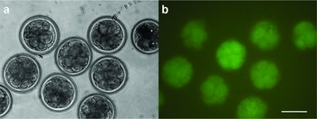
Embryos derived from the transfected cells completely expressed GFP at the 2-cell, 4-cell and 8–16-cell stages without mosaicism (Fig. 3), which corresponds with previous studies (Lai et al., Reference Lai, Park, Cheong, Kuhholzer, Samuel, Bonk, Im, Rieke, Day, Murphy, Carter and Prather2002; Hyun et al., Reference Hyun, Lee, Kim, Kim, Lee, Nam, Jeong, Kim, Yeom, Kang, Han, Lee and Hwang2003; Yin et al., Reference Yin, Lee, Yu, Choi, Koo, Kwon, Lee, Cho, Jin, Kim, Shin, Kim, Kim and Kong2007; Hong et al., Reference Hong, Kim, Jang, Oh, Park, Kang, Koo, Kim, Kwon, Koo, Ra, Kim, Ko and Lee2009b). These results suggest that the GFP gene could be used as a valuable marker in canine iSCNT studies.
Expression of GFP in cultured embryos after iSCNT. (a) Visible light image. (b) Fluorescence image. Scale bars, 200 μm. (See online for a colour version of this figure.)

In summary, our results demonstrated that, following successful isolation of canine transgenic cells and their use as nuclear transfer donors, iSCNT embryos showed stable GFP expression and developed to the 8–16-cell stage. To our knowledge, this report is the first report to describe canine transgenic cloned embryo production by the iSCNT approach. Once the efficiency is improved, it will be a powerful research tool for the in vitro bioassay of transgenic donor cells in canines.
Acknowledgements
We acknowledge Dr Barry D. Bavister and Ma Ninia Gomez for their valuable assistance in the editing of this manuscript. This study was financially supported by NRF (#M10625030005–10N2503300510), SNU foundation (Benefactor; RNL BIO), Nestlé Purina PetCare, the Research Institute for Veterinary Science, and BK21 programme for Veterinary Science.