Implications
In this study, we compared the reproductive performance between two chicken lines divergently selected for abdominal fat content, including reproductive traits, plasma concentrations of reproductive hormones and genes involved in the reproduction. We found lean birds have better reproductive performance compared with the fat ones. Our research is helpful for current broiler breeding programs and for improving the understanding of the relationship between obesity and reproduction in birds.
Introduction
Obesity has been associated with reproductive dysfunction in many species at different levels, including reproductive hormone levels, oocyte development, semen quality, etc. In humans, it is reported that obese men have lower sperm count and total testosterone (T) (Jensen et al., Reference Jensen, Andersson, Jorgensen, Andersen, Carlsen, Petersen and Skakkebaek2004), and obese women have poor conception and implantation rates (Brewer and Balen, Reference Brewer and Balen2010). In mice, obesity negatively affects the female reproductive capacity by reducing the number of oocytes and preantral follicles (Sagae et al., Reference Sagae, Menezes, Bonfleur, Vanzela, Zacharias, Lbaczeuski, Franci and Sanvitto2012). The adverse impact of obesity on reproduction has also been reported in domestic livestock. Arnett et al. (Reference Arnett, Holland and Totusek1971) found that obese cows lost more calves at parturition, and produced less milk than normal cows. In leptin-resistant obese sows, the corpora lutea are less functional during the estrous cycle and early pregnancy, which may contribute to their low reproductive efficiency (Astiz et al., Reference Astiz, Gonzalez-Bulnes, Perez-Solana, Sanchez-Sanchez and Torres-Rovira2013).
The effects of obesity on reproduction in chicken have been the subject of research interest for many years. It has been shown that diet-induced obesity has no significant effect on testes weight, semen volume or sperm density in male White Leghorn cocks (Parker and Arscott, Reference Parker and Arscott1972). In females, initial egg production under ad libitum feeding was lower in the fat line (Whitehead et al., Reference Whitehead, Armstrong and Herron1990). Compared with ad libitum feeding, food restriction enhanced egg production in both lines (Hocking and Whitehead, Reference Hocking and Whitehead1990). The ovaries of fat line females fed ad libitum had a high proportion of atretic yellow follicles and there was more extensive atresia in small white follicles of fat line birds even when food was severely restricted (Hocking and Whitehead, Reference Hocking and Whitehead1990). Recently, it has also been demonstrated that diet-induced obesity compromises the ovarian function of hens by altering ovarian hormone production and exerting proapoptotic effects on granulosa cells (Walzem and Chen, Reference Walzem and Chen2014). Although the influences of obesity on reproduction in male fowl have been studied by feeding them high-energy rations, as far as we know, there is no report of the effects of obesity on genetically obese male broilers. The effects of genetic obesity on the reproduction of female broilers have not yet been studied comprehensively.
Therefore, in this study, the reproductive performance, including the reproductive traits, reproductive hormones and the transcription of genes associated with reproduction, were compared, between the genetically lean and fat lines established by divergent selection for abdominal fat content at Northeast Agricultural University (NEAUHLF).
Material and methods
Ethics statement
All animal work was conducted according to the guidelines for the care and use of experimental animals established by the Ministry of Science and Technology of the People’s Republic of China (Approval no.: 2006-398) and approved by the Laboratory Animal Management Committee of the NEAUHLF.
Animals
The NEAU broiler lines divergently selected for abdominal fat content (NEAUHLF) have been selected since 1996 using abdominal fat percentage (AFP) and plasma very low-density lipoprotein (VLDL) concentration as selection criteria (Guo et al., Reference Guo, Sun, Shang, Leng, Wang, Wang and Li2011). The 0 generation (G0) of the two lines came from the same grandsire line originating from the Arbor Acres breed, which was then divided into two lines according plasma VLDL concentration at 7 weeks. From G1 to G18, plasma VLDL concentrations were measured for all male birds at 7 weeks, and AFP of the male birds in the first hatch was measured after slaughter at 7 weeks. Sibling birds from the families with lower (lean line) or higher (fat line) AFP than the average value of the population were selected as candidates for breeding, considering the BW at 7 weeks and plasma VLDL concentration of male birds in the second hatch and egg production of female birds in both hatches. A significant difference in AFP at 7 weeks of age has been found between the two lines since G4. Selection was continued for 18 generations. Birds from G14 to G18 were used in the current study.
For males (females), the birds of two lines had access to feed and water ad libitum from hatching to 7 weeks (from hatching to 2 weeks), and were equally feed restricted from 8 weeks to 56 weeks of age (from 3 weeks to 56 weeks of age) according to the guidelines given in Arbor Acres Plus Broiler Parent Stock Management Handbook (2014). All birds were kept in the same environmental conditions.
Measurement of carcass traits
Birds from the second hatch of G18 were slaughtered at 7, 14, 25, 30, 45 or 56 weeks of age. BW and abdominal fat weight (AFW) were measured, and AFW was calculated as a percentage of BW (AFP). In all, 30 males (15 from the lean line and 15 from the fat line) and 30 females (15 from the lean line and 15 from the fat line) were used at each of the ages described above.
Reproductive trait determination in lean and fat lines
Ages at first egg and total egg numbers in lean and fat female birds
The lean and fat lines’ ages at first egg (AFE) and total egg numbers (EN) from the first egg to 40 weeks of age in both hatches of every generation were calculated. To determine AFE and EN, a total of 2641 (1303 from the lean line and 1338 from the fat line) and 2117 (1108 from the lean line and 1009 from the fat line) female birds from five generations (G14 to G18) were used, respectively.
Testicular weights and semen quality characteristics of lean and fat male birds
Testicular weight (TW) was measured and TW was calculated as a percentage of BW (TWP) after the chicken from the second hatch were slaughtered at 7, 14, 25, 30, 45 and 56 weeks of age in G18. In all, 30 males (15 from the lean line and 15 from the fat line) were used at each age. The semen quality characteristics, including semen volume, semen pH, sperm concentration, percentage of motile spermatozoa and percentage of spermatozoa with an abnormal morphology, were determined at 30, 45 and 56 weeks of age in G18. In all, 60 males (30 from the lean line and 30 from the fat line) were used and the ejaculate of each male was collected three times at each age. The procedures were performed as described in detail by Peters et al. (Reference Peters, Shoyebo, Ilori, Ozoje, Ikeobi and Adebambo2008) and carried out by the same experienced person to avoid subjective assessments. In brief, the semen of each male was collected three times a week by the back massage method. The semen volume was measured by a graduated tube, and the semen pH was detected by the pH-indicator paper ranging from 6.4 to 8.0. Then, the semen was immediately diluted by 1 μl of semen with 199 μl of 0.75% NaCl, and submitted for examining the following sperm characteristics. The sperm concentration was detected under the microscope (40×10) by counting the spermatozoa using the hemocytometer. To determine the sperm motility, a drop of semen was placed onto a microscope slide by the micropipette, and then spread by a glass cover slip, to assess the percentage of progressing motile sperm under a microscope (40×10). The sperm with abnormal morphology was observed by staining the slides of sperm samples with 0.5% gentian violet solution under the microscope (40×10), and a minimum of 500 sperms were counted on each slide to calculate the abnormal sperm per sample.
Fertility and hatchability of lean and fat lines
In every generation, eggs from the first hatch were collected for 15 to 18 days after two rounds of artificial insemination following family establishment (one sire: four dams), and eggs from the second hatch were collected for another 15 to 18 days after the first hatch. The lean and fat lines’ egg fertility and hatchability in both two hatches of every generation were calculated. A total of 1240 individuals from five generations (G14 to G18) were used (620 from the lean line and 620 from the fat line).
Blood sampling and measurement of reproductive hormones in plasma
All birds were fasted for 10 h, and venous blood samples were collected before slaughtering in 1.5 ml tubes containing EDTA-Na2 at 7, 14, 25, 30, 45 and 56 weeks of age in G18. In all, 60 females (30 from the lean line and 30 from the fat line) and 60 males (30 from the lean line and 30 from the fat line) from the second hatch were used at each of the age. After centrifugation, the plasma was separated and stored at −20°C for later analysis. The plasma concentrations of reproductive hormones, including FSH, LH and estrogen (E2) in the lean and fat females, and FSH, LH and T in the lean and fat males, were measured by radioimmunoassay (RIA) using RIA kits (Beifang Biotechnology Institution, Beijing, China) with previously described methods (Goldsmith and Follett, Reference Goldsmith and Follett1983; Dorgan et al., Reference Dorgan, Fears, McMahon, Friedman, Patterson and Greenhut2002). Detailed information of the RIA kits used in the present study were provided in the Supplementary Table S1.
Tissue collection and RNA extraction
The hypothalamus, pituitary and ovary were collected from each female and the hypothalamus, pituitary and right testis of each male in G18 immediately after slaughtering at 7, 14, 25, 30, 45 or 56 weeks of age. In all, 30 females (15 from the lean line and 15 from the fat line) and 30 males (15 from the lean line and 15 from the fat line) were used at each of the ages tested. All the tissues were washed with 0.75% NaCl solution and then snap frozen in liquid nitrogen and stored at −80°C until RNA extraction. Total RNA was extracted from the frozen tissues using TRIzol Reagent (Invitrogen, Carlsbad, CA, USA), according to the manufacturer’s protocol, and diluted in nuclease-free water. RNA quality was assessed by visualization of the 18S and 28S ribosomal RNA bands on a denaturing formaldehyde agarose gel. Only RNA with a 28S:18S ratio between 1.8 and 2.1 was used for reverse transcription. Then, the RNA was stored at −80°C for further analysis.
First-strand complementary synthesis and quantitative real-time PCR
Complementary DNA (cDNA) was synthesized from 1 µg of total RNA using the ImProm-II™ Reverse Transcription System (Promega, Madison, WI, USA), according to the manufacturer’s instructions. Gene expression was detected with quantitative real-time PCR (qPCR) on a 7500 Real-time PCR System (Applied Biosystems, Foster City, CA, USA). Each qPCR reaction contained 3.6 μl of FastStart Universal SYBR Green Master Mix (Roche, Indianapolis, IN, USA), 0.2 μl (10 mM) of each primer, 1 μl of cDNA and H2O to a final volume of 10 μl. The thermal cycling conditions included an initial hold at 95°C for 10 min and 40 cycles at 95°C for 15 s and 60°C for 1 min. All the samples were assayed in triplicate. The target genes were gonadotropin-releasing hormone 1(GNRH1) in the hypothalamus, FSHβ polypeptide (FSHB) and LHβ polypeptide (LHB) in the pituitary, and FSHB, LHB, estrogen receptor 1 (ESR1) and insulin receptor (INSR) in the ovary or testis. The PCR efficiency of GNRH1, FSHB, LHB, ESR1, INSR and non-POU domain containing, octamer-binding (NONO) were 98.96%, 96.30%, 103.18%, 96.45%, 102.65% and 98.68%, respectively. The expression of each target gene was normalized to the expression of the reference gene, NONO. The relative fold differences in expression were calculated using the
 $$2^{{{\minus}\Delta C_{T} }} $$
method (Schmittgen and Livak, Reference Schmittgen and Livak2008), in which ΔC
T
=C
T
(target gene)−C
T
(NONO). The primer sequences are given in the Supplementary Table S2.
Statistical analysis
All the data were tested for normality. The specific statistical methods used were as follows. (1) Data for BW, AFW, AFP, TW, TWP and gene expression levels, which were normally or approximately normally distributed and had small sample numbers (n=15), were analyzed with a t test. The results of the analysis of those data are presented as means and SEM. (2) Data for AFE, EN, semen quality characteristics, fertility, hatchability and plasma reproductive hormonal levels, which were normally or approximately normally distributed and had larger sample numbers (n⩾30), were assessed with a GLM procedure. Model (1) was used for the analysis of AFE, EN, fertility and hatchability, which fitted line (L), generation (G) and hatch (H) as fixed effects. Model (2) was used for the analysis of the semen quality characteristics and plasma reproductive hormonal levels, which fitted with the line (L) as fixed effects, as follows:
 $$Y\,{\equals}\,\mu {\plus}L{\plus}G{\plus}H{\plus}F{\rm }\left( L \right){\plus}D{\rm }\left( {F,\,L} \right){\plus}e$$
 $$Y\,{\equals}\,\mu {\plus}L{\plus}F\left( L \right){\plus}D\left( {F,\,L} \right){\plus}e$$
In both model (1) and model (2), Y was the dependent variable, μ was the population mean, F (L) was a random effect of the family nested within the line, D (F, L) was a random effect of the dam nested within the line and the family and e was the random error. The results of the analysis of these data are presented as least squares means and SEM. All the statistical analyses were performed with the JMP 11.0 software (SAS Institute Inc., Cary, NC, USA), with a statistical significance level of P<0.05.
Results
Carcass traits of lean and fat birds
To clarify the background of the lean and fat females and males used in this study, BW and AFW of each female and male from G18 were measured and AFP was calculated at 7, 14, 25, 30, 45 and 56 weeks of age. As shown in the Supplementary Tables S3 and S4, AFW and AFP were significantly higher in the fat females and males than in the lean ones (P<0.05 or P<0.01). However, there was no significant difference in BW between the lean and fat females or males at any time points.
Reproductive traits in lean and fat lines
Ages at first egg and total egg numbers of lean and fat females
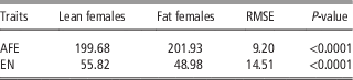
Age at first egg and EN of the lean and fat females from G14 to G18 were analyzed. As shown in Table 1, the average AFE of the lean line was 2 days earlier than that of the fat line (P<0.01). The average egg production of the lean line was 13.96% higher than that of the fat line (P<0.01).
Comparison of age at first egg (AFE) and egg numbers (EN) between lean and fat female broilers divergently selected for abdominal fat content

RMSE=root mean square error.
The P-value refers to the significance of lean females v. fat females.
Testicular weights and semen quality characteristics of lean and fat males
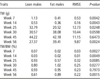
Testicular weights and TWP were compared in the lean and fat males of G18 at different stages of their growth and development (7, 14, 25, 30, 45 and 56 weeks of age). As shown in Table 2, TW and TWP of the lean males were markedly higher than those of the fat males at 7, 14 and 25 weeks of age (P<0.05 or P<0.01). Interestingly, we also noted that, in contrast, the fat males had greater TW and TWP (P<0.01) at 56 weeks of age.
Comparison of absolute and relative testicular weights (TW) between lean and fat male broilers divergently selected for abdominal fat content

RMSE=root mean square error.
The P-value refers to the significance of lean males v. fat males.
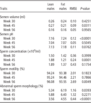
The semen quality characteristics of the lean and fat males from G18 at 30, 45 and 56 weeks of age were measured. As shown in Table 3, the lean males had a greater semen volume than the fat males at 45 weeks of age (P<0.05). The semen pH of the fat males was higher than that of lean males at 30 weeks of age (P<0.01). Larger numbers of sperm were observed in the lean males at 45 weeks of age (P<0.01). We also found that the lean males had a higher percentage of motile sperm at 56 weeks of age (P<0.01) and fewer sperm with an abnormal morphology at both 30 and 56 weeks of age (P<0.05).
Comparison of semen quality between lean and fat male broilers divergently selected for abdominal fat content

RMSE=root mean square error.
The P-value refers to the significance of lean males v. fat males.
Fertility and hatchability of lean and fat lines
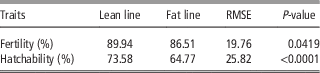
The fertility and hatchability of two lines were compared. As shown in Table 4, fertility and hatchability were both significantly higher in the lean line than in the fat line (P<0.05 and P<0.01, respectively).
Comparison of fertility and hatchability between lean and fat broiler lines divergently selected for abdominal fat content

RMSE=root mean square error.
The P-value refers to the significance of lean line v. fat line.
Plasma reproductive hormonal concentrations of lean and fat birds
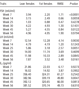
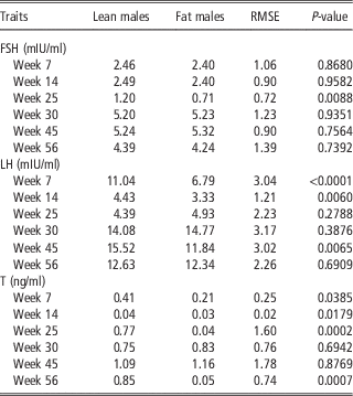
Given the differences in the reproductive traits of the lean and fat birds, the plasma concentrations of FSH, LH and E2 of the females (Table 5) and FSH, LH and T of the males (Table 6) in G18 were assayed with RIA method. The plasma concentrations of FSH were significantly higher in the lean females than in the fat females at 7, 14 and 45 weeks of age (P<0.05). The plasma levels of LH were markedly higher in the lean females than in the fat females at 25 and 56 weeks of age (P<0.05), as were the levels of E2 at 30 weeks of age (P<0.01). The plasma concentrations of FSH were significantly higher in the lean males than in the fat males at 25 weeks of age (P<0.01). The lean males had markedly higher plasma levels of LH at 7, 14 and 45 weeks of age (P<0.01) and of T at 7, 14, 25 and 56 weeks of age (P<0.05) than the fat males.
Comparison of reproductive hormones between lean and fat female broilers divergently selected for abdominal fat content

E2=estrogen; RMSE=root mean square error.
The P-value refers to the significance of lean females v. fat females.
Comparison of reproductive hormones between lean and fat male broilers divergently selected for abdominal fat content

T=testosterone; RMSE=root mean square error.
The P-value refers to the significance of lean males v. fat males.
Reproduction-associated gene expression in lean and fat birds
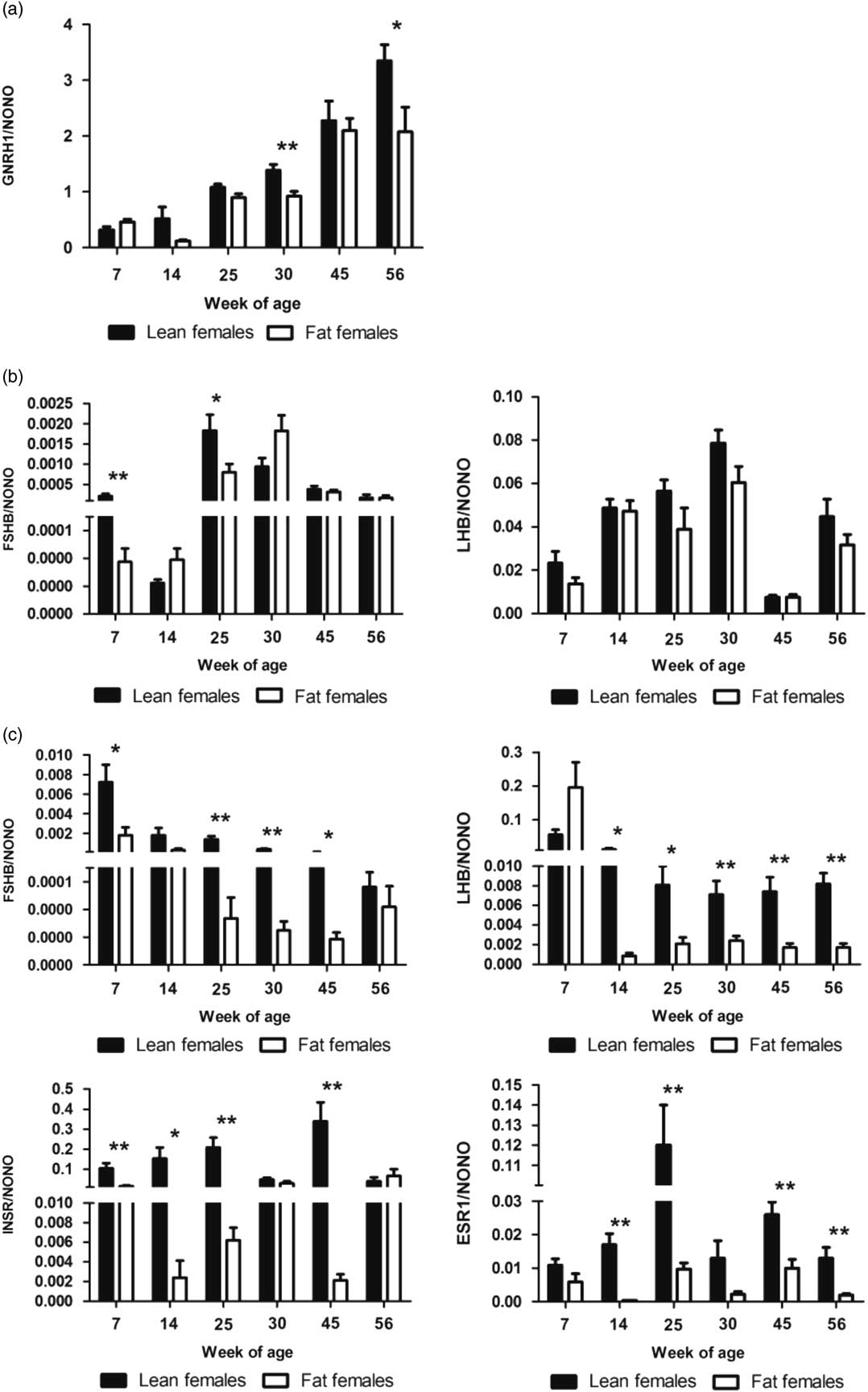
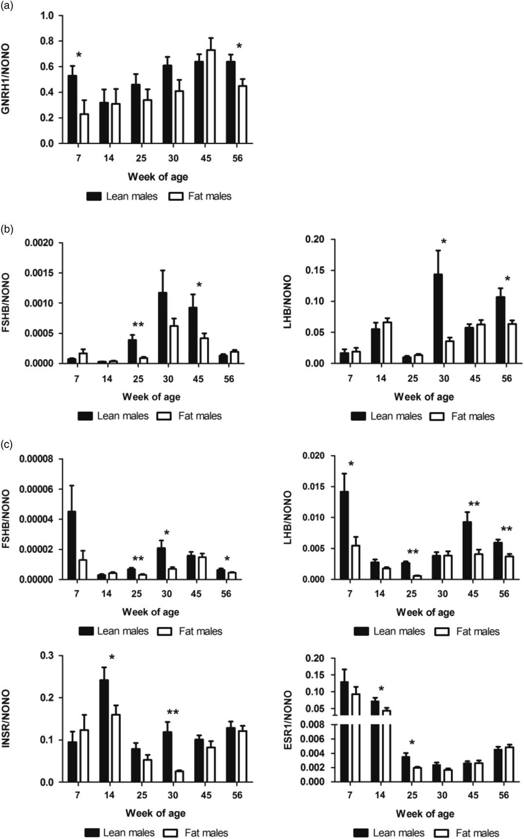
We examined the relative mRNA abundance of genes associated with reproduction in the hypothalamus, pituitary and ovary or testis tissues of the birds from G18 at 7, 14, 25, 30, 45 and 56 weeks of age. As shown in Figure 1a, the relative expression of GNRH1 was significantly higher in the hypothalamus of the lean females than in those of the fat females at 30 and 56 weeks of age (P<0.05 or P<0.01). In the pituitary gland, FSHB expression at 7 and 25 weeks of age was much higher in the lean females than in the fat females (P<0.05 or P<0.01), whereas LHB expression did not differ between the lean and fat females (Figure 1b). The expression levels of FSHB at 7, 25, 30 and 45 weeks of age, LHB at 14, 25, 30, 45 and 56 weeks of age, INSR at 7, 14, 25 and 45 weeks of age, and ESR1 at 14, 25, 45 and 56 weeks of age were significantly higher in the ovaries of the lean females than in those of the fat females (P<0.05 or P<0.01) (Figure 1c). The relative hypothalamic expression of GNRH1 was significantly higher in the lean males than in the fat males at 7 and 56 weeks of age (P<0.05) (Figure 2a). In the pituitary, FSHB expression at 25 and 45 weeks of age and LHB expression at 30 and 56 weeks of age was much higher in the lean males than in the fat males (P<0.05 or P<0.01) (Figure 2b). In the testis, FSHB at 25, 30 and 56 weeks of age, LHB at 7, 25, 45 and 56 weeks of age, INSR at 14 and 30 weeks of age and ESR1 at 14 and 25 weeks of age were markedly higher in the lean males than in the fat males (P<0.05 or P<0.01) (Figure 2c).
Comparison of the relative mRNA abundance of genes in the hypothalamus–pituitary–ovary involved in reproduction between lean and fat female broilers divergently selected for abdominal fat content. (a) Gonadotropin-releasing hormone 1 (GNRH1) in the hypothalamus; (b) FSHβ polypeptide (FSHB) and LHβ polypeptide (LHB) in the pituitary; (c) FSHB, LHB, insulin receptor (INSR) and estrogen receptor 1 (ESR1) in the ovary. The expression levels of each gene were determined by real-time PCR, and the expressed RNA levels were normalized to non-POU domain containing, octamer-binding gene (NONO). Results are given with the mean value and standard error (n=15). The black columns represent the lean line and the white columns represent the fat line. Significant difference: *P<0.05, **P<0.01.

Comparison of the relative mRNA abundance of genes in the hypothalamus-pituitary-testis involved in reproduction between lean and fat male broilers divergently selected for abdominal fat content. (a) Gonadotropin-releasing hormone 1 (GNRH1) in the hypothalamus; (B) FSHβ polypeptide (FSHB) and LHβ polypeptide (LHB) in the pituitary; (c) FSHB, LHB, insulin receptor (INSR) and estrogen receptor 1 (ESR1) in the testis. The expression levels of each gene were determined by real-time PCR, and the expressed RNA levels were normalized to non-POU domain containing, octamer-binding gene (NONO). Results are given with the mean value and standard error (n=15). The black columns represent the lean line and the white columns represent the fat line. Significant difference: *P<0.05, **P<0.01.

Discussion
In this study, genetically lean and fat birds with similar BWs and significantly different abdominal fat deposition were used, and they differed significantly in reproductive performance, including in their reproductive traits, sex hormones and the expression of reproductive-associated genes.
Comparison of the reproductive performance between lean and fat female birds
Age at first egg and EN are important egg production traits in chicken. Our results show that lean birds had earlier onset of egg production (AFE) and larger EN than fat females. Previous studies in chicken (Bornstein et al., Reference Bornstein, Plavnik and Lev1984) and quails (Yang et al., Reference Yang, Suh, Choi, Shin, Han and Lee2013) found that adequate BW and fatness were required at the onset of laying, but that obesity played a negative role in egg production. Hocking and Whitehead (Reference Hocking and Whitehead1990) found that fat hens had few yellow follicles and a high rate of atresia at first egg, and Walzem and Chen (Reference Walzem and Chen2014) reported that diet-induced obesity compromised ovarian function in hens. Obesity in humans and mice also negatively affects oocyte quality and/or maturation and the numbers of oocytes and follicles produced (Brewer and Balen, Reference Brewer and Balen2010; Sagae et al., Reference Sagae, Menezes, Bonfleur, Vanzela, Zacharias, Lbaczeuski, Franci and Sanvitto2012). The delayed onset of egg production observed in the fat line may be the result of slower follicular development and/or maturation caused by lower sex hormone levels and/or lower relative expression of reproduction-associated genes, which further reduce the numbers of eggs produced by fat females. The mechanisms underlying the adverse effects of obesity on egg production in chicken requires further clarification.
Fertility and hatchability are the main indicators of reproductive efficiency in chicken. We found that fertility and hatchability were higher in the lean line than in the fat line. Both paternal and maternal factors can affect fertility (Brillard, Reference Brillard2003). In chicken, the maternal factors of egg quality and the ability of the oviduct to store and maintain sperm after each ovulation are also important to fertility (Brillard, Reference Brillard2003). Based on the results of the present study, we speculate that higher levels of reproductive hormones, which affect ovulation and the development of the female reproductive tract, contributed to the higher fertility of the lean line. Hatchability is strongly influenced by incubation conditions and egg quality, which was affected by the quality of oocytes. In chicken, obesity has been shown to increase the likelihood of poor-quality oocytes (Walzem and Chen, Reference Walzem and Chen2014). Therefore, considering the same incubation conditions, lower levels of reproductive hormones and weaker expression of genes related to follicular development and maturation in the fat females may lead to poorer oocyte quality and thus lower hatchability in the fat line. The paternal factors affecting fertility and hatchability will be discussed in the second part of discussion.
Given the differences in the reproductive traits of the two female chicken lines, we measured the plasma concentrations of FSH, LH, and E2 at different ages, because they play important roles in the regulation of female reproduction. Specifically, FSH, which is mainly secreted by the pituitary gland, regulates follicular development in the ovary and stimulates the secretion of E2 and progesterone (Shanmugam et al., Reference Shanmugam, Pandita and Palta2010). Luteinising hormone, which is also produced by the pituitary gland, promotes ovulation and E2 secretion coordinately with FSH in the female (Norris and Carr, Reference Norris and Carr2013). Unlike mammals, E2 is mainly secreted by the theca cells in chicken, rather than by the granulosa cells , and plays an important role in the development of the oviduct and in the synthesis of egg yolk proteins by the liver (Li et al., Reference Li, Leghari, He, Zeng, Mi and Zhang2014). In the present study, the average plasma concentrations of FSH, LH and E2 were lower in the fat females than in the lean females, which could have impaired the functions of the gonads and resulted in poorer reproductive performance.
Then we examined whether the expression of genes associated with reproductive traits differed between the two lines. Gonadotropin-releasing hormone 1 plays a critical role in regulating the secretion of FSH and LH in birds, including chicken (Dunn and Sharp, Reference Dunn and Sharp1999). Our results showed higher expression of GNRH1 in the hypothalamus of lean females, which may partially explain higher plasma concentrations of FSH and LH in lean female birds observed in our study.
The synthesis of FSH and LH is stimulated by GnRH through the transcriptional and posttranscriptional regulation of their β subunits (Haisenleder et al., Reference Haisenleder, Dalkin, Ortolano, Marshall and Shupnik1991; Dalkin et al., Reference Dalkin, Burger, Aylor, Haisenleder, Workman, Cho and Marshall2001). We found gene expression levels of FSHB and LHB in the pituitary were consistent with plasma concentrations of FSH and LH in the current study.
We also found higher levels of FSHB, LHB, INSR and ESR1 in the ovary of lean females. The higher expression levels of FSHB and LHB in the lean females may have positively affected their reproductive performance by promoting ovarian development. Insulin plays an important role in female reproduction, affecting steroidogenesis and ovulation, mainly mediated by INSR, which is widely distributed in the ovary (Poretsky et al., Reference Poretsky, Cataldo, Rosenwaks and Giudice1999). Thus, the lower expression of INSR may have a negative effect on the steroidogenesis and ovulation. Estrogen plays an important role in the development of the oviduct and the synthesis of egg yolk proteins by the liver (Li et al., Reference Li, Leghari, He, Zeng, Mi and Zhang2014), and functions by binding its receptor (Liu et al., Reference Liu, Li, Gilbert, Xiao, Zhao, Wang, Yin and Zhu2015). The higher ESR1 expression detected in the ovaries of the lean females may lead to further effects on the reproductive performance by activating the functions of E2.
Comparison of the reproductive performance between lean and fat male birds
The seminiferous tubules and Leydig cells are the two main functional components of the testes. Accordingly, the primary functions of the testes are sperm production and T secretion (McCullagh, Reference McCullagh1948). Therefore, TW and TWP were measured and compared in this study. Interestingly, we found that the lean males had higher TW and TWP than the fat males in the early stages of growth (7, 14 and 25 weeks of age), but lower TW and TWP at 56 weeks of age. The relationship between testis weight and specific cell numbers in the testis has been studied. In Japanese quail, Ahmed et al. (Reference Ahmed, Nagaoka, Fayez, Abdel-Daim, Samir and Watanabe2015) reported that after P-Nitrophenol administration, testis weight and plasma concentration of T decreased. The histology of the testes showed hypocellularity of spermatogenic cell and spermatocyte and a decrease of appearance of spermatids and spermatozoa population. Although in another study on quails exposed to clothianidin, the results showed unchanged relative testis weights with a decreased number of germ cells by histology (Tokumoto et al., Reference Tokumoto, Danjo, Kobayashi, Kinoshita, Omotehara, Tatsumi, Hashiguchi, Sekijima, Kamisoyama, Yokoyama, Kitagawa and Hoshi2013). Combined with the results in our current study, we think lean line male birds have a better development of testes.
Semen quality is an important trait in male reproductive performance. Therefore, the semen quality in the lean and fat males was measured in this study. The values for semen volume, semen pH, sperm concentration, percentage of motile spermatozoa and percentage of spermatozoa with an abnormal morphology in the NEAUHLF lines were similar to those in previously published reports (Parker and Arscott, Reference Parker and Arscott1972; Kelso et al., Reference Kelso, Cerolini, Speake, Cavalchini and Noble1997). Our results showed that the lean males had higher sperm concentrations and larger numbers of motile and morphologically normal sperm than the fat males. The semen quality of male broiler breeders is reported to be affected by breed, age, feed and environmental factors, including temperature and humidity (Karaca et al., Reference Karaca, Parker and McDaniel2002). When the lean and fat males were maintained under the same feed and management conditions, the poorer development of the testes and lower reproductive hormone levels and related gene expression in the fat males was probably associated with their poorer semen quality, which further negatively affected the fertility and hatchability in the fat line.
We also measured the plasma concentrations of FSH, which regulates male testicular development and promotes the growth and maturation of sperm (Franchimont et al., Reference Franchimont, Chari and Demoulin1975); LH, which acts on Leydig cells to promote the secretion of androgens in males (Franchimont et al., Reference Franchimont, Chari and Demoulin1975); and T, which is secreted by Leydig cells and is essential for the growth and maintenance of the entire reproductive system (except for the Leydig cells themselves) (McCullagh, Reference McCullagh1948) at different ages. In this study, the average plasma concentrations of FSH, LH and T were higher in the lean males, which may have better promoted their testis development, spermatogenesis and steroidogenesis. Similar results have been reported in obese men (Jensen et al., Reference Jensen, Andersson, Jorgensen, Andersen, Carlsen, Petersen and Skakkebaek2004) and mice (Swerdloff et al., Reference Swerdloff, Batt and Bray1976).
The mRNA expression of these genes was also determined in the male birds. The expression of GNRH1 in the hypothalamus and FSHB and LHB in the pituitary gland was higher in the lean males, which may have led to the higher plasma concentrations of FSH and LH in the lean males.
The expression of the FSHB, LHB, INSR and ESR1 genes in the testis was determined. The expression of these genes was lower in the fat males than in the lean males, which may have contributed to their poorer semen quality by affecting their testicular development. In males, INSR plays a critical role in the regulation of male reproduction, probably by modulating spermatogenesis (Walters et al., Reference Walters, Allan and Handelsman2012). The lower expression of INSR mRNA in the fat males suggests that excessive fat deposition impairs spermatogenesis and further reduces sperm concentrations, partly through an INSR-mediated mechanism. It is well documented that spermatogenesis is regulated by gonadotropins and T, the effects of which are modulated by complex interrelationships with other factors, including E2 (Chimento et al., Reference Chimento, Sirianni, Casaburi and Pezziv2014), which is also a receptor-mediated hormone. Estrogen receptor 1 is a classical E2 receptor and its deletion (as in ESR1-knockout animals) reduces fertility, possibly by affecting spermatogenesis (Hess et al., Reference Hess, Bunick, Lee, Bahr, Taylor, Korach and Lubahn1997). The lower expression of ESR1 mRNA in fat males may be associated with their poorer semen quality via the dysregulation of spermatogenesis.
In summary, lean birds showed better reproductive performance, and because unique divergent chicken lines were used in this study, these results imply that selecting against abdominal fat deposition positively affects the reproductive performance of birds, possibly by influencing the expression of reproductive hormones and the genes associated with reproduction. We hope that these results will provide useful information for current broiler breeding programs and improve our understanding of the relationship between obesity and reproduction in birds.
Acknowledgments
The authors gratefully acknowledge the members of the poultry breeding group at the Northeast Agricultural University for their help in managing the birds and collecting the data. This research was supported by the National 863 Project of China (no. 2013AA102501), the China Agriculture Research System (no. CARS-42), University Nursing Program for Young Scholars with Creative Talents in Heilongjiang Province (UNPYSCT-2016003) and Heilongjiang Postdoctoral Financial Assistance (no. LBH-TZ0612).
Supplementary material
To view supplementary material for this article, please visit https://doi.org/10.1017/S1751731117002658