Coronary arterial fistula is a rare congenital vascular malformation. It consists of an abnormal communication between the coronary artery and one of the cardiac chambers or vessels adjacent to the heart. Patients with coronary arterial fistula may develop congestive heart failure,Reference Starc, Bowman and Hordof 1 , Reference Said 2 myocardial insufficiency,Reference Said 2 infective endocarditis,Reference Said 2 dysrhythmia,Reference Said 2 or rupture of the fistula, especially if it is aneurysmal.Reference Bauer, Allmendinger, Flaherty, Owlia, Rossi and Chen 3
We report an unusual case of a progressive aneurysmal dilation after transcatheter closure of coronary arterial fistula distally at the exit point that was successfully treated by a second occlusion device implanted at the entry point.
Case report
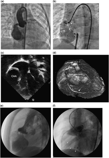
A 3-year-old boy presented with heart failure and with cardiomegaly on chest X-ray. A continuous murmur was audible at the right lower sternal border. Coronary arterial fistula was diagnosed by cross-sectional echocardiography, following which an ascending aortogram confirmed a large right coronary arterial fistula to mid right ventricular cavity. The fistula-feeding artery was tortuous and aneurysmal in appearance in its middle and distal segments (Fig 1a). No normal native right coronary artery or side branches could be identified, and the left coronary artery was normal. The diameter of the aneurysm was 25×38 mm and the exit point measured 4 mm. Transcatheter closure was successfully performed using Amplatzer Duct Occluder I 8/6 (St Jude Medical, Saint Paul, Minnesota, United States of America) to close the fistulous tract at its right ventricular exit. There was no shunt into the right ventricle after device implantation. However, there was opacification of a short tortuous segment of the right coronary artery branch (Fig 1b). The patient was discharged home without complications. However, he was lost to follow-up.
( a ) Ascending aortogram in antero-posterior projection showing a large right coronary arterial fistula with fusiform aneurysmal dilatation and draining into the right ventricle. ( b ) Selective right coronary angiogram in left anterior oblique projection showing the Amplatzer Duct Occluder I (black arrow) placed at the right ventricular exit. Slight opacification of a part of a tortuous right coronary artery branch is also present (white arrows). ( c ) Apical four-chamber view at the level of mitral valve orifice demonstrating a large right coronary artery aneurysm (CAA) compressing the right ventricle. ( d ) Volume rendered CT image of the right coronary artery (RCA) aneurysm and the distal right coronary artery (arrow). ( e ) Selective angiogram in lateral projection with partial occlusion using a 6 Fr balloon wedge catheter at the proximal part of the coronary artery showing minimal flow to the dilated aneurysmal right coronary artery. ( f ) Selective left coronary artery angiography in lateral projection showed cross-filling of the posterior descending artery and the distal right coronary artery (arrows).

After 3 years, he was referred by the local paediatric cardiologist for further clinical assessment. He had remained asymptomatic and the electrocardiogram showed normal sinus rhythm with non-specific ST-T wave changes. Echocardiography demonstrated a large aneurysm of the previous right coronary artery fistula (Fig 1c). The previous device was seated well distally without a residual shunt. The left ventricular systolic function was preserved. Cardiac CT showed a large aneurysmal blind ending pouch measuring 31×42 mm with compression of the right ventricle. Compared with the previous measurements before the first intervention, the diameter of the aneurysm had increased. The right coronary artery branches were still poorly identified. However, there was evidence of opacification of the posterior descending artery and the distal right coronary artery (Fig 1d).
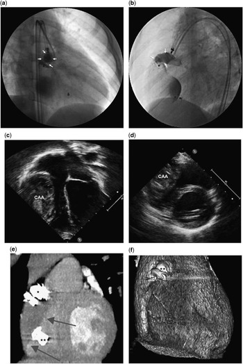
To prevent possible aneurysmal rupture, a repeat cardiac catheterisation was performed under general anaesthesia with access of both the femoral arteries with a view to occluding the fistula proximally. Heparin 100 U/kg was administered intravenously at the start of the procedure. Selective right coronary angiogram was performed with partial occlusion of the entry point with a 6 Fr balloon wedge catheter at the proximal part of the aneurysm (Fig 1e). There were no ST-T wave changes on electrocardiography during the 15-minute test occlusion, although very small myocardial branches were opacified proximally. The distal right coronary artery was not opacified. The diameter of the origin of the right coronary artery was 6 mm, whereas the maximum diameter of the aneurysm was 30×55 mm. Selective left coronary angiography confirmed a normal left coronary artery system with cross-filling of the distal right coronary artery, similar to that seen on the cardiac CT (Fig 1d and f). The inflow into the aneurysm from the proximal right coronary artery was then occluded with a 12-mm Amplatzer Vascular Plug II (St Jude Medical; Fig 2a and b). The haemodynamics and the electrocardiogram remained stable throughout the procedure. He was discharged home 2 days later on 5 mg/kg of oral aspirin.
Selective right coronary artery (RCA) angiography in right anterior oblique ( a ) and lateral ( b ) projection immediately after deployment of Amplatzer Vascular Plug II proximally (arrow). Tiny residual flow is still visible. Organised clot formation was demonstrated inside the aneurysm in ( c ) apical four-chamber and in ( d ) parasternal short-axis echocardiographic views. ( e ) Coronal oblique reformatting demonstrated a thrombosed aneurysm (arrows) with two devices in the proximal (star) and distal part (stars). ( f ) Volume rendered CT image showed device proximally (stars) and reduction of the aneurysm after vascular plug implantation.

Follow-up echocardiogram 2 weeks later showed that the device was seated well at the proximal right coronary artery entry point with no residual shunt. The aneurysm was obliterated by organised thrombus (Fig 2c and d). The ventricular function remained unchanged and the electrocardiogram was normal.
The patient was well 7 months later, and cardiac CT showed complete thrombosis with almost complete disappearance of the aneurysm (Fig 2e and f). Aspirin was discontinued at that stage.
Discussion
It has been recommended that closure of coronary arterial fistulas should be performed before the age of 20 years, to reduce the surgical morbidity and the incidence of long-term complications.Reference Cheung, Au, Cheung, Chiu and Lee 4 This was based on the natural history studies showing an increasing incidence of complications beyond 20 years of age.Reference Liberthson, Sagar and Berkoben 5 Coronary arterial fistulas have been closed with devices for many years as an acceptable alternative to surgery.Reference Qureshi and Tynan 6 Closure of the fistulas distally at the exit point has been recommended to avoid occlusion of the more proximal normal myocardial branches. This recommendation has been considered appropriate for those proximal fistulas,Reference Gowda, Latson, Kutty and Prieto 7 which have dilated feeding vessels without aneurysms and which are usually the side branches of the native coronary arteries. However, when the feeding artery to the fistula is of a distal type, in occluding these, it may not be possible to avoid occlusion of the normal myocardial branches, if the occlusion is performed more proximally. Such fistulas present a clinical and technical challenge. These patients with large distal coronary fistulas may be at risk of coronary events after closure, irrespective of the age of intervention.Reference Gowda, Forbes and Singh 8
The decision to intervene in patients with large distal coronary fistula remains controversial. The site of device closure in the proximal variety of coronary fistulas depends on the presence of aneurysms. Some of these fistulas can be closed at the exit point, whereas others can be closed at the entry point close to the native coronary artery. In our patient, our preference was catheter closure as the risk of transcatheter treatment is comparable to surgery.Reference Cheng 9 As for the closure technique, it was decided initially to occlude the right ventricular exit point to preserve any possible perfusion from the normal right coronary artery branches that may arise more proximally. Generally with distal occlusion, thrombus may extend more proximally, and therefore our belief was that the dilated aneurysm of the right coronary artery should reduce in size after closure, whereas the normal right coronary artery branches should improve in size with increasing flow through them. Until more recently, there has been no agreement on the site of closure of such fistulas.Reference Gowda, Latson, Kutty and Prieto 7 , Reference Gowda, Forbes and Singh 8
In large distal type of coronary arterial fistulas, after catheter or surgical intervention, coronary artery occlusion,Reference Gowda, Forbes and Singh 8 , Reference Hiraishi, Misawa and Horiguchi 10 or the development of discrete stenosis with perfusion defects,Reference Hiraishi, Misawa and Horiguchi 10 or persistent dilatation of the conduit vessel,Reference Liberthson, Sagar and Berkoben 5 have been reported. Our patient had a coronary arterial fistula that was of proximal type. The aneurysm was from a side branch of the native right coronary artery. The distal native right coronary artery was opacified from the left coronary artery on angiography almost back to its origin. After occlusion of the aneurysm distally, instead of regression, the aneurysm progressively enlarged. This is an unusual unreported complication. It is not clear what the mechanism of this enlargement is. It may be owing to high expansion forces within the aneurysm, transmitted from the aortic pressure, or poor development or unusual remodelling of the native right coronary artery branches. The right coronary artery branches of this patient that were opacified proximally appeared small, although they were connected to the right coronary artery aneurysm more proximally. If surgery had been considered, it would have needed closure of the fistula, as well as resection of the aneurysm. The conventional surgical approach of closing these fistulas at the exit point could have resulted in a similar complication to that noted after the initial intervention. The alternative surgical treatment that could have been used would be to occlude both the entry and exit points, which was ultimately performed by the catheter technique in our patient.
The enlarging aneurysm was considered to have the potential for spontaneous rupture. To prevent this possible complication in the future, it was decided to obliterate the aneurysm by occluding the entry point. Although temporary balloon occlusion testing does not guarantee that there will not be myocardial ischaemia,Reference Liberthson, Sagar and Berkoben 5 it is the only test available during an intervention. In our patient, there was no evidence of myocardial ischaemia during such balloon test occlusion. An Amplatzer Vascular Plug II was used to occlude the fistula proximally, because of its ability to adapt itself to the vascular anatomy and with a stable radial force lessening the possibility of migration. The device was oversized by 50% in relation to the vessel to ensure its stability in a high pressure state.
Faced with a similar anatomy, our recommendation now is to approach transcatheter closure with occlusion of both the entry and exit points to exclude the aneurysm, as recommended by Gowda.Reference Gowda, Forbes and Singh 8
Conclusion
Our patient has demonstrated a rare complication of progressive aneurysmal dilation after distal catheter closure of a large proximal type of coronary arterial fistula. Catheter closure of such coronary arterial fistulas at both the entry and exit points of the aneurysm is recommended.
Acknowledgements
The authors would like to acknowledge Suvipaporn Siripornpitak, MD, for providing computed tomography images for this report and Yuttapong Wongsawasdiwat, MD, for referring the patient for further management.
Financial Support
This report does not involve human and/or animal experimentation. Patient and parent inform consent has been officially signed.
Conflicts of Interest
None.
Ethical Standards
This report does not involve human and/or animal experimentation. Patient and parent’s inform consent has been officially signed.