Case summary
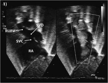
A 4.2 kg term male was born with prenatal diagnosis of transposition of the great arteries and started on prostaglandin E1. Postnatal transthoracic 2-D echocardiogram confirmed the same diagnosis and also showed a large superior sinus venosus defect (6 mm), with partially anomalous pulmonary venous return of the right upper and middle lobe pulmonary veins entering the superior vena cava between the azygos vein and right atrium (Fig 1), patent foramen ovale, and a large patent ductus arteriosus.Reference Fyler, Buckley and Hellenbrand 1
Transthoracic echocardiogram showing a high sinus venosus defect. RA=right atrium; RUPV=right upper lobe pulmonary vein; SVD=sinus venosus defect; SVC=superior vena cava.

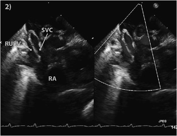
The patient demonstrated adequate mixing, and at 4 days of age he was taken for complete repair. Following sternotomy, the ductus was divided and cardiopulmonary bypass was established with the aorta and right atrial cannulation. Following cardioplegic arrest, an arterial switch operation with Lecompte manoeuvre was completed. Using intermittent circulatory arrest, a lateral right atriotomy was extended into the superior vena cava to the level of azygos vein where multiple small pulmonary venous branches were identified, draining the right upper and middle lobes. An oval patch of lightly tanned autologous pericardium was used to baffle the anomalous pulmonary veins to the left atrium. The atriotomy and lateral superior vena cava opening were closed with a second piece of pericardium. The patent foramen ovale was primarily closed. The patient was weaned from cardiopulmonary bypass uneventfully. Operative epicardial and discharge transthoracic echocardiogram showed good ventricular function, laminar flow in the superior vena cava, no residual intracardiac shunts, and an unobstructed pulmonary venous baffle (Fig 2). The patient was extubated on the second postoperative day and discharged 8 days later without complication. At 6 months’ follow-up, he continued to do well without evidence of systemic or pulmonary venous obstruction.
Postoperative echocardiogram showing normal drainage of pulmonary veins into the left atrium. RA=right atrium; RUPV=right upper lobe pulmonary vein; SVC=superior vena cava.

Discussion
Transposition of the great arteries has been reported to occur concomitantly with many congenital heart malformations, including partially and total anomalous pulmonary venous return.Reference Gontijo, Fantini, Barbosa, Gomes, Gutierrez and Vrandecic 2 , Reference Talwar, Rajashekar, Reedy, Choudhary and Airan 3 However, to the best of our knowledge, complete neonatal repair of transposition of the great arteries associated with sinus venosus defect and partially anomalous pulmonary venous return has not been reported.
Our patient had excellent mixing from a large atrial communication, as well as oxygenated pulmonary venous blood returning from the superior vena cava to the right atrium and systemic right ventricle, and therefore did not require an atrial septostomy.
Careful preoperative consideration of this patient’s anatomy was the single most important component in developing the operative plan, most importantly, the pulmonary venous anatomy in relation to the superior vena cava, azygos vein, pulmonary artery, and right atrial junction. We felt that magnetic resonance imaging was not necessary. However, in athymic patients or those with poor acoustic windows, additional imagingReference Grosse-Wortmann, Al-Otay and Goo 4 may allow for improved diagnostic accuracy.
Our dilemma was that the initial management of the sinus venosus defect at the time of arterial switch operation is because of the potential early and late physiological effects of a large left-to-right shunt and/or anatomic consequences from sinus node dysfunction, superior venal cava, and/or pulmonary vein stenosis. Our options were partial or full closure of the sinus venosus defect, leaving the anomalous veins to the superior vena cava with or without pulmonary venous banding, Warden procedure, a complete baffling procedure using one or two patches, or no intervention. No intervention may be preferable if the sinus venosus defect was small and the anomalous pulmonary vein(s) enter high in the superior vena cava; however, disadvantages include the additional haemodynamic burden of left-to-right shunting acutely after an arterial switch operation and the need for late reoperation for sinus venosus defect closure and pulmonary vein baffling. Closing the sinus venosus defect either fully or partially and leaving the pulmonary veins to the right atrium is a viable option, provided there is no significant pulmonary venous burden; however, preoperative surgery would likely be required. In our experience, the Warden procedure is not preferred in neonates or children owing to an increased risk for superior vena cava stenosis.Reference Walker, Mayer, Alexander, Walsh and Berul 5
In our experience, sinus node dysfunction following repair of sinus venosus defects in children is uncommon.Reference Walker, Mayer, Alexander, Walsh and Berul 5 – Reference Stewart, Bailliard and Kelle 7 A lateral atriotomy extending across the cavoatrial junction avoids the sinoatrial blood supply, preserving nodal function. Cardiopulmonary bypass with intermittent circulatory arrest was used to optimise visualisation and avoid cannulation and distortion of the superior vena cava.
In young neonates with a sinus venosus defect, in combination with other complex forms of congenital heart disease, and a significant shunt burden, complete repair should be considered at the initial operation. Limitations of a complete neonatal repair include the risks: of sinus node dysfunction, pulmonary vein stenosis, baffle obstruction, and superior vena cava stenosis. Continued longer term surveillance is required to assure un-obstructed pulmonary and systemic venous pathways.
Financial Support
This research received no specific grant from any funding agency, commercial, or not-for-profit sectors.
Conflicts of Interest
None.