





Introduction
Anisakid nematodes are widely distributed parasites of marine mammals, occurring throughout the world’s oceans. Their life cycles are complex: planktonic or semi-planktonic crustaceans function as the first intermediate hosts, whereas fish and molluscs act as transport or paratenic hosts. The adult stages develop in marine mammals, mainly cetaceans for species of the genera Anisakis, Skrjabinisakis and Pseudoterranova, and pinnipeds for Phocanema and Contracaecum species (Køie et al., Reference Køie, Berland and Burt1995; Køie and Fagerholm, Reference Køie and Fagerholm1995; Mattiucci et al., Reference Mattiucci, Cipriani, Levsen, Paoletti and Nascetti2018).
The integration of molecular and genetic tools into the study of anisakid morphospecies has substantially refined their systematics and taxonomy, elucidated phylogenetic relationships and enabled the recognition of sibling species as well as the description of new taxa (Mattiucci and Nascetti, Reference Mattiucci and Nascetti2008; Mattiucci et al., Reference Mattiucci, Cipriani, Webb, Paoletti, Marcer, Bellisario, Gibson and Nascetti2014, Reference Mattiucci, Cipriani, Levsen, Paoletti and Nascetti2018). More recently, a taxonomic revision within the family Anisakidae lead to a reorganization at the genus level and the resurrection of some genera, such as Skrjabinisakis, and Phocanema (Safonova et al., Reference Safonova, Voronova and Vainutis2021; Takano and Sata, Reference Takano and Sata2022; Bao et al., Reference Bao, Giulietti, Levsen and Karlsbakk2023). These recent studies have also expanded our understanding on the ecology, geographical distribution and host associations of anisakid nematodes (e.g. Mattiucci and Nascetti, Reference Mattiucci and Nascetti2008; Cavallero et al., Reference Cavallero, Nadler, Paggi, Barros and D’Amelio2011; Klimpel et al., Reference Klimpel, Kuhn, Busch, Karl and Palm2011; Kuhn et al., Reference Kuhn, Cunze, Kochmann and Klimpel2016; Li et al., Reference Li, Lü, Nadler, Gibson, Zhang, Chen, Zhao and Guo2018; Mattiucci et al., Reference Mattiucci, Cipriani, Levsen, Paoletti and Nascetti2018; Shamsi et al., Reference Shamsi, Sprohnle-barrera and Shafaet Hossen2019).
Following recent reclassifications within the family Anisakidae, 6 nominal species of Anisakis (A. simplex (s.s.), A. pegreffii, A. berlandi, A. typica (s.l.), A. ziphidarum and A. nascettii) and 3 species of Skrjabinisakis (S. physeteris, S. paggiae and S. brevispiculata) are currently recognized as parasites of cetaceans at the adult stage (Mattiucci and Nascetti, Reference Mattiucci and Nascetti2008; Cavallero et al., Reference Cavallero, Nadler, Paggi, Barros and D’Amelio2011; Mattiucci et al., Reference Mattiucci, Cipriani, Webb, Paoletti, Marcer, Bellisario, Gibson and Nascetti2014, Reference Mattiucci, Cipriani, Levsen, Paoletti and Nascetti2018; Di Azevedo et al., Reference Di Azevedo, Carvalho and Iñiguez2017; Shamsi, Reference Shamsi2021). Pseudoterranova ceticola is also regarded as a valid anisakid species (Deardorff and Overstreet, Reference Deardorff and Overstreet1981; Gibson, Reference Gibson, Stone, Platt and Khalil1983; Bao et al., Reference Bao, Olsen, Levsen, Cipriani, Giulietti, Storesund, García-seoane and Karlsbakk2022). In contrast, Pseudoterranova kogiae, the type species of this genus, originally described by Johnston and Mawson (Reference Johnston and Mawson1939) and later considered valid by Gibson (Reference Gibson, Stone, Platt and Khalil1983), remains a species inquirenda.
The pygmy sperm whale, Kogia breviceps (de Blainville, 1838), and the dwarf sperm whale, Kogia sima (Owen, 1866), are the only 2 extant species of the family Kogiidae within the superfamily Physeteroidea (Heyning, Reference Heyning1997; Rice, Reference Rice1998; Plön et al., Reference Plön, Best, Duignan, Lavery, Bernard, Van Waerebeek and Baker2023). Despite their broad distribution in tropical and warm-temperate waters of the Atlantic, Pacific and Indian oceans (McAlpine, Reference McAlpine, Würsig, Thewissen and Kovacs2018), kogiid whales remain among the least studied cetaceans. Their elusive behaviour has limited direct observations, and current knowledge of their biology, ecology and distribution is largely based on stranded individuals (Plön, Reference Plön2004; Staudinger et al., Reference Staudinger, McAlarney, McLellan and Pabst2013; Moura et al., Reference Moura, Acevedo-trejos, Tavares, Meirelles, Silva, Oliveira, Santos, Wickert, Machado, Siciliano and Merico2016; Maggioni et al., Reference Maggioni, Pupillo, Rota, Ramazzotti, Martín, Poli, Dumortier, Méndez-fernández, Torres-pereira, Sá, ten Doeschate, Brownlow, Covelo, López, Galimberti and Valsecchi2026). Kogiid whales act as definitive hosts for anisakid nematodes, harbouring a distinct parasite assemblage, including Skrjabinisakis brevispiculata, S. paggiae and 2 Pseudoterranova species, namely P. kogiae and P. ceticola. These parasites have been reported from definitive, intermediate and paratenic hosts across tropical and temperate waters of the Atlantic and Indo-Pacific Oceans, largely overlapping with the distribution of their kogiid hosts and extending from approximately 50°S to 50–60°N latitude (Gibson and Harris, Reference Gibson and Harris1979; Deardorff and Overstreet, Reference Deardorff and Overstreet1981; Abollo et al., Reference Abollo, López, Gestal, Benavente and Pascual1998; Santos and Lodi, Reference Santos and Lodi1998; González-Solís et al., Reference González-Solís, Vidal-Martínez, Antochiw-Alonso and Ortega-Argueta2006; Colón-Llavina et al., Reference Colón-Llavina, Mignucci-Giannoni, Mattiucci, Paoletti, Nascetti and Williams2009; Di Azevedo et al., Reference Di Azevedo, Knoff, Carvalho, Mello, Lopes Torres, Gomes and Iñiguez2015, Reference Di Azevedo, Carvalho and Iñiguez2017; Kuhn et al., Reference Kuhn, Cunze, Kochmann and Klimpel2016; Mattiucci et al., Reference Mattiucci, Cipriani, Levsen, Paoletti and Nascetti2018; Shamsi et al., Reference Shamsi, Sprohnle-barrera and Shafaet Hossen2019; Hernández-Olascoaga et al., Reference Hernández-Olascoaga, Guillén-hernández and Díaz-gamboa2023; Cipriani et al., Reference Cipriani, Palomba, Giulietti, Aco-Alburqueque, Andolfi, Ten Doeschate, Brownlow, Davison and Mattiucci2024; Seade et al., Reference Seade, Conga, Santos, Moura, de Farias, Silva, Costa, Amaral, de Souza, Emin-lima, Scofield, Riet-correa, Cerqueira and Bezerra Júnior2025). Parasites have been widely used to investigate ecology, feeding habits and trophic interactions in cetaceans (Cipriani et al., Reference Cipriani, Palomba, Giulietti, Marcer, Mazzariol, Santoro, Albuquerque, Covelo, López, Santos, Pierce, Brownlow, Davison, McGovern, Frantzis, Alexiadou, Højgaard, Mikkelsen, Paoletti, Nascetti, Levsen and Mattiucci2022; Saldaña et al., Reference Saldaña, López, López, Covelo, Remesar, Martínez-calabuig, García-dios, Morrondo, Díez-baños and Panadero2022; Ramilo et al., Reference Ramilo, Rodríguez, López, González, López, Pierce, Pascual and Abollo2024). In poorly known hosts such as kogiid whales, the study of anisakid biodiversity may therefore provide additional information on host ecology, feeding behaviour and parasite transmission in oceanic and deep-water food webs. The aim of the present study was to characterize the diversity of anisakid nematodes in pygmy sperm whales stranded along the Spanish Atlantic coast.
Materials and methods
Sample collection and morphological analysis
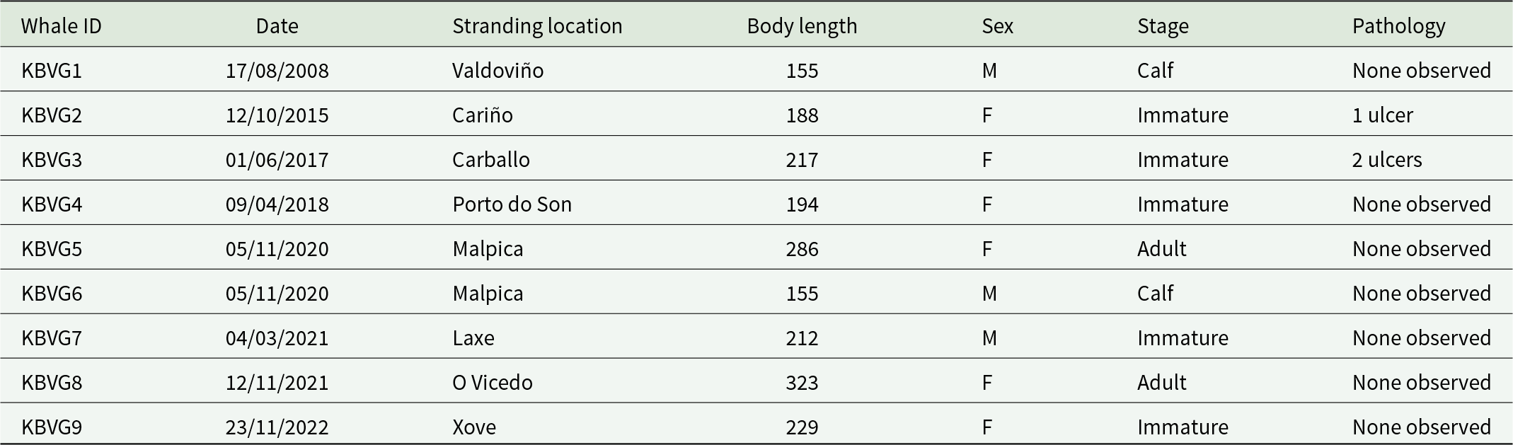
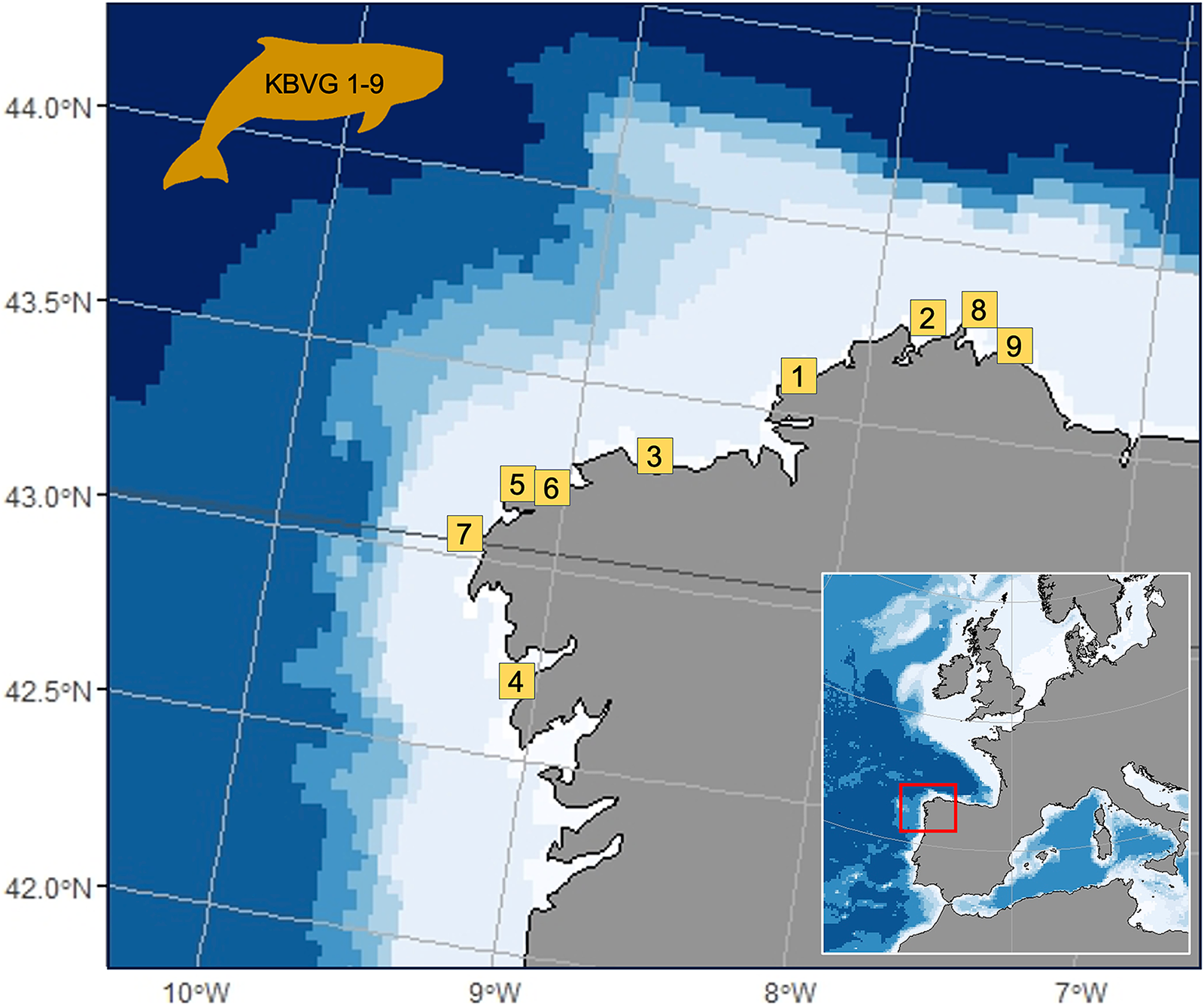
Nine pygmy sperm whales stranded along the Galician coast (north-western Spain) between 2008 and 2022 were examined (see Table 1 and Figure 1 for details). The individuals originated from the regional stranding network, and necropsies were performed by authorized CEMMA (Coordinadora para o Estudo dos Mamíferos Mariños) personnel following standardized protocols (Kuiken and Garcia-Hartmann, Reference Kuiken and Garcia-Hartmann1991; Geraci and Lounsbury, Reference Geraci and Lounsbury2005). Total length and individual characteristics are reported in Table 1.
Summary of stranded pygmy sperm whale specimens analysed in this study, including stranding date and area, body length (cm), sex, ontogenetic stage and observed pathological conditions

Table 1 Long description
Each row lists a whale ID with stranding date, stranding location, body length in centimeters, sex, ontogenetic stage, and pathology. IDs run from KBVG1 to KBVG9, with dates from 17/08/2008 to 23/11/2022 and locations Valdovino, Carino, Carballo, Porto do Son, Malpica, Laxe, O Vicedo, and Xove. Body length ranges from 155 to 323. Sex includes three males and six females. Stages include two calves, five immature, and two adult. Pathology is recorded as none observed for KBVG1, KBVG4, KBVG5, KBVG6, KBVG7, KBVG8, and KBVG9. Ulcers are recorded for KBVG2 with 1 ulcer and KBVG3 with 2 ulcers.
Sampling locations of the pygmy sperm whale specimens examined in this study. Numbered labels correspond to specimen codes KBVG1 and KBVG9; detailed information for each specimen is provided in Table 1.

Figure 1 Long description
The stranding locations of pygmy sperm whales along the Galician coast in northwestern Spain are displayed. Numbered markers from 1 to 9 indicate specific stranding sites along the coastline. An inset map of Europe highlights the region of interest with a red box. The main map includes latitude and longitude lines, ranging from approximately 42.0 degrees N to 44.0 degrees N and 10.0 degrees W to 7.0 degrees W. The whale icon labeled ′KBVG 1-9′ is positioned in the upper left, representing the study area for the specimens. The map uses a gradient to depict ocean depth or another variable, with darker shades offshore and lighter shades near the coast.
Nematode parasites were recovered from the forestomach and the cardiac and pyloric regions of the stomach. From each host, a representative subsample of nematodes was opportunistically collected from all accessible gastric regions, including worms free in the lumen, associated with prey remains and attached to the gastric mucosa, in order to maximize the representativeness of the parasite assemblage. The overall infection levels were not quantified. Specimens were preserved frozen or in 70% ethanol and shipped to the laboratory of Sapienza University of Rome (Italy) for further analyses. A total of 500 nematodes were randomly selected from the material recovered from the stranded pygmy sperm whales and measured for total length (mm). The specimens were examined under a light microscope for preliminary assignment to genus level based on general diagnostic morphological traits, primarily the ventriculus shape and the spicule size in males (Di Azevedo et al., Reference Di Azevedo, Carvalho and Iñiguez2017; Mattiucci et al., Reference Mattiucci, Cipriani, Levsen, Paoletti and Nascetti2018). The anterior and posterior ends of the nematodes were then excised and mounted on glass slides in lactophenol for detailed morphological examination under an optical microscope, while the remaining body fragments were preserved in 70% ethanol for molecular analyses.
Detailed morphological screening was based on the diagnostic characters provided by Mattiucci et al. (Reference Mattiucci, Cipriani, Levsen, Paoletti and Nascetti2018) for Anisakis and Skrjabinisakis, and by Deardorff and Overstreet (Reference Deardorff and Overstreet1981), as revised by Bao et al. (Reference Bao, Olsen, Levsen, Cipriani, Giulietti, Storesund, García-seoane and Karlsbakk2022), for Pseudoterranova. The characters examined included the morphology of the cephalic end, the length and shape of the ventriculus, the presence or absence of an intestinal caecum, the length and shape of the spicules, the ratio between right and left spicule lengths, the morphology of the male caudal end and the arrangement of caudal papillae. These features allowed a further assignment of specimens to morphotypes. Sex was determined whenever possible based on diagnostic morphological characters, including the presence of spicules in males and the vulva and/or ovaries in females. Mature adults were distinguished from pre-adults by the presence of fully developed caudal papillae and spicules in males, and eggs in females.
Genetic identification and phylogenetic analysis of anisakid parasites
A subsample comprising over 250 specimens, representatives of all the nematode morphospecies from the examined pygmy sperm whales, was genetically identified by sequence analysis of the mitochondrial cytochrome c oxidase subunit II gene (mtDNA cox2), and the complete internal transcribed spacer (ITS) region of the nuclear ribosomal DNA – including ITS-1, 5.8S and ITS-2 – and partial large subunit ribosomal DNA (LSU or 28S rDNA).
Genomic DNA was extracted from approximately 2 mg of tissue per specimen using the Quick-gDNA Miniprep Kit (Zymo Research), following the manufacturer’s protocol. The mtDNA cox2 gene was amplified by polymerase chain reaction (PCR) using the primers 211 F (5’-TTTTCTAGTTATATAGATTGRTTTYAT-3’) and 210 R (5′-CACCAACTCTTAAAATTA TC-3′) (Nadler and Hudspeth, Reference Nadler and Hudspeth2000). PCR was carried out according to the procedures described by Mattiucci et al. (Reference Mattiucci, Cipriani, Webb, Paoletti, Marcer, Bellisario, Gibson and Nascetti2014). The ITS region rDNA (ITS-1, 5.8S rDNA and ITS-2) was amplified using the primers NC5 (5′-GTA GGT GAA CCT GCG GAA GGA TCA TT-3′) and NC2 (5′-TTA GTT TCT TTT CCT CCG CT-3′) (Zhu et al., Reference Zhu, D’Amelio, Paggi and Gasser2000). PCR was carried out according to the procedures reported in Zhu et al. (Reference Zhu, D’Amelio, Paggi and Gasser2000). Partial sequences of the 28S rDNA were amplified using primers 28SF (5′-AGCGGAGGAAAAGAAACTAA-3′) and 28SR (5′-ATCCGTGTTTCAAGACGGG-3′) (Li et al., Reference Li, Lü, Nadler, Gibson, Zhang, Chen, Zhao and Guo2018), according to the protocol described by Li et al. (Reference Li, Lü, Nadler, Gibson, Zhang, Chen, Zhao and Guo2018). PCR products were purified and sequenced by Biofab (Rome, Italy). Sequences were assembled with ChromasPro 2.1.5 (Technelysium Pty Ltd., Tewantin, Australia) and aligned using ClustalX 2.0 (Larkin et al., Reference Larkin, Blackshields, Brown, Chenna, McGettigan, McWilliam, Valentin, Wallace, Wilm, Lopez, Thompson, Gibson and Higgins2007). Alignments were inspected manually using BioEdit (Hall, Reference Hall1999) to identify nucleotide substitutions, and informative sites were noted. Sequence identity was verified using the Nucleotide Basic Local Alignment Search Tool (BLAST; www.ncbi.nlm.nih.gov/BLAST).
Phylogenetic analyses based on the mtDNA cox2, ITS rDNA and 28S rDNA sequences were carried out using the method implemented in BEAST v1.10.4. The best-fit nucleotide substitution model for each gene dataset was selected using the Akaike Information Criterion (AIC) in jModelTest v2.1.7, resulting in a GTR + I + G model for mtDNA cox2, GTR + G for ITS rDNA and HKY + G for 28S rDNA. A strict molecular clock and a Yule speciation process were applied as tree priors. The analysis was run for 10 million generations, sampling every 10 000 generations. Convergence and effective sample sizes (>200) were assessed using Tracer v1.7. The first 10% of trees were discarded as burn-in, and the remaining trees were summarized with TreeAnnotator v1.10.4 and visualized in FigTree v1.4.2. The outgroups and their GenBank reference numbers are reported in Figures 2–4.
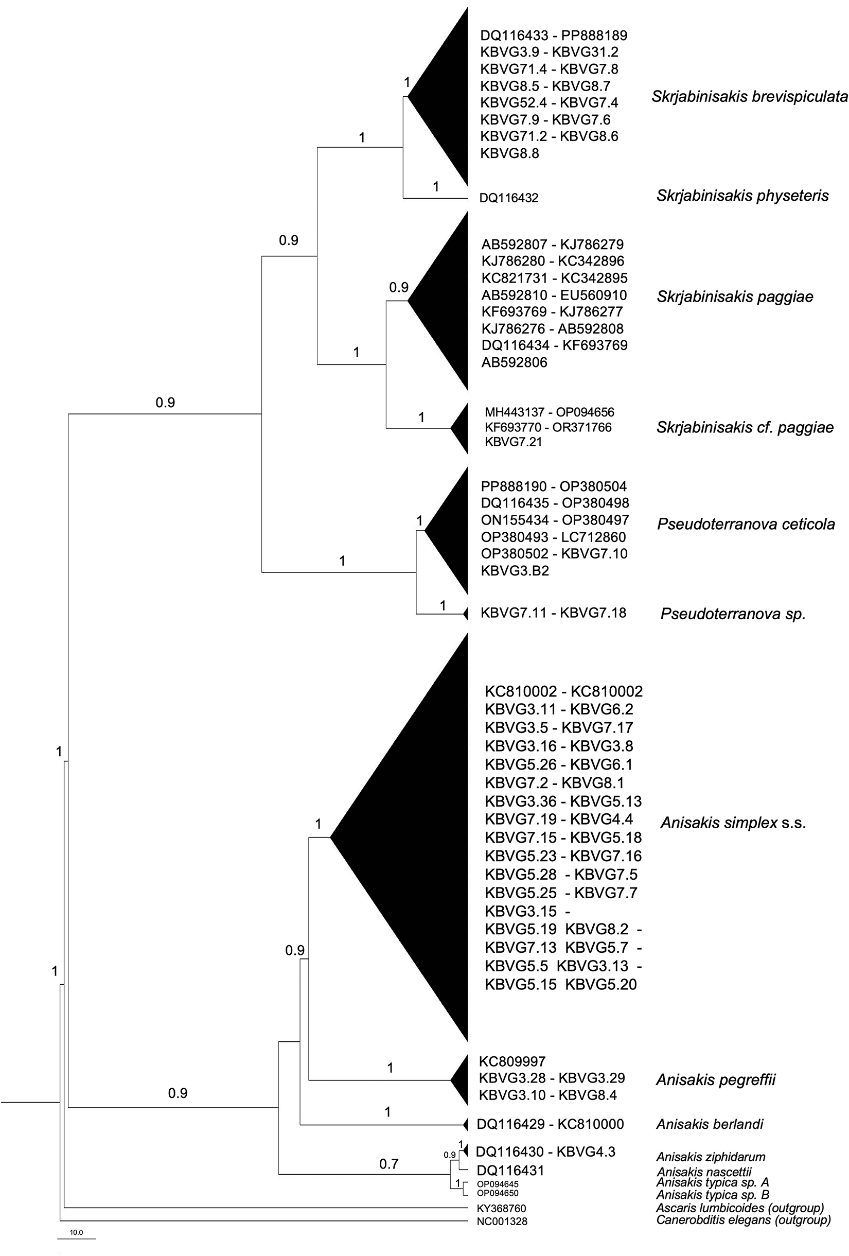
Bayesian inference (BI) phylogenetic tree based on mtDNA cox2 sequences of anisakid nematodes recovered from stranded pygmy sperm whales (Kogia breviceps) examined in this study and reference sequences retrieved from GenBank. Numbers at nodes indicate posterior probability values; values <0.70 are not shown. Newly generated sequences are indicated by specimen codes. Outgroup taxa and GenBank accession numbers are shown in the figure.

Figure 2 Long description
Bayesian inference phylogenetic tree oriented vertically, depicting the evolutionary relationships among anisakid nematodes based on mtDNA cox2 sequences. The tree includes several clades, each represented by triangles, with labels indicating species or groups. Posterior probability values are shown at the nodes, with values less than 0.70 omitted. The tree starts with a node labeled DQ116433 - PP885189, leading to a clade labeled Skrjabinisakis brevispiculata. Below, a node labeled DQ194242 leads to Skrjabinisakis physeteris. Further down, a node labeled AB592697 - KU752976 leads to Skrjabinisakis paggiae, followed by another node labeled MH931107 - OP966856 leading to Skrjabinisakis cf. paggiae. The next node, labeled PP885190 - OP536404, leads to Pseudoterranova ceticola and another node labeled KBVG17.11 - KBVG17.18 leads to Pseudoterranova sp. The tree continues with a node labeled KCB10002 - KCB10002 leading to Anisakis simplex s.s., followed by a node labeled KCB9997 - KEVG3.29 leading to Anisakis pegreffii. The final node, labeled DQ116429 - KC809943, leads to Anisakis berlandi. Outgroup taxa are indicated at the bottom, with GenBank accession numbers provided. The scale bar at the bottom represents genetic distance.
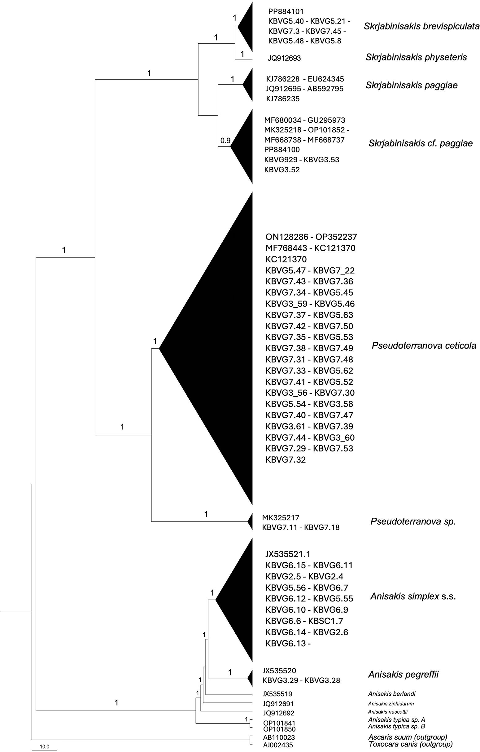
Bayesian inference phylogenetic tree based on ITS rDNA sequences of anisakid nematodes recovered from stranded pygmy sperm whales (Kogia breviceps) examined in this study and reference sequences retrieved from GenBank. Numbers at nodes indicate posterior probability values; values <0.70 are not shown. Newly generated sequences are indicated by specimen codes.

Figure 3 Long description
The tree is oriented vertically, with branches extending from top to bottom. At the top, the tree splits into several branches, each labeled with different species names and specimen codes. The first major branch includes Skjabinaskis brevispiculata, Skjabinaskis physeteris, Skjabinaskis paggiae and Skjabinaskis cf. paggiae, with posterior probability values indicated at the nodes. The next major branch represents Pseudoterranova ceticola, with a large cluster of specimen codes listed. Below this, another branch labeled Pseudoterranova sp. is shown. Further down, Anisakis simplex s.s. is represented, followed by Anisakis pegreffii at the bottom. Posterior probability values are shown at various nodes, with values less than 0.70 not displayed. The tree includes newly generated sequences indicated by specimen codes and reference sequences retrieved from GenBank.
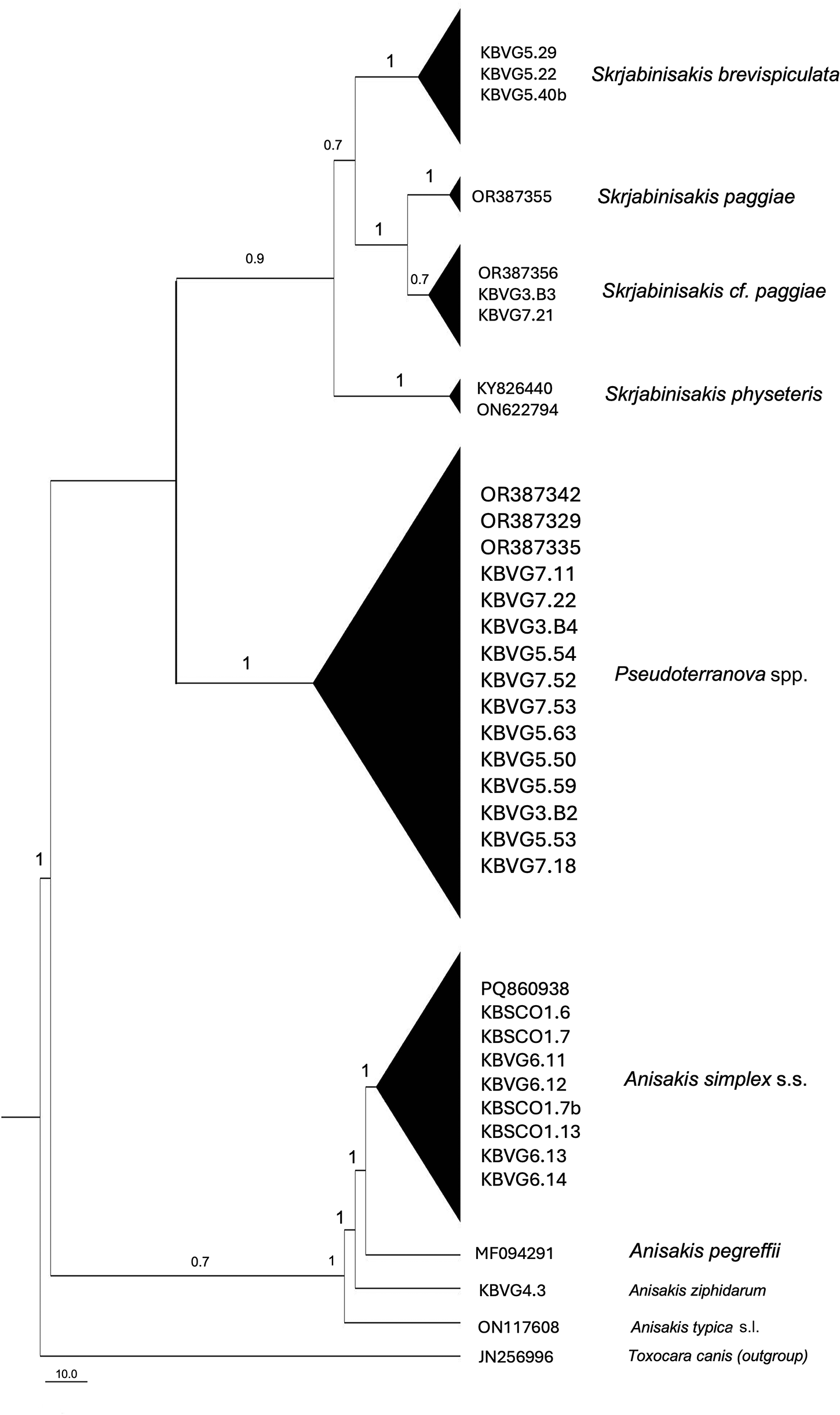
Bayesian inference phylogenetic tree based on 28S rDNA sequences of anisakid nematodes recovered from stranded pygmy sperm whales (Kogia breviceps) examined in this study and reference sequences retrieved from GenBank. Numbers at nodes indicate posterior probability values; values <0.70 are not shown. Newly generated sequences are indicated by specimen codes. Outgroup taxa and GenBank accession numbers are shown in the figure.

Figure 4 Long description
The phylogenetic tree is oriented vertically, displaying evolutionary relationships among anisakid nematodes based on 28S rDNA sequences. The tree begins at the top with Skjabinisascaris brevispiculata, represented by KBVG5.29, KBVG5.22 and KBVG5.40b, with a posterior probability value of 1. Below, Skjabinisascaris paggiae is shown with OR387356 and Skjabinisascaris cf. paggiae with OR387396, KBVG5.38 and KBVG7.21, both having a posterior probability of 1. Skjabinisascaris physeteris is represented by KY926440 and ON622794, also with a posterior probability of 1. Further down, Pseudoterranova spp. includes OR387342, OR387329, OR387335, KBVG7.11, KBVG7.22, KBVG3.84, KBVG5.54, KBVG7.52, KBVG7.62, KBVG5.63, KBVG5.50, KBVG5.59, KBVG3.52, KBVG5.53 and KBVG7.18, with a posterior probability of 1. Anisakis simplex s.s. is represented by PBQ69083, KBSCO1.6, KBSCO1.17, KBCVG6.11, KBCVG6.12, KBSCO1.7b, KBCVG6.13, KBCVG6.14, with a posterior probability of 1. Anisakis pegreffii is shown with MF952491 and KBVG4.3, followed by Anisakis physeteris, Anisakis typica s.s. and Toxocara canis as outgroups, with a posterior probability of 0.7. The tree includes posterior probability values at nodes, indicating the confidence in the branching events, with values less than 0.70 not shown.
Results
Morphological assignment of anisakids
Specimen details and observed pathology are summarized in Table 1. Based on the morphological examination of 500 randomly selected nematodes recovered from the stranded pygmy sperm whales, 3 main morphotypes were recognized: Skrjabinisakis spp., characterized by a short ventriculus; Pseudoterranova, showing the typical intestinal caecum extending along the ventriculus; and Anisakis simplex (s.l.), distinguished by a long, curved ventriculus.
A summary of the relative frequencies of the anisakid morphotypes recovered from pygmy sperm whales, together with their mean body length, length range, developmental stage composition and sex ratio, is reported in Table 2. Additional host-specific data on the distribution of anisakid morphotypes are provided in Supplementary Table 6.
Summary of the relative frequencies of anisakids morphotypes recovered from pygmy sperm whales, including mean body length (mm), length range (mm), proportion of developmental stages (adult and pre-adult, %) and sex ratio (M: F)

Table 2 Long description
Species are listed with specimens number, relative frequency, mean length in millimeters, length range in millimeters, adult and pre adult percentages, and sex ratio. Pseudoterranova spp. had 202 specimens, mean length 19.5, length range 9 to 28, 90 percent adult and 10 percent pre adult, and sex ratio 1 to 1. A. simplex (s.l.) had 170 specimens, mean length 29.4, length range 8 to 68, 40 percent adult and 60 percent pre adult, and sex ratio listed as NS. S. brevispiculata had 98 specimens, mean length 42.6, length range 12 to 79, 90 percent adult and 10 percent pre adult, and sex ratio 1 to 1. S. paggiae had 22 specimens, mean length 20.4, length range 14 to 30, 50 percent adult and 50 percent pre adult, and sex ratio 1 to 1.
Specimens assigned to P. ceticola were comparatively small-bodied at the adult stage relative to the other morphotypes and displayed a prominent intestinal caecum extending along the ventriculus. Males showed approximately 40 precloacal papillae and 5 postcloacal papillae (or 6 when considering the double papilla), consistent with the description provided by Deardorff and Overstreet (Reference Deardorff and Overstreet1981). A single male specimen exhibited an intestinal caecum, 65–70 precloacal papillae and 6 postcloacal papillae, closely resembling the description of P. kogiae as reported by Johnston and Mawson (Reference Johnston and Mawson1939). Almost all Pseudoterranova spp. specimens (>90%) displayed adult characters.
Among the specimens with Skrjabinisakis-like morphology, 22 displayed a ventriculus with a marked median constriction, closely resembling the violin-shaped ventriculus typical of S. paggiae (Mattiucci et al., Reference Mattiucci, Cipriani, Levsen, Paoletti and Nascetti2018), whereas the remaining 98 exhibited a short ventriculus consistent with the description of S. brevispiculata (Mattiucci et al., Reference Mattiucci, Cipriani, Levsen, Paoletti and Nascetti2018). Male specimens of both species were characterized by short and approximately equal spicules.
Most individuals belonging to the A. simplex (s.l.) morphotype were immature (60%), while few specimens exceeding 50 mm in length (18, representing 15%) showed fully developed adult characters, such as spicules in males and ovaries containing eggs in females.
Genetic identification of anisakids
Readable sequences for at least 1 marker were obtained from 181 out of 250 analysed specimens, likely due to suboptimal preservation of the nematode material (Table 3). Although not all 3 markers were successfully amplified for all specimens and species, for some taxa sequences were obtained across all loci, allowing unambiguous multilocus identification (Tables 3 and 4).
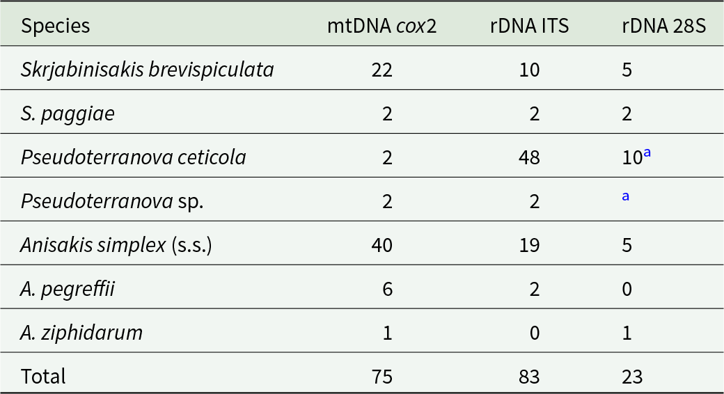
Number of sequences obtained for each anisakid species and genetic marker (mtDNA cox2, rDNA ITS and rDNA 28S) from pygmy sperm whales

Table 3 Long description
Counts of sequences are listed by species across three genetic markers: mtDNA cox2, rDNA ITS, and rDNA 28S. Skrjabinisakis brevispiculata has 22 for mtDNA cox2, 10 for rDNA ITS, and 5 for rDNA 28S. S. paggiae has 2 for mtDNA cox2, 2 for rDNA ITS, and 2 for rDNA 28S. Pseudoterranova ceticola has 2 for mtDNA cox2, 48 for rDNA ITS, and 10 marked with an asterisk for rDNA 28S. Pseudoterranova sp. has 2 for mtDNA cox2, 2 for rDNA ITS, and an asterisk in the rDNA 28S column with no number shown. Anisakis simplex (s.s.) has 40 for mtDNA cox2, 19 for rDNA ITS, and 5 for rDNA 28S. A. pegreffii has 6 for mtDNA cox2, 2 for rDNA ITS, and 0 for rDNA 28S, and A. ziphidarum has 1 for mtDNA cox2, 0 for rDNA ITS, and 1 for rDNA 28S. Totals are 75 for mtDNA cox2, 83 for rDNA ITS, and 23 for rDNA 28S.
Notes: Species identification was inferred based on GenBank BLAST comparisons and sequence alignment analyses.
a Indicates that 28S rDNA sequences were identical between Pseudoterranova ceticola and Pseudoterranova sp.
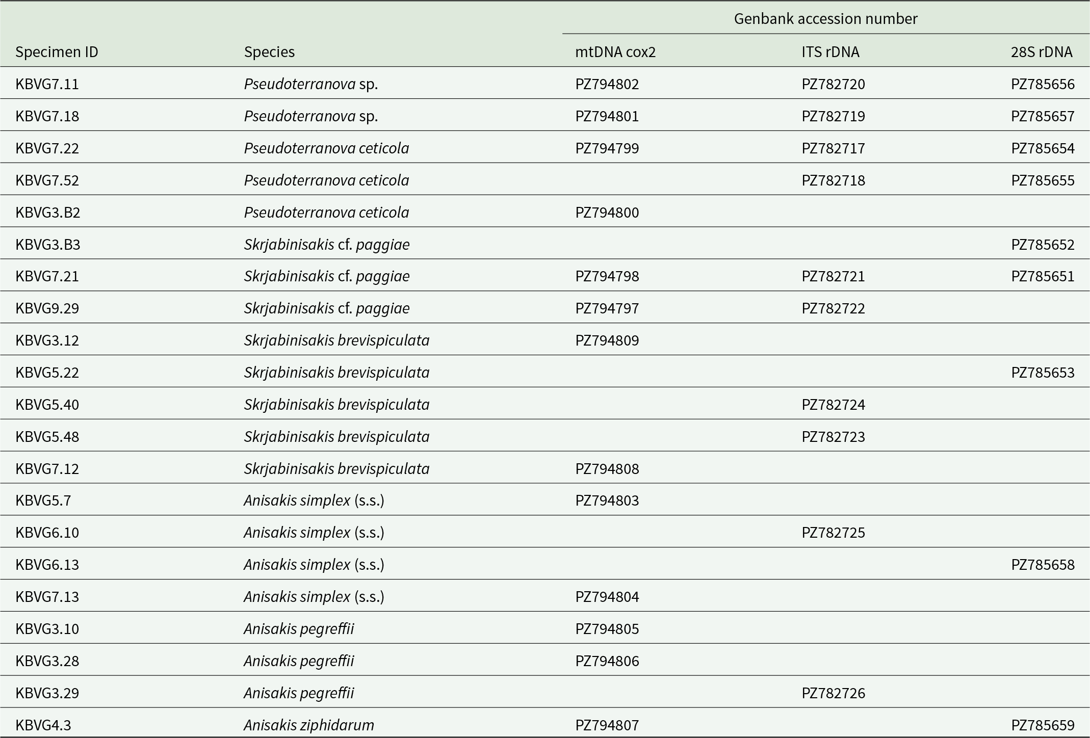
GenBank accession numbers of sequences generated in this study for each specimen and genetic marker (mtDNA cox2, ITS rDNA and 28S rDNA)

Table 4 Long description
Columns list Specimen ID, Species, and Genbank accession number entries for mtDNA cox2, ITS rDNA, and 28S rDNA. Pseudoterranova sp. includes KBVG7.11 with PZ794802, PZ782720, and PZ785656, and KBVG7.18 with PZ794801, PZ782719, and PZ785657. Pseudoterranova ceticola includes KBVG7.22 with PZ794799, PZ782717, and PZ785654, KBVG7.52 with ITS rDNA PZ782718 and 28S rDNA PZ785655, and KBVG3. B2 with mtDNA cox2 PZ794800. Skrjabinisakis cf. paggiae includes KBVG3. B3 with 28S rDNA PZ785652, KBVG7.21 with PZ794798, PZ782721, and PZ785651, and KBVG9.29 with PZ794797 and PZ782722. Skrjabinisakis brevispiculata includes KBVG3.12 with mtDNA cox2 PZ794809, KBVG5.22 with 28S rDNA PZ785653, KBVG5.40 with ITS rDNA PZ782724, KBVG5.48 with ITS rDNA PZ782723, and KBVG7.12 with mtDNA cox2 PZ794808. Anisakis simplex (s.s.) includes KBVG5.7 with mtDNA cox2 PZ794803, KBVG6.10 with ITS rDNA PZ782725, KBVG6.13 with 28S rDNA PZ785658, and KBVG7.13 with mtDNA cox2 PZ794804. Anisakis pegreffii includes KBVG3.10 with mtDNA cox2 PZ794805, KBVG3.28 with mtDNA cox2 PZ794806, and KBVG3.29 with ITS rDNA PZ782726. Anisakis ziphidarum includes KBVG4.3 with mtDNA cox2 PZ794807 and 28S rDNA PZ785659.
Partial mtDNA cox2 sequences (580 bp) obtained from 2 specimens displaying a Skrjabinisakis morphotype with an A. paggiae-type ventriculus showed 98.6% identity with sequences deposited in GenBank as S. paggiae (OR371766), whereas 22 sequences with a S. brevispiculata-type ventriculus matched S. brevispiculata at 99.6% (KJ786283). These same specimens showed, at the ITS rDNA, a 99.7% identity with the sequences of S. paggiae deposited in GenBank (PP884100), and 10 specimens had 100% identity with S. brevispiculata (PP884101). At the 28S rDNA marker, 2 sequences matched 100% with a GenBank sequence of S. cf. paggiae (OR387340), while 3 matched 100% with S. brevispiculata (OR387356).
Among the 46 sequences at mtDNA cox2 obtained from nematodes with A. simplex s.l. complex morphotype, 40 matched A. simplex (99.8%, KT852513) and 6 matched A. pegreffii (99.8%, OQ919779). A total of 19 ITS rDNA sequences obtained from nematodes with A. simplex (s.l.) morphotype were identical (100%) and matched 100% with A. simplex (s.s.) (PP884102), while 2 matched 100% with A. pegreffii (OM279537). At the 28S rDNA marker, 5 sequences matched 100% with the A. simplex (s.s.) sequence deposited in GenBank (PQ860935).
The mtDNA cox2 sequence of a single pre-adult specimen not morphologically assigned showed 100% identity with A. ziphidarum (DQ116430, GenBank). Also, the 28S rDNA sequence obtained from this individual matched 100% with an A. ziphidarum (s.s.) sequence deposited in GenBank (OR387346).
MtDNA cox2 sequences from 2 specimens with a Pseudoterranova morphotype showed 99.4% identity with P. ceticola (OP380507), whereas 2 morphologically similar specimens displayed a lower similarity, with only 90% identity to the same P. ceticola reference sequence. Out of the ITS rDNA sequences obtained from nematodes morphologically assigned to Pseudoterranova, 48 matched P. ceticola at 99.8% identity (OP352243), whereas the remaining 2 matched at 100% with a deposited sequence (MK325217) reported as Anisakis sp. B. At the 28S rDNA locus, 10 sequences morphologically assigned to Pseudoterranova showed 100% identity with the P. ceticola sequence deposited in GenBank (OR387340).
A subset of the sequences generated in this study were deposited in GenBank. The corresponding GenBank accession numbers, organized by species and loci (cox2 mtDNA, ITS rDNA and 28S rDNA), are reported in Table 4.
Phylogenetic analyses
The phylogenetic analyses were based on mtDNA cox2, ITS rDNA and 28S rDNA sequences generated in this study, together with additional reference sequences retrieved from GenBank for each marker (see Figures 2–4; Supplementary Table 5).
The phylogenetic analysis based on the mtDNA cox2 gene (Figure 2) revealed that the specimens morphologically assigned to Pseudoterranova are divided into 2 distinct clades, both supported by high posterior probability values. The first lineage included 2 sequences corresponding morphologically to P. ceticola (KBVG7.22 and KBVG3.B2), clustering together with multiple P. ceticola sequences available in GenBank (Figure 2; Table 4). The second lineage comprised 2 novel sequences (here referred to as Pseudoterranova sp., i.e. specimens KBVG7.11 and KBVG7.18) for which no diagnostic morphological characters could be investigated, as both specimens were females (Figure 2).
Similarly, the phylogenetic analysis of the ITS rDNA region showed the presence of 2 distinct lineages within Pseudoterranova. One clade included 30 newly generated sequences from the pygmy sperm whales examined in this study, including male specimens with morphological features consistent with P. ceticola, together with sequences previously deposited in GenBank and identified as P. ceticola (Figure 3; Table 4 and Supplementary Table 5). The second lineage included 2 sequences obtained in this study (here referred to as Pseudoterranova sp.; specimens KBVG7.11 and KBVG7.18), as well as 1 sequence available in GenBank originally reported as Anisakis sp. and detected in a pigmy sperm whale from Australian waters (GenBank accession no. MK325217) (Table 4).
In contrast, phylogenetic analysis based on the 28S rDNA dataset showed that all Pseudoterranova sequences obtained in this study clustered within a single clade, without separation into the lineages observed in the cox2 and ITS datasets (Figure 4).
The phylogenetic analysis of cox2 mtDNA (Figure 2) also revealed that S. paggiae (s.l.) specimens divided into 2 closely related but distinct clades, even supported by high posterior probability values. One clade included 2 novel sequences generated in this study from pygmy sperm whales (KBVG7.21 and KBVG9.29), together with multiple S. paggiae sequences available in GenBank, including specimens referred to as S. cf. paggiae (OR371766) from Diretmus argenteus in Canary Island waters or ‘S. paggiae-like’ collected in K. sima stranded on Brazilian coasts (KF693770) (Di Azevedo et al., Reference Di Azevedo, Carvalho and Iñiguez2017) (Figure 2, Supplementary Table 5). The second clade included S. paggiae sequences originating from several geographic regions, including sequences obtained from adult specimens collected in Kogia sima in Brazil (KF693769), and from the same host along Southern Philippine’s coast (KC342895) (Figure 2, Supplementary Table 5).
Also, the phylogenetic analysis of ITS rDNA seems to support the existence of 2 distinct clades within S. paggiae (s.l.) (Figure 3). One clade comprised 2 newly generated sequences from the pygmy sperm whales examined in this study (KBVG7.21 and KBVG9.29), together with S. paggiae sequences previously deposited in GenBank, including an adult specimen obtained from a pygmy sperm whale stranded on Australian coasts (MK325218) (Figure 3, Table 4). The second clade was formed only by S. paggiae sequences available in GenBank, obtained from adult specimens from pygmy sperm whale in NE Atlantic (JQ912695) and Philippine waters (KJ786228) (Figure 3, Table 4). The phylogenetic analysis based on the 28S rDNA dataset revealed 2 clades. One included a sequence identified as S. paggiae by Bao et al. (Reference Bao, Giulietti, Levsen and Karlsbakk2023) (OR387355) from D. argenteus off the Canary Islands, whereas the second clade comprised 2 sequences obtained in this study (KBVG7.21 and KBVG3.B3) together with a specimen identified as S. cf. paggiae (OR387356) also originating from D. argenteus in Canary Islands waters (Table 4 and Supplementary Table 5).
Sequences obtained for S. brevispiculata at the mtDNA cox2, ITS rDNA and 28S markers consistently clustered within a clade including reference sequences of S. brevispiculata previously deposited in GenBank (Figures 2–4). Likewise, cox2, ITS rDNA and 28S sequences of A. simplex (s.s.) and A. pegreffii grouped in well-supported clades with the corresponding reference sequences of both species available in GenBank (Figures 2–4).
Additional host-specific data on the distribution of genetically identified anisakid species for each whale are provided in Supplementary Table 7.
Comparative sequence alignments
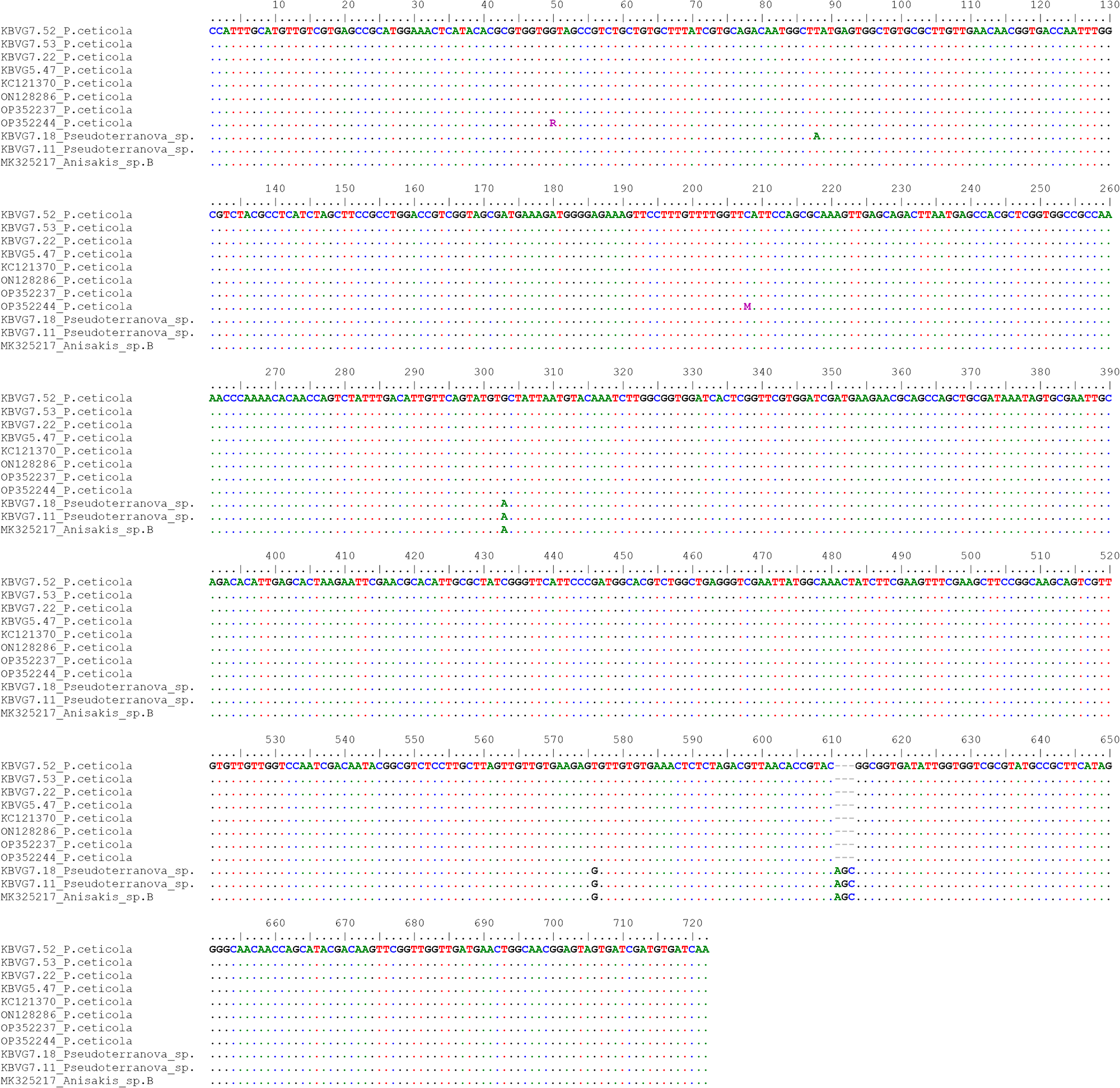
Sequence alignments of ITS rDNA, 28S rDNA and mtDNA cox2 are presented for taxa showing unexpected clustering in the phylogenetic trees. Comparative analysis of the ITS rDNA sequences obtained in this study for Pseudoterranova together with reference sequences retrieved from GenBank (Figure 5, Supplementary Table 5) revealed fixed nucleotide substitutions at 2 diagnostic positions (303 and 576) and a 3-base indel distinguishing 2 sequence groups (611–613), with nucleotide positions here and below referring to the numbering used in the corresponding sequence alignments. In particular, the indel corresponded to the triplet AGC in Pseudoterranova sp. sequences.
Sequence alignment of the ITS rDNA gene, including sequences of Pseudoterranova generated in this study (KBVG7.52 – KBVG7.53 – KBVG7.22 – KBVG5.47 – KBVG7.18 – KBVG7.11) and reference sequences retrieved from GenBank (Supplementary Table 5). Dots indicate identical nucleotides, whereas dashes represent gaps.

Figure 5 Long description
A multiple sequence alignment of ITS rDNA gene sequences is presented in several horizontal blocks. Each block contains rows of nucleotide letters aligned beneath a numbered position scale. The position scale runs across the top of each block, marking nucleotide positions incrementally. Sequence identifiers appear along the left side of each row, including labels such as KBVG7.52, KBVG7.53, KBVG7.22, KBVG5.47, KBVG7.18 and KBVG7.11, alongside reference sequence labels. Dots appear throughout the rows to indicate positions where nucleotides are identical to the reference. Dashes appear at certain positions to represent gaps in the alignment. At specific positions along the alignment, individual nucleotide letters are visually distinct from the surrounding dots, drawing attention to substitution sites. Markers are visible above the alignment at positions 303 and 576, indicating diagnostic nucleotide substitution sites. A further marked region appears at positions 611 to 613, corresponding to a three-base indel site. The letters at the indel region differ between sequence groups, with the triplet A G C present in Pseudoterranova sp. rows. The alignment blocks progress sequentially from lower position numbers at the top of the image to higher position numbers toward the bottom, covering the full length of the aligned gene region.
One group included sequences generated in this study that matched the morphological description of P. ceticola, together with several P. ceticola sequences retrieved from GenBank. The second group comprised 2 specimens identified as Pseudoterranova sp. (KBVG7.11 and KBVG7.18), which were identical to a sequence deposited in GenBank as Anisakis sp. B in a pygmy sperm whale from Australian waters (GenBank accession no. MK325217) (Figure 5, Supplementary Table 5).
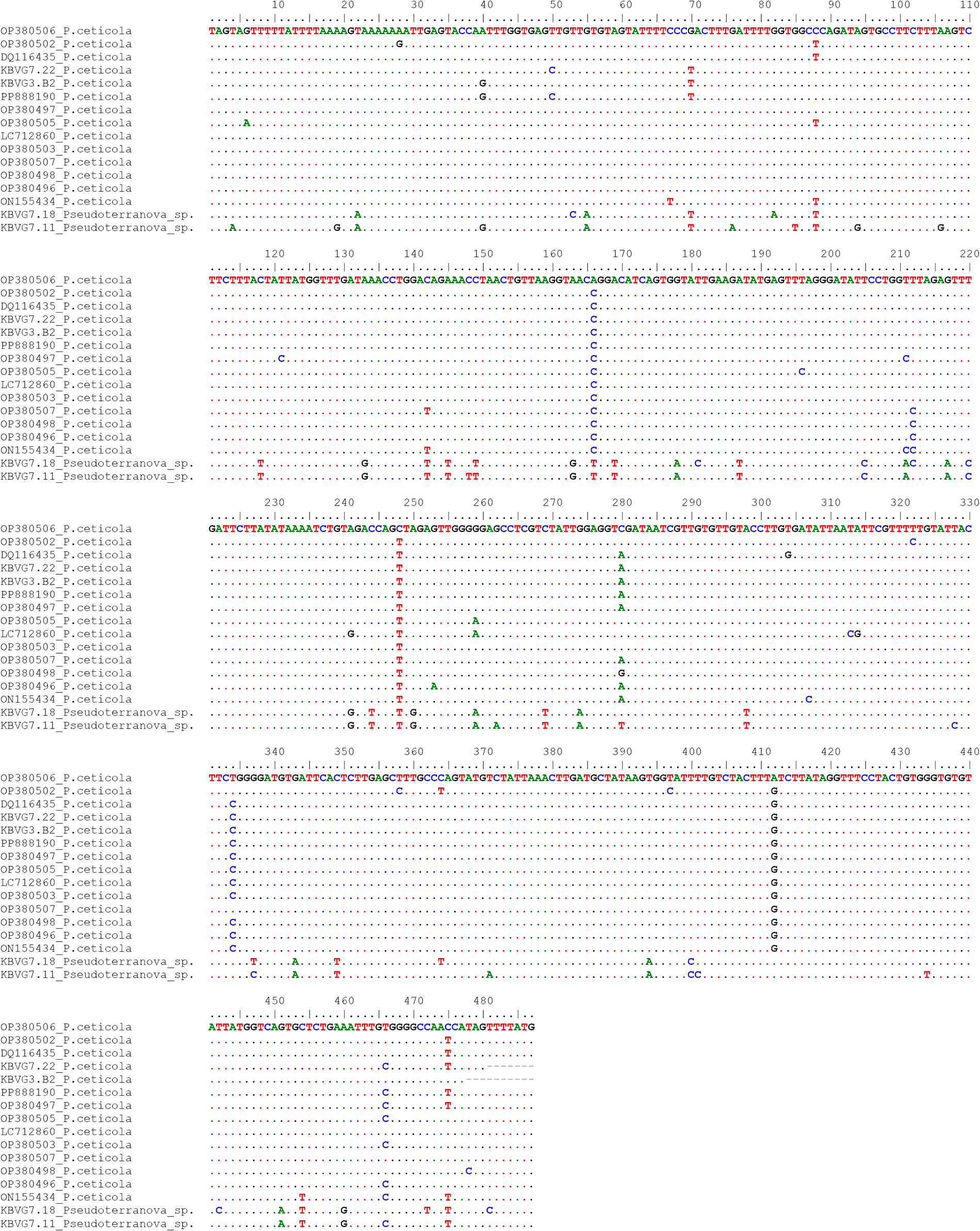
Similarly, the alignment of mtDNA cox2 sequences of Pseudoterranova obtained in this study with reference sequences retrieved from GenBank (Figure 6, Supplementary Table 5) revealed 22 single nucleotides separating 2 sequence groups. One group included specimens obtained in this study and morphologically identified as P. ceticola (KBVG7.22 and KBVG3.B2), which showed high sequence similarity to P. ceticola sequences deposited in GenBank (Figure 6, Supplementary Table 5). The second group comprised 2 sequences identified as Pseudoterranova sp. (KBVG7.11 and KBVG7.18). No mtDNA cox2 sequences corresponding to this genotype were available in GenBank for comparison.
Sequence alignment of the mtDNA cox2 gene, including sequences of Pseudoterranova (KBVG7.22 – KBVG3.B2 – KBVG7.18 – KBVG7.11) and P. Ceticola (KBVG7.22 and KBVG3.B2) generated in this study, as well as reference sequences retrieved from GenBank (Supplementary Table 5). Dots indicate identical nucleotides, whereas dashes represent gaps.

Figure 6 Long description
Sequence alignment of mtDNA genes from Pseudoterranova species. Sample IDs such as KBVG7.22, KBVG3. B2, KBVG7.18 and KBVG7.11 are listed on the left. Nucleotide positions are numbered across the top, ranging from 1 to 600. Sequences are arranged in rows, with dots indicating identical nucleotides and dashes representing gaps. Colored letters (A, C, G, T) highlight differences between sequences. The alignment reveals conserved regions and variable sites, showing SNP patterns and differences between groups. The color-coding aids in identifying nucleotide variations across the samples.
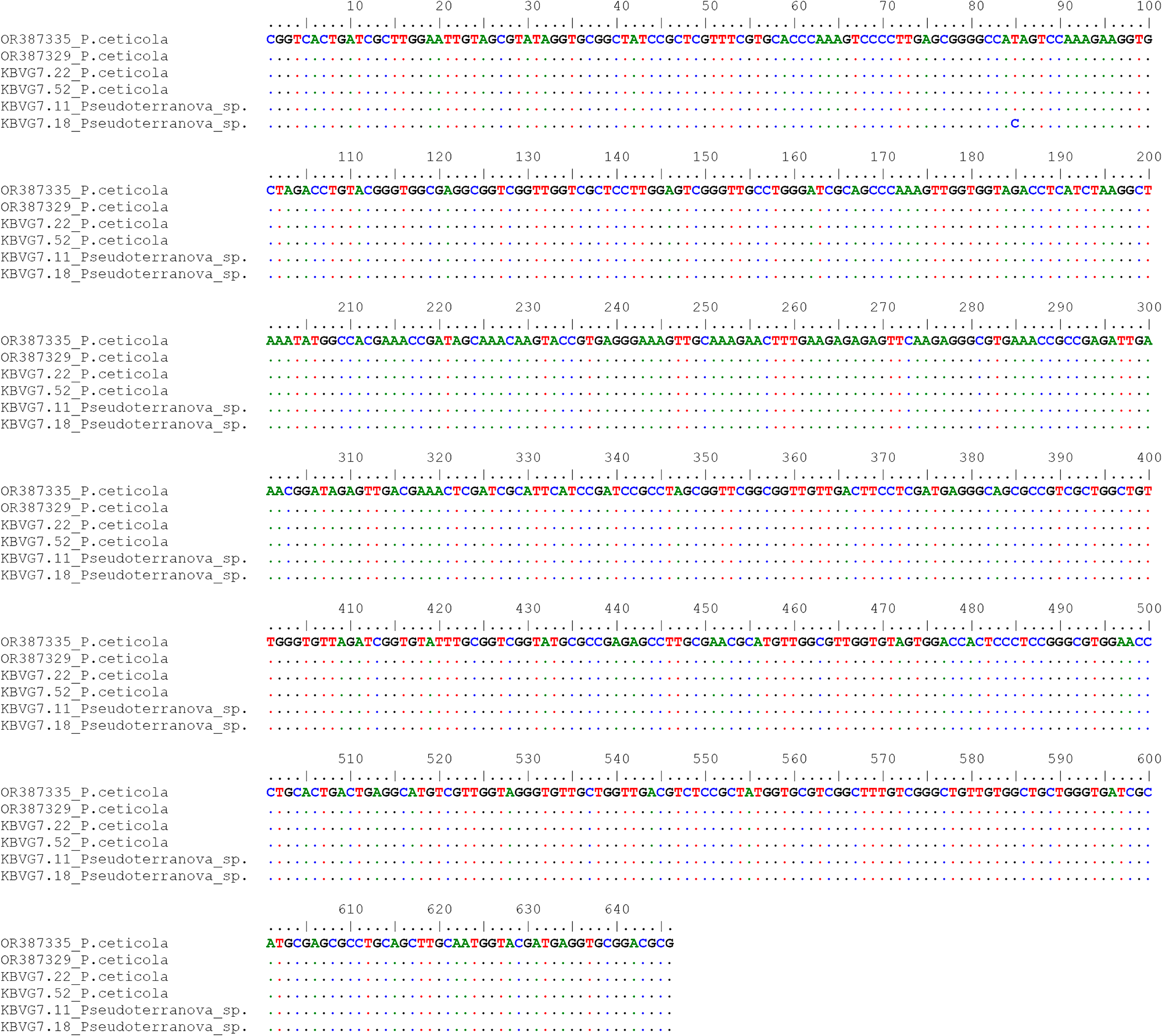
Analysis of the 28S rDNA sequence alignment, including 10 Pseudoterranova sequences generated in this study together with reference sequences retrieved from GenBank (Supplementary Table 5), showed no nucleotide variation among them, with all sequences being 100% identical. The only exception was specimen KBVG7.18, which displayed a single substitution at position 85.
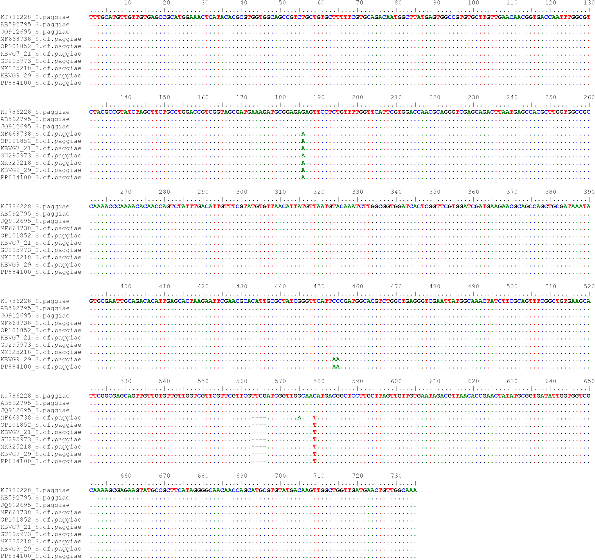
Comparative analysis of ITS rDNA sequences obtained in this study for S. paggiae, together with reference sequences retrieved from GenBank (Figure 8, Supplementary Table 5), revealed 2 sequence groups separated by fixed nucleotide substitutions at 2 diagnostic positions (186 and 579) and a 4-base indel (563-566). One group included only S. paggiae sequences retrieved from GenBank (Figure 8, Supplementary Table 5), whereas the other group comprised 2 specimens obtained in this study (KBVG7.21 and KBVG9.29) together with GenBank sequences reported as S. cf. paggiae (Figure 8, Supplementary Table 5). At the indel position, S. paggiae showed the sequence TTCG, whereas a gap was present in S. cf. paggiae (Figure 8).
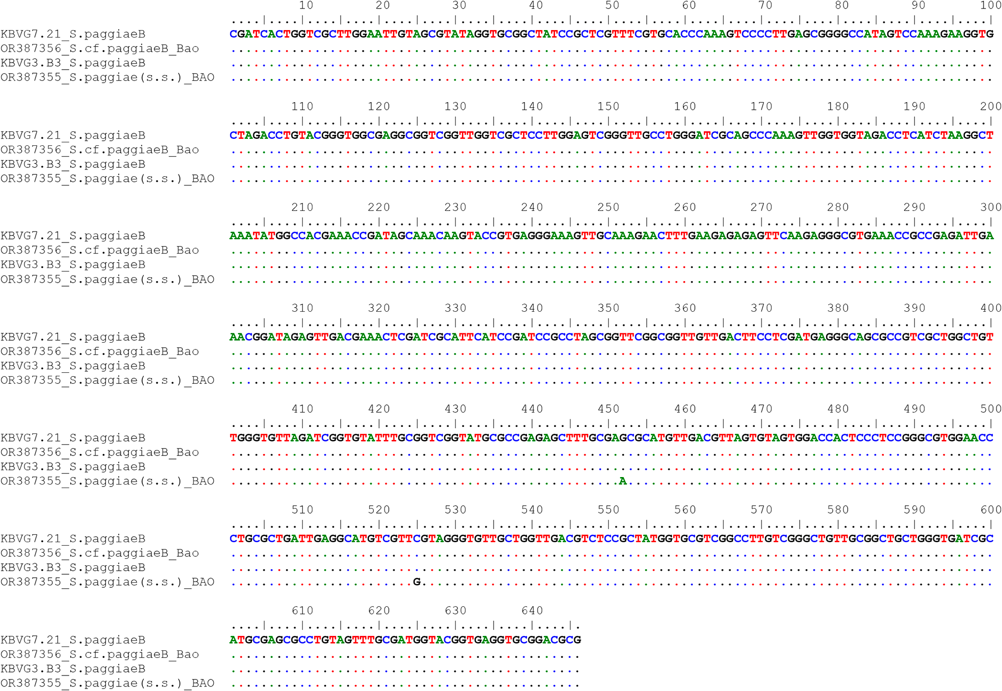
At the 28S rDNA locus, alignment of the S. paggiae sequence dataset generated in this study with reference sequences retrieved from GenBank revealed fixed nucleotide substitutions at 2 diagnostic positions separating 2 sequence groups (Figure 9). The 2 28S sequences obtained in this study (KBVG7.21 and KBVG3.B3) showed 100% identity with sequences reported as S. cf. paggiae by Bao et al. (Reference Bao, Olsen, Levsen, Cipriani, Giulietti, Storesund, García-seoane and Karlsbakk2022) (Figure 9, Supplementary Table 5). In contrast, a sequence identified as S. paggiae (OR387355) differed by 2 nucleotide substitutions (452 and 525).
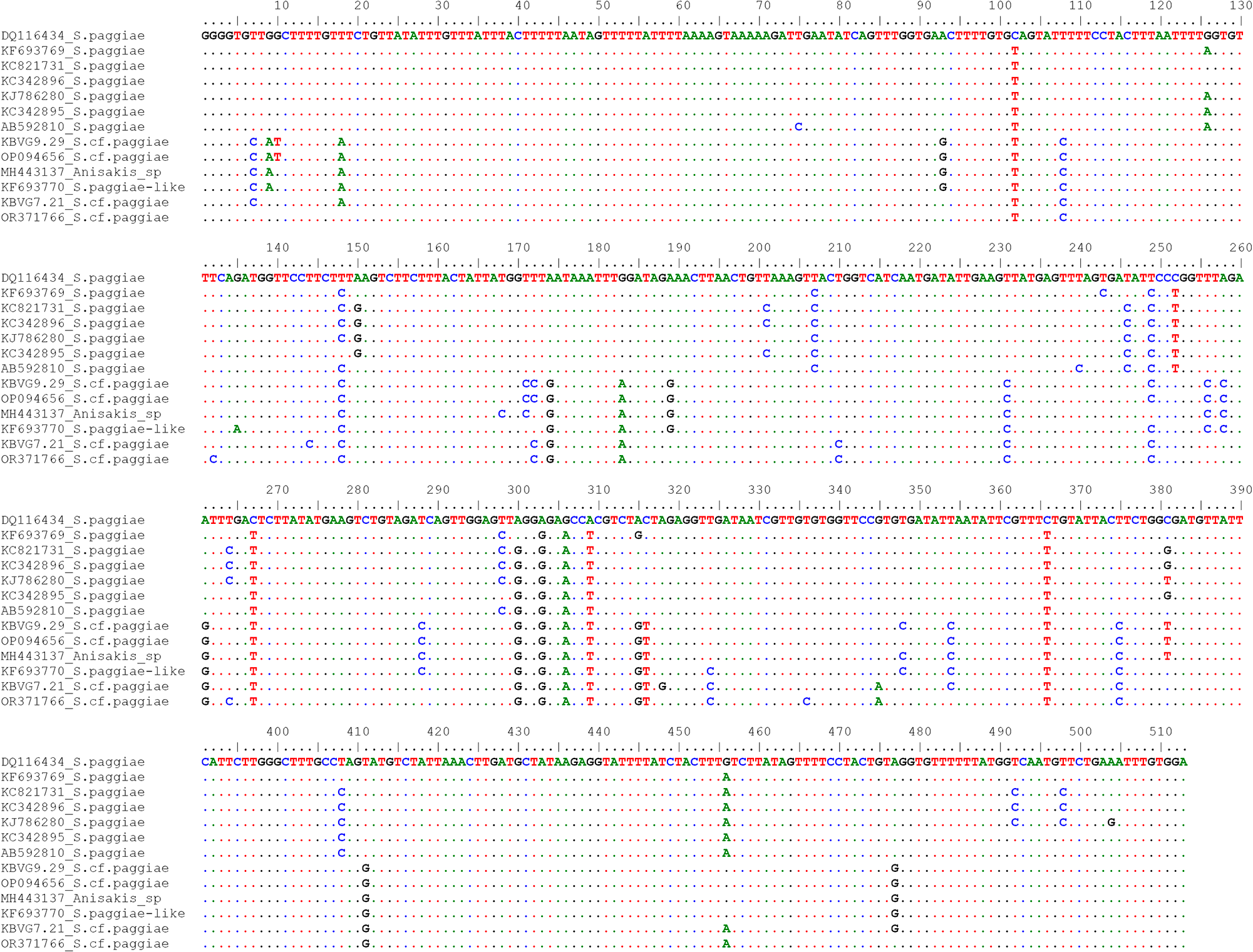
Consistently, the alignment of S. paggiae mtDNA cox2 sequences obtained in this study with reference sequences retrieved from GenBank (Figure 10, Supplementary Table 5) revealed that 9 diagnostic nucleotide positions (positions 108, 174, 183, 231, 252, 261, 306, 375 and 411) distinguished 2 sequence groups. One group comprised the 2 sequences obtained in this study (KBVG7.21 and KBVG9.29), together with GenBank sequences reported as S. cf. paggiae (OR371766) and S. paggiae-like (KF693770). The second group included several GenBank sequences identified as S. paggiae, including the sequence generated from the holotype (DQ116434)(Figure 10, Supplementary Table 5).
Discussion
Kogiid whales are elusive and poorly studied cetaceans, recognized as specific hosts of several anisakid species. This study, reporting novel data on the anisakid species diversity in 9 pygmy sperm whales (Kogia breviceps) stranded along the Galician coast (north-western Spain), suggests that the diversity of anisakid parasites in this host may be higher than previously recognized. Among the species identified here, Skrjabinisakis brevispiculata, S. paggiae and Pseudoterranova ceticola were originally described from kogiid whales and are considered host-specific at the adult stage, with adult records restricted to pygmy and dwarf sperm whales (Abollo and Pascual, Reference Abollo and Pascual2002; González-Solís et al., Reference González-Solís, Vidal-Martínez, Antochiw-Alonso and Ortega-Argueta2006; Mattiucci and Nascetti, Reference Mattiucci and Nascetti2008; Cavallero et al., Reference Cavallero, Nadler, Paggi, Barros and D’Amelio2011; Quiazon et al., Reference Quiazon, Santos and Yoshinaga2013; Di Azevedo et al., Reference Di Azevedo, Carvalho and Iñiguez2017; Shamsi et al., Reference Shamsi, Sprohnle-barrera and Shafaet Hossen2019; Irigoitia et al., Reference Irigoitia, Palomba, Braicovich, Lanfranchi, Denuncio, Gana, Mattiucci and Timi2021; Hernández-Olascoaga et al., Reference Hernández-Olascoaga, Guillén-hernández and Díaz-gamboa2023). By contrast, Anisakis simplex (s.s.), A. pegreffii and A. ziphidarum are common but non-host-specific parasites of kogiid and appear to have reduced fitness or incomplete development in this whale species (Santoro et al., Reference Santoro, Di Nocera, Iaccarino, Cipriani, Guadano Procesi, Maffucci, Hochscheid, Blanco, Cerrone, Galiero, Nascetti and Mattiucci2018; Cipriani et al., Reference Cipriani, Palomba, Giulietti, Marcer, Mazzariol, Santoro, Albuquerque, Covelo, López, Santos, Pierce, Brownlow, Davison, McGovern, Frantzis, Alexiadou, Højgaard, Mikkelsen, Paoletti, Nascetti, Levsen and Mattiucci2022, Reference Cipriani, Palomba, Giulietti, Aco-Alburqueque, Andolfi, Ten Doeschate, Brownlow, Davison and Mattiucci2024).
Previous studies reported genetic variation within P. ceticola and S. paggiae (Di Azevedo et al., Reference Di Azevedo, Carvalho and Iñiguez2017; Bao et al., Reference Bao, Giulietti, Levsen and Karlsbakk2023), suggesting that the taxonomic boundaries of these taxa may not yet be fully resolved. In this study, multilocus analyses of mtDNA cox2, ITS rDNA and 28S rDNA provide further evidence that specimens currently assigned to P. ceticola and S. paggiae include distinct genetic lineages. These lineages may represent additional, closely related taxa within Pseudoterranova and Skrjabinisakis that are not yet formally recognized.
Phylogenetic analyses of mtDNA cox2 and ITS rDNA revealed 2 closely related but clearly distinct phylogenetic lineages among specimens morphologically assigned to Pseudoterranova (Figures 2 and 3). Both lineages included adult-stage specimens occurring sympatrically within the same definitive hosts, suggesting limited gene flow between the 2 taxa in this host, likely reflecting reproductive isolation. Sequence alignments at both loci identified diagnostic nucleotide differences supporting this separation (Figures 5 and 6). One lineage corresponded to P. ceticola and matched the diagnostic morphological features as described in Deardorff and Overstreet (Reference Deardorff and Overstreet1981). The second lineage matched a sequence deposited in GenBank as Anisakis sp. B from a pygmy sperm whale in Australian waters (MK325217) (Figure 3), but its species-level morphology could not be assessed because mature males were not available. The 28S rDNA sequence showed little or no variation among Pseudoterranova specimens, despite clear differentiation of the same individuals at the ITS rDNA and mtDNA cox2 loci (Figures 5–7). A single specimen (KBVG7.18) displayed 1 nucleotide substitution in the 28S rDNA sequence and also showed variation at the other markers. Overall, these findings support the highly conserved nature of 28S rDNA in the Pseudoterranova taxa analysed here and indicate its limited value for this species delimitation.
Sequence alignment of the partial large subunit 28s rDNA gene, including sequences of Pseudoterranova generated in this study (KBVG7.22 – KBVG7.52 – KBVG7.11 – KBVG7.18) and reference sequences retrieved from GenBank (Supplementary Table 5). Dots indicate identical nucleotides, whereas dashes represent gaps.

Figure 7 Long description
A multiple sequence alignment of the partial large subunit 28s rDNA gene is shown. Sequences are labeled with accession or sample names such as MK325215P.ceticola and RSV7.11Pseudoterranova_sp. The alignment is organized in rows with nucleotide position numbers at the top, ranging approximately from 1 to 610. Colored letters represent nucleotides, with dots indicating identical nucleotides and dashes representing gaps. The layout includes multiple rows separated by line breaks, each labeled with accession or sample names. Key takeaway: the alignment illustrates the degree and pattern of similarity across different samples, highlighting where differences occur.
The type species of the genus Pseudoterranova is P. kogiae, and it was originally described from a pygmy sperm whale in Australian waters (Johnston and Mawson, Reference Johnston and Mawson1939), whereas P. ceticola was first described from K. sima stranded in Mississippi, USA (Deardorff and Overstreet, Reference Deardorff and Overstreet1981). In the original description of P. ceticola, Deardorff and Overstreet (Reference Deardorff and Overstreet1981) reported 38–50 precloacal and 5 postcloacal papillae, as observed in the male specimens assigned here to this species. In contrast, P. kogiae was described as having 65–70 precloacal and 6 postcloacal papillae. Additionally, P. kogiae was reported to possess 3 transverse rows of plectanes near the posterior cloacal lip (Johnston and Mawson, Reference Johnston and Mawson1939), although these structures may have been based on an incorrect illustration possibly referring to a Skrjabinisakis species (Bao et al., Reference Bao, Olsen, Levsen, Cipriani, Giulietti, Storesund, García-seoane and Karlsbakk2022).
In the present material, all male specimens examined morphologically displayed approximately 40 precloacal papillae and 5 postcloacal papillae (or 6 when considering the double papilla), and 19 of these were genetically confirmed as P. ceticola, including specimens (KBVG7.22 and KBVG7.53).
Since its original description, P. kogiae has not been reported again. Gibson (Reference Gibson, Stone, Platt and Khalil1983) retained its validity as the type species of the genus, but no genetic data are currently available for this taxon. However, an ITS rDNA sequence of an unidentified Anisakis sp. from an Australian pygmy sperm whale, for which no morphological description was provided (Shamsi et al., Reference Shamsi, Sprohnle-barrera and Shafaet Hossen2019), showed 100% identity with 2 sequences obtained here from specimens with clear Pseudoterranova morphology. Bao et al. (Reference Bao, Olsen, Levsen, Cipriani, Giulietti, Storesund, García-seoane and Karlsbakk2022), in their genetic and morphological characterization of the larval stage of P. ceticola, reported that the specimen identified as Anisakis sp. by Shamsi et al. (Reference Shamsi, Sprohnle-barrera and Shafaet Hossen2019) differed from P. ceticola reference sequences by a unique 3 bp insertion and 2 nucleotide substitutions in the ITS rDNA region. The same differences separated the 2 genotypes identified in the present study and matched with Anisakis sp. (Figure 5), suggesting that this lineage likely represents a distinct Pseudoterranova taxon. Its genetic correspondence with the Australian sequence, together with its Pseudoterranova morphology and the origin of the Australian specimen from the type locality of P. kogiae, suggests that this genotype could correspond to this species. However, all specimens clustering within this second Pseudoterranova clade (here interpreted as a potential sibling lineage of P. ceticola) were females and therefore lacked the male diagnostic characters required for direct comparison with the original description of P. kogiae by Johnston and Mawson (Reference Johnston and Mawson1939). The only male Pseudoterranova specimen showing more than 60 precloacal papillae, a feature consistent with P. kogiae, had degraded DNA and could not be sequenced at any of the loci analysed.
A pattern of genetic heterogeneity was observed in Skrjabinisakis paggiae. Phylogenetic analyses based on mtDNA cox2, ITS rDNA and 28S rDNA revealed 2 closely related but distinct lineages among specimens morphologically assigned to S. paggiae (Figures 2–4). The sequences generated in the present study clustered exclusively within 1 of these lineages, together with several GenBank sequences reported as S. paggiae-like and Skrjabinisakis cf. paggiae (KF693770 and OR371766, respectively) (Figures 2–4; Table 4). The second lineage consisted exclusively of GenBank sequences identified as S. paggiae, including the sequence generated from the holotype used in the original species description by Mattiucci et al. (Reference Mattiucci, Nascetti, Dailey, Webb, Barros, Cianchi and Bullini2005), later sequenced by Valentini et al. (Reference Valentini, Mattiucci, Bondanelli, Webb, Mignucci-Giannoni, Colón-Llavina and Nascetti2006) (DQ116434). This pattern was consistently supported by sequence alignments across all analysed loci. Alignments of ITS rDNA, 28S rDNA and mtDNA cox2 sequences (Figures 8–10) revealed fixed nucleotide differences between the 2 lineages, including substitutions and indels. Although the number of diagnostic differences was limited, comparable levels of interspecific divergence have been reported between closely related anisakid species. For example, 2 fixed SNPs at the mtDNA cox2 locus have been used to discriminate closely related species such as A. simplex (s.s.) and A. pegreffii (Mattiucci et al., Reference Mattiucci, Cipriani, Webb, Paoletti, Marcer, Bellisario, Gibson and Nascetti2014), and similar differences were reported between A. typica A and B (Cipriani et al., Reference Cipriani, Palomba, Giulietti, Marcer, Mazzariol, Santoro, Albuquerque, Covelo, López, Santos, Pierce, Brownlow, Davison, McGovern, Frantzis, Alexiadou, Højgaard, Mikkelsen, Paoletti, Nascetti, Levsen and Mattiucci2022). However, some positions appearing diagnostic in the present dataset may prove polymorphic when more specimens are analysed, and additional specimens would be needed to assess whether these differences remain diagnostic across a broader dataset.
Sequence alignment of the ITS rDNA gene including sequences of S. paggiae generated in this study (KBVG7.21 – KBVG9.29) and reference sequences retrieved from GenBank (Supplementary Table 5). Dots indicate identical nucleotides, whereas dashes represent gaps.

Figure 8 Long description
Sequence alignment of the ITS rDNA gene, including sequences generated in this study (KBVG7.21 to KBVG9.29) and reference sequences from GenBank. Dots indicate identical nucleotides, while dashes represent gaps. The alignment is organized in multiple rows with sequence identifiers on the left. A reference or consensus line is at the top, with position tick marks across the top. The alignment shows mostly conserved positions with occasional mismatches and indels. Highlighted columns indicate variant positions. Example sequences include ′TTTG′ and ′TTCG′. Color coding is used to represent different nucleotides, aiding in visual differentiation of sequence variations.
Sequence alignment of the partial large subunit 28s rDNA gene obtained using BioEdit (Hall, Reference Hall1999), including sequences of S. paggiae generated in this study (KBVG7.21 – KBVG3.B3) and reference sequences retrieved from GenBank (Supplementary Table 5). Dots indicate identical nucleotides, whereas dashes represent gaps.

Figure 9 Long description
A technical, data-focused sequence alignment figure presented in a plain monospaced layout with no decorative elements. Five sequences are stacked in labeled rows across multiple blocks covering nucleotide positions 1 to 640. The sequence labels visible are KBVG7.21, S. paggiae, KBVG3. B3, S. cf. paggiae Bao and OR387355 S. paggiae Bao. Position numbers run along the top of each block at intervals, marking the nucleotide coordinates. Dots appear throughout the alignment to indicate nucleotide positions that are identical to the reference, while dashes mark gap positions. Individual nucleotide letters A, C, G and T are shown at positions where sequences differ from one another. At position 452, a substitution is visible where OR387355 S. paggiae Bao carries a different nucleotide compared to KBVG7.21 and KBVG3. B3, which appear identical to S. cf. paggiae Bao across that region. At position 525, a second substitution is similarly visible in the OR387355 S. paggiae Bao sequence relative to the other sequences. The sequences KBVG7.21 and KBVG3. B3 show near-identical nucleotide characters to S. cf. paggiae Bao throughout the aligned region, with only dots visible between them across most positions. The alignment is arranged in successive horizontal blocks, each covering a segment of the full 1 to 640 position range, with all five sequence rows present in each block. Nucleotide characters are visually distinguished by differing letter styling across the rows. No background imagery, icons, or stylistic flourishes are present.
Sequence alignment of the mtDNA cox2 gene obtained using BioEdit (Hall, Reference Hall1999), including sequences of S. paggiae generated in this study (KBVG9.29 – KBVG7.21) and reference sequences retrieved from GenBank (Supplementary Table 5). Dots indicate identical nucleotides, whereas dashes represent gaps.

Taken together, the molecular differences and consistent phylogenetic clustering support the presence of distinct evolutionary lineages within S. paggiae s.l., suggesting that this taxon may represent a complex of closely related or cryptic taxa. Although the specimens analysed here corresponded to a single genotype, i.e. S. cf. paggiae, comparisons with GenBank sequences indicate that both genotypes can occur in sympatry in the same host species or geographic area. This has been reported for adult S. paggiae (KF693769) and ‘S. paggiae-like’ (KF693770) from Kogia sima stranded along the Brazilian coast (Di Azevedo et al., Reference Di Azevedo, Carvalho and Iñiguez2017), and for larval S. paggiae (OR387355) and S. cf. paggiae (OR387356) from mesopelagic fish off the Canary Islands (Bao et al., Reference Bao, Giulietti, Levsen and Karlsbakk2023). These findings warrant further integrative investigation combining detailed morphological re-examination with broader molecular sampling.
Several male specimens assigned to Skrjabinisakis cf. paggiae were recovered in the present study. However, a formal morphological description was not undertaken because specimens of the nominal species S. paggiae were not available for direct comparison. A targeted morphological study comparing both lineages will be needed to support formal species delimitation.
Ecological insights
The randomly selected nematode samples examined in this study provide insight into anisakid assemblage infecting pygmy sperm whales from the Galician coast. Within the analysed material, Pseudoterranova spp. was the most common taxon (41%), followed by A. simplex (s.l.) (35%), S. brevispiculata (20%) and S. paggiae s.l. (4%) (Table 2). Because total parasite burdens were not estimated at necropsy, these values should be interpreted as relative frequencies within the analysed subsample.
Pseudoterranova spp. specimens were mostly mature adults. Adult specimens of Pseudoterranova spp. have previously been documented in kogiid whales stranded along the Iberian coasts of the north-eastern Atlantic (Abollo et al., Reference Abollo, Gestal and Pascual2001; Abollo and Pascual, Reference Abollo and Pascual2002), in Scottish waters (Cipriani et al., Reference Cipriani, Palomba, Giulietti, Aco-Alburqueque, Andolfi, Ten Doeschate, Brownlow, Davison and Mattiucci2024), off South Africa (Gibson and Harris, Reference Gibson and Harris1979), in tropical waters of the central western Atlantic, including the Caribbean Sea and the coast of Florida (Gunter and Overstreet, Reference Gunter and Overstreet1974; Valentini et al., Reference Valentini, Mattiucci, Bondanelli, Webb, Mignucci-Giannoni, Colón-Llavina and Nascetti2006; Colón-Llavina et al., Reference Colón-Llavina, Mignucci-Giannoni, Mattiucci, Paoletti, Nascetti and Williams2009; Cavallero et al., Reference Cavallero, Nadler, Paggi, Barros and D’Amelio2011), off the Brazilian coast (Santos and Lodi, Reference Santos and Lodi1998; Di Azevedo et al., Reference Di Azevedo, Knoff, Carvalho, Mello, Lopes Torres, Gomes and Iñiguez2015, Reference Di Azevedo, Carvalho and Iñiguez2017; Seade et al., Reference Seade, Conga, Santos, Moura, de Farias, Silva, Costa, Amaral, de Souza, Emin-lima, Scofield, Riet-correa, Cerqueira and Bezerra Júnior2025), in Mexican waters (Deardorff and Overstreet, Reference Deardorff and Overstreet1981; González-Solís et al., Reference González-Solís, Vidal-Martínez, Antochiw-Alonso and Ortega-Argueta2006; Hernández-Olascoaga et al., Reference Hernández-Olascoaga, Guillén-hernández and Díaz-gamboa2023), in Atlantic Canada waters (McAlpine et al., Reference McAlpine, Murison and Hoberg1997) and in warm waters off Australia (Shamsi et al., Reference Shamsi, Sprohnle-barrera and Shafaet Hossen2019). In contrast, larval stages have so far been detected exclusively in mesopelagic and bathypelagic fish inhabiting tropical and warm-temperate regions (reviewed by Bao et al., Reference Bao, Olsen, Levsen, Cipriani, Giulietti, Storesund, García-seoane and Karlsbakk2022). In fact, Pseudoterranova spp. life cycle is presumably related to deep-sea waters (Bao et al., Reference Bao, Olsen, Levsen, Cipriani, Giulietti, Storesund, García-seoane and Karlsbakk2022).
Anisakis simplex (s.l.) represented the second most prevalent anisakid group identified in the analysed whales, consisting of mixed infections of A. simplex (s.s.) (87%) and A. pegreffii (13%). The coexistence of these 2 species is consistent with their known geographic distribution, as the north-eastern Atlantic represents a sympatric area of the 2 taxa (Mattiucci et al., Reference Mattiucci, Cipriani, Levsen, Paoletti and Nascetti2018, Reference Mattiucci, Palomba, Belli, Aco-Alburqueque, Cipriani, Roca-gerones, Santoro, Webb and Nascetti2025; Ramilo et al., Reference Ramilo, Rodríguez, López, González, López, Pierce, Pascual and Abollo2024). In this region, adult stages of both species are commonly reported in cetaceans, particularly mysticetes and delphinids (Højgaard, Reference Højgaard1999; Ugland et al., Reference Ugland, Strømnes, Berland and Aspholm2004; Mattiucci et al., Reference Mattiucci, Cipriani, Levsen, Paoletti and Nascetti2018; Lakemeyer et al., Reference Lakemeyer, Siebert, Abdulmawjood, Ryeng, IJsseldijk and Lehnert2020; Pons-Bordas et al., Reference Pons-Bordas, Hazenberg, Hernandez-Gonzalez, Pool, Covelo, Sánchez-Hermosín, López, Saavedra, Fraija-Fernández, Fernández and Aznar2020; Ryeng et al., Reference Ryeng, Lakemeyer, Roller, Wohlsein and Siebert2021; Cipriani et al., Reference Cipriani, Palomba, Giulietti, Marcer, Mazzariol, Santoro, Albuquerque, Covelo, López, Santos, Pierce, Brownlow, Davison, McGovern, Frantzis, Alexiadou, Højgaard, Mikkelsen, Paoletti, Nascetti, Levsen and Mattiucci2022), whereas their third-stage larvae (L3) are widespread among numerous fish and cephalopod hosts (Abollo et al., Reference Abollo, Gestal and Pascual2001; Mattiucci et al., Reference Mattiucci, Cipriani, Levsen, Paoletti and Nascetti2018; Cipriani et al., Reference Cipriani, Palomba, Giulietti, Bao, Mattiucci and Levsen2021), which act as intermediate or paratenic hosts and represent the main transmission pathway to definitive hosts.
In the pygmy sperm whales examined in this study, most specimens assigned to A. simplex (s.l.) were immature, accounting for 60% of the individuals, whereas adults represented 40%. The mean body length was 29.4 mm, with most specimens exceeding 50 mm showing fully developed adult characters. Kogiid whales likely do not represent hosts in which A. simplex (s.l.) has high fitness, as previously suggested (Santoro et al., Reference Santoro, Di Nocera, Iaccarino, Cipriani, Guadano Procesi, Maffucci, Hochscheid, Blanco, Cerrone, Galiero, Nascetti and Mattiucci2018; Cipriani et al., Reference Cipriani, Palomba, Giulietti, Aco-Alburqueque, Andolfi, Ten Doeschate, Brownlow, Davison and Mattiucci2024). Previous studies have shown a relationship between parasite body size and reproductive fitness in Anisakis species (Ugland et al., Reference Ugland, Strømnes, Berland and Aspholm2004; Gomes et al., Reference Gomes, Quiazon, Kotake, Fujise, Ohizumi, Itoh and Yoshinaga2021). In particular, A. simplex (s.s.) reaches larger body sizes and higher egg production in suitable definitive hosts, such as killer whales and mysticetes, whereas smaller individuals are typically observed in dolphins (Ugland et al., Reference Ugland, Strømnes, Berland and Aspholm2004; Gomes et al., Reference Gomes, Quiazon, Kotake, Fujise, Ohizumi, Itoh and Yoshinaga2021). Kogiid whales may acquire A. simplex (s.l.) through ingestion of infected fish and cephalopods; however, infections in these hosts appear to rarely progress to fully mature adults (Santoro et al., Reference Santoro, Di Nocera, Iaccarino, Cipriani, Guadano Procesi, Maffucci, Hochscheid, Blanco, Cerrone, Galiero, Nascetti and Mattiucci2018; Cipriani et al., Reference Cipriani, Palomba, Giulietti, Marcer, Mazzariol, Santoro, Albuquerque, Covelo, López, Santos, Pierce, Brownlow, Davison, McGovern, Frantzis, Alexiadou, Højgaard, Mikkelsen, Paoletti, Nascetti, Levsen and Mattiucci2022, Reference Cipriani, Palomba, Giulietti, Aco-Alburqueque, Andolfi, Ten Doeschate, Brownlow, Davison and Mattiucci2024). This pattern suggests that kogiids likely function as occasional or suboptimal hosts for members of the A. simplex species complex. Interestingly, 4 of the 9 analysed pygmy sperm whales, representing calves or the smallest immature individuals, were infected almost exclusively with A. simplex (s.l.) (supplementary tables 6 and 7). This parasite assemblage may reflect a diet more oriented towards pelagic fish and squids, which are commonly infected by these parasite species in north-eastern Atlantic waters off Spain (Ross, Reference Ross1979; Klages et al., Reference Klages, Cockcroft and Best1989; Santos et al., Reference Santos, Pierce, López, Reid, Ridoux and Mente2006; Taylor et al., Reference Taylor, Baird, Barlow, Dawson, Ford, Mead, Notarbartolo Di Sciara, Wade and Pitman2012; Plön, Reference Plön, Hackländer and Zachos2022). Indeed, A. simplex (s.l.) has been reported at high infection levels in several pelagic fish and squid species from this same area (Abollo et al., Reference Abollo, Gestal and Pascual2001; Levsen et al., Reference Levsen, González, Mattiucci, Cipriani, Paoletti, Gay, Højgaard, Joensen, Hastie, Bao, MacKenzie, Pierce, Karl, Ostermeyer, Buchmann, Bušelić, Mladineo and Pascual2018; Santos et al., Reference Santos, Matos, Guardone, Golden, Armani, Caldeira and Vieira-pinto2022; Ramilo et al., Reference Ramilo, Rodríguez, Pascual, González and Abollo2023, Reference Ramilo, Rodríguez, López, González, López, Pierce, Pascual and Abollo2024). Pelagic fish have also been occasionally reported among the prey of pygmy sperm whales in the north-eastern Atlantic (Desportes, Reference Desportes1985; Santos et al., Reference Santos, Pierce, López, Reid, Ridoux and Mente2006). Calves and immature individuals are frequently represented among coastal strandings and have been suggested to occur in coastal waters (Plön et al., Reference Plön, Oosthuizen, Best, Cockcroft, Kemper and Bernard2025). They may also be less capable of sustained deep dives than adults, which typically forage on prey from deep offshore and slope environments (Spitz et al., Reference Spitz, Cherel, Bertin, Kiszka, Dewez and Ridoux2011; McAlpine, Reference McAlpine, Würsig, Thewissen and Kovacs2018). Consequently, younger individuals may rely more heavily on pelagic prey occurring in shallower or neritic habitats during early-life stages.
Although S. brevispiculata and S. paggiae (likely also S. cf. paggiae) are considered specific anisakid parasites of kogiid whales, they accounted for only a minor fraction of the nematodes collected in the present study. Nevertheless, adult male and female S. brevispiculata specimens were present, and their body size was consistent with the original species description (Mattiucci et al., Reference Mattiucci, Nascetti, Dailey, Webb, Barros, Cianchi and Bullini2005), confirming that this parasite can develop to fully mature adults in pygmy sperm whales. To date, S. brevispiculata has been documented in kogiids from warm and temperate waters of both the Atlantic and Pacific Oceans, including records from the Spanish coasts of the north-eastern Atlantic (Abollo et al., Reference Abollo, López, Gestal, Benavente and Pascual1998; Mattiucci et al., Reference Mattiucci, Paggi, Nascetti, Abollo, Webb, Pascual, Cianchi and Bullini2001; Cipriani et al., Reference Cipriani, Palomba, Giulietti, Aco-Alburqueque, Andolfi, Ten Doeschate, Brownlow, Davison and Mattiucci2024). In contrast, larval stages of this species have rarely been reported, and only at low infection levels, in fish from the North Atlantic (reviewed in Mattiucci et al., Reference Mattiucci, Cipriani, Levsen, Paoletti and Nascetti2018), with occasional records also from northern seas. More recently, S. brevispiculata has also been detected in mesopelagic fish from tropical sectors of the Indian Ocean, suggesting that myctophids may act as intermediate or paratenic hosts, either directly as prey of kogiids or indirectly via cephalopods, which constitute an important component of the kogiid diet (Cabrera-Gil et al., Reference Cabrera-Gil, Deshmukh, Cervera-estevan, Fraija-Fernández, Fernández and Aznar2018).
The detection of S. paggiae s.l. nematodes confirms the occurrence of this taxon at the adult stage in pygmy sperm whales from the north-eastern Atlantic, as previously documented (Cipriani et al., Reference Cipriani, Palomba, Giulietti, Aco-Alburqueque, Andolfi, Ten Doeschate, Brownlow, Davison and Mattiucci2024). Interestingly, the ITS rDNA sequence of the single S. paggiae specimen previously reported from a pygmy sperm whale stranded in Scottish waters (GenBank reference number PP884100) matched the taxon here identified as S. cf. paggiae. This finding suggests that this genotype is present in kogiid whales from this geographic area. Adult specimens of S. paggiae s.l. have been recorded in pygmy sperm whales stranded in warm tropical waters of the central Atlantic, including the Caribbean Sea and the Florida coast, as well as in the southwestern Pacific near New Caledonia (Mattiucci et al., Reference Mattiucci, Nascetti, Dailey, Webb, Barros, Cianchi and Bullini2005; Cavallero et al., Reference Cavallero, Nadler, Paggi, Barros and D’Amelio2011; Garrigue et al., Reference Garrigue, Derville, Bonneville, Brisset, Bustamante, Cleguer, Clua, Dabin, Fiat, Justine, Machful, Mai, Plichon, Portal, Sidobre, Steel, Vivier and Vourey2023). Records of larval stages are comparatively scarce but indicate a broader host range. Larvae attributed to S. paggiae s.l. have been identified in the common fangtooth (Anoplogaster cornuta) from the Irminger Sea in the north-eastern Atlantic (Klimpel et al., Reference Klimpel, Kuhn, Busch, Karl and Palm2011), in the European hake Merluccius merluccius from Atlantic waters off Morocco and Mauritania (Mattiucci et al., Reference Mattiucci, Abaunza, Ramadori and Nascetti2004), and in the swordfish Xiphias gladius from equatorial Atlantic waters, where they occurred along with S. physeteris and S. brevispiculata (Garcia et al., Reference Garcia, Santos, Damiano, Nascetti and Mattiucci2008).
The results obtained indicate a distinct biodiversity of anisakid nematode species associated with pygmy sperm whales, and most likely with kogiid whales in general. Further genetic and morphological investigations will be necessary to confirm the existence of 2 sibling lineages within the Skrjabinisakis paggiae complex and to clarify the taxonomy, distribution and diagnostic features of the 2 Pseudoterranova species identified here. Despite their relatively limited representation among cetaceans, kogiid whales appear to harbour a higher diversity of anisakid parasites than more common and widely distributed cetacean hosts, most likely associated with deep-water environments. The relationships between host and parasite observed in pygmy sperm whales and several anisakid species (i.e. S. brevispiculata, S. paggiae and P. ceticola) likely involve multiple intermediate and paratenic hosts and reflect a long coevolutionary history of adaptations to deep-sea habitats with limited anthropogenic disturbance. These associations between hosts and parasites also suggest a strong adaptation of these anisakids to mesopelagic and bathypelagic ecosystems.
The pygmy sperm whale typically inhabits offshore habitats, diving at depths of 500–1000 m along the continental slopes and deep shelf areas, feeding predominantly on cephalopods, with fish and crustaceans constituting secondary prey (Ross, Reference Ross1979; Klages et al., Reference Klages, Cockcroft and Best1989; Santos et al., Reference Santos, Pierce, López, Reid, Ridoux and Mente2006; Taylor et al., Reference Taylor, Baird, Barlow, Dawson, Ford, Mead, Notarbartolo Di Sciara, Wade and Pitman2012; Plön, Reference Plön, Hackländer and Zachos2022). The detection of larval stages of S. paggiae in mesopelagic fish (Klimpel et al., Reference Klimpel, Kuhn, Busch, Karl and Palm2011), S. brevispiculata in myctophids (Cabrera-Gil et al., Reference Cabrera-Gil, Deshmukh, Cervera-estevan, Fraija-Fernández, Fernández and Aznar2018) and P. ceticola in mesopelagic and bathypelagic fish species (Bao et al., Reference Bao, Olsen, Levsen, Cipriani, Giulietti, Storesund, García-seoane and Karlsbakk2022) further indicates that these anisakid species are linked to food webs operating in deep-water, mesopelagic and bathypelagic environments.
Gastric ulcers associated with aggregations of Anisakis simplex (s.l.) larvae are commonly reported in cases of heavy infections in delphinids and mysticetes, whereas such lesions appear to be comparatively uncommon in physeterids (Smith, Reference Smith1989; Abollo et al., Reference Abollo, López, Gestal, Benavente and Pascual1998; Hrabar et al., Reference Hrabar, Bočina, Gudan Kurilj, Đuras and Mladineo2017; Shamsi et al., Reference Shamsi, Sprohnle-barrera and Shafaet Hossen2019; Lakemeyer et al., Reference Lakemeyer, Siebert, Abdulmawjood, Ryeng, IJsseldijk and Lehnert2020; Pons-Bordas et al., Reference Pons-Bordas, Hazenberg, Hernandez-Gonzalez, Pool, Covelo, Sánchez-Hermosín, López, Saavedra, Fraija-Fernández, Fernández and Aznar2020; Gomes et al., Reference Gomes, Quiazon, Kotake, Fujise, Ohizumi, Itoh and Yoshinaga2021; Ryeng et al., Reference Ryeng, Lakemeyer, Roller, Wohlsein and Siebert2021; Cipriani et al., Reference Cipriani, Palomba, Giulietti, Aco-Alburqueque, Andolfi, Ten Doeschate, Brownlow, Davison and Mattiucci2024; Seade et al., Reference Seade, Conga, Santos, Moura, de Farias, Silva, Costa, Amaral, de Souza, Emin-lima, Scofield, Riet-correa, Cerqueira and Bezerra Júnior2025). Only sporadic gastric lesions were observed among the 9 pygmy sperm whales analysed, and minor ulcerative lesions were recorded in only 2 immature individuals (Table 1). Recently, Seade et al. (Reference Seade, Conga, Santos, Moura, de Farias, Silva, Costa, Amaral, de Souza, Emin-lima, Scofield, Riet-correa, Cerqueira and Bezerra Júnior2025) reported several gastric lesions in a dwarf sperm whale, Kogia sima, associated with infection by multiple anisakid nematodes. Future investigations based on pathological examinations of gastric lesions in cetaceans infected with anisakid nematodes may help clarify whether specific parasite species are more frequently associated with ulcerative lesions. In particular, the targeted identification of nematodes embedded within these lesions could provide insights into potential differences in pathogenicity among anisakid species and their interaction with the physiological or health status of the host.
Conclusions
Kogiid whales remain poorly studied, and this limited knowledge also extends to their parasite fauna. The analysis of anisakid infections in 9 stranded pygmy sperm whales (Kogia breviceps) from the Galician coast, north-western Spain, provides new insight into their parasite diversity. The combined use of morphological observations and genetic markers revealed a greater diversity of anisakid nematodes in this host species than previously recognized. In particular, the findings suggest that Skrjabinisakis paggiae may represent a complex of closely related or cryptic species. Similarly, 2 Pseudoterranova genotypes were identified in syntopy within a single host, corresponding to P. ceticola and a second taxon likely representing P. kogiae, based on genetic correspondence with a sequence previously reported from an adult specimen in the type locality in Australian waters. The present findings highlight a distinctive pattern of anisakid diversity and host–parasite associations in kogiid whales, suggesting a parasite assemblage highly specific to this taxon and likely reflecting long-term co-evolutionary processes. Overall, pygmy sperm whales appear to represent an important definitive host supporting a diverse anisakid community within mesopelagic and deep-sea food webs. Improving our understanding of the host–parasite relationships involving koogids may also provide valuable ecological insights into the natural history of these elusive cetaceans, particularly in the context of increasing anthropogenic pressures and ongoing biodiversity loss in marine ecosystems.
Supplementary material
The supplementary material for this article can be found at https://doi.org/10.1017/S0031182026102844.
Data availability statement
All data generated or analysed during this study are included in this article. DNA sequences generated are deposited on GenBank under accession numbers reported in results, and available: https://www.ncbi.nlm.nih.gov/genbank/.
Acknowledgements
The authors are grateful to all those involved in the recovery, necropsy and sampling of stranded marine mammals, as well as in the collection of parasitological material. Special thanks are extended to the staff and collaborators of CEMMA (Coordinadora para o Estudo dos Mamíferos Mariños) for their dedication and support throughout the field and laboratory activities associated with this study.
ChatGPT (OpenAI; GPT-5.6; accessed April–August 2026) was used during manuscript preparation solely for English-language editing, including corrections of grammar, spelling and typographical errors, and improvements to sentence clarity and readability. All scientific content, interpretation and conclusions were developed by the authors, who reviewed and approved the final text and take full responsibility for its content. All scientific content, data analysis, interpretation of results and conclusions were conceived, verified and approved by the authors.
Author contributions
P.C.: Conceptualization, study design and methodology, parasite inspection and morphological analyses, data analysis, writing of the original draft and manuscript revision. M.P.: Molecular analyses, genetic analyses, writing and manuscript revision. L.G.: Genetic analyses, writing, and manuscript revision. A.S.: Sampling, parasite inspection and morphological analyses and manuscript revision. P.Co: Sampling, manuscript writing, and revision. A.L.: Coordination of sampling activities, manuscript writing and revision. R.P.: Manuscript writing and revision. SM: Manuscript writing and revision.
Financial support
The Galician stranding network has the support of the Biodiversity Foundation of the Spanish Ministry for Ecological Transition, with the Revargal project, which is financed by the European Union – NextGenerationEU, through the Recovery, Transformation and Resilience Plan; and also has the support of the Xunta de Galicia – Galician Ministry of the Environment.
Competing interests
None.
Ethical standards
All samples were collected from dead animals necropsied by the Galician stranding network, with legal permits from the Spanish Ministry for Ecological Transition and the Galician Ministry of the Environment.



 Open access

