INTRODUCTION
Babesia gibsoni is a tick-transmitted haemoprotozoan parasite that causes piroplasmosis in wild and domestic canids. The most characteristic clinical symptoms of the disease are fever, marked anaemia with icterus, haemoglobinuria, inappetence, marked thirst, weakness, prostration, marked splenomegaly, hepatomegaly, and death (Levine, 1985; Boozer and Macintire, 2003). Young and adult dogs are prone to the disease, but young dogs often develop more serious disease than older dogs when the organism is transmitted by ticks (Bose et al. 1995; De Waal, 2000; Muhlnickel et al. 2002). Canine B. gibsoni infection is endemic in many parts of Asia, Australia, Africa, the Middle East, and North America (Birkenheuer et al. 2003 b; Boozer and Macintire, 2003). Such worldwide distribution could be attributed to the ease in the mobility (import-export) of dogs and the presence of various tick vectors. Control of canine B. gibsoni infection is apparently difficult because of the ubiquitous nature of the carrier-tick population and the indefinite tick-mammal-tick feeding cycle of the infected ticks (Shaw, Day and Breitschwerdt, 2001; Caccio et al. 2002).
The most common diagnostic tool for B. gibsoni infection is the direct identification of the intra-erythrocytic parasite in thin blood smears. However, the low level of parasitaemia hampers the detection of the parasites, especially during the chronic stage of infection (Yamane et al. 1993 a,b). The molecular amplification of Babesia DNA using the polymerase chain reaction (PCR), is likely to be more sensitive than visualization of parasites in light microscopy (Bose et al. 1995; Birkenheuer, Levy and Breitschwerdt, 2003 a). The indirect fluorescent antibody test (IFAT) using B. gibsoni-infected erythrocytes has been used for serodiagnosis because it can identify, in particular, the chronically infected dogs that have few, if any, detectable parasites in the blood (Yamane et al. 1993 a,b). Although IFAT is regarded to be highly sensitive, one drawback for the use of this diagnostic tool is its moderate specificity. This is partly because of the tendency for antigenic cross-reactions with other closely related Babesia species or normal dog erythrocytes; added to that, IFAT also has the limitation of its subjective estimation of the intensity of the fluorescence (Yamane et al. 1993 b; Bose et al. 1995; Birkenheuer et al. 2003 a). Recently, the enzyme-linked immunosorbent assay (ELISA) with recombinant Babesia antigens has been reported to be highly specific, sensitive, and economical with regard to materials (Fukumoto et al. 2001; Verdida et al. 2004). However, the above tests are still time-consuming and labour-intensive. Moreover, these tests require professional personnel and special laboratory materials and equipment, which make them unsuitable for clinical applications. Therefore, there is a need to develop a rapid and simple method that is suitable for use at clinical sites for the diagnosis of B. gibsoni infection.
Recently, the use of the immunochromatographic test (ICT) has been reported for diagnosis of infectious diseases, and is commercially available (Mills et al. 1999; Chandler, Gurmin and Robinson, 2000; Reithinger et al. 2002; Richardson et al. 2002; Mohebali, Taran and Zarei, 2004). The ICT is a nitrocellulose membrane (NC)-based immunoassay method. This method is as simple as loading the sample as soon as the test strip is available, and results can be evaluated by the naked eye within a few minutes. In the present study, we developed an ICT using recombinant P50, an immunodominant surface antigen of B. gibsoni for the diagnosis of canine babesiosis.
MATERIALS AND METHODS
Parasite
The NRCPD strain of B. gibsoni was maintained in Beagles medium as described previously (Fukumoto et al. 2001).
Preparation of recombinant P50t
A truncated P50 gene without fragments encoding a signal peptide and a hydrophobic C-terminus was cloned and highly expressed in Escherichia coli as a fusion protein with glutathione S-transferase (GST) (Verdida et al. 2004). The GST-P50t was purified with Glutathione-Sepharose 4B (Amersham Pharmacia Biotech, USA). To remove the GST affinity tail from the fusion protein, thrombin protease was used to cleave the P50t from the GST-Sepharose 4B complex according to the manufacturer's instructions (Amersham Pharmacia Biotech, USA). The purified P50t without GST was dialysed against 5 mM sodium phosphate buffer at pH 6·8. A high yield of highly purified recombinant P50t was thus obtained and used as antigens for both conjugation and immobilization (see below).
Conjugation of BgP50t with gold colloid
The recombinant P50t (75 μg/ml) was mixed gently with 50 nm gold colloid (British BioCell International, SDX, UK) (1[ratio ]8 v/v) at pH 6·8 and incubated at room temperature for 10 min. Then, for blocking, 0·05% polyethylene glycol 20000 (PEG) and 1% bovine serum albumin (BSA) were added and mixed gently. After centrifugation at 4000 g for 30 min 90% of the supernatant was discarded. The pellet was then resuspended by sonication and washed with PBS containing 0·05% BSA and 0·05% PEG. After centrifugation, the pellet was resuspended in PBS with 0·05% BSA and 0·05% PEG. The concentration of the conjugate was adjusted until the absorbance at 520 nm reached 5·0. The conjugated antigen was diluted in 10 mM Tris-HCl (pH 8·2) with 5% sucrose, sprayed evenly for adsorption on a 160-mm borosilicate glass-fibre paper (Schleicher and Schuell, NH, USA), and dried in a vacuum overnight.
Mouse anti-BgP50t IgG antibody
Five BALB/c female mice were inoculated intraperitoneally with 500 μl of GST-P50t (200 μg/ml) mixed with an equal volume of Freund's complete adjuvant (DIFCO Laboratories, MI, USA). Three booster immunizations were given at an interval of 14 days with 0·5 ml of GST-P50t antigen mixed with an equal volume of Freund's incomplete adjuvant (DIFCO). Ten days after the third booster immunization, the specific immunoglobulin G (IgG) antibody titre was detected by IFAT and ELISA. The serum was collected when the titre was more than 1[ratio ]12000. The total IgG was then purified with the Econo-Pac® Protein A Kit (Bio-Rad Laboratories, CA, USA).
Immobilization of P50t and anti-P50t IgG antibody on a nitrocellulose membrane (NC)
Both the capture antigen, P50t (75 μg/ml), and mouse anti-P50t IgG antibody (2·0 mg/ml) were jetted linearly as a test line and control line, respectively, on an NC cast on a plastic backing (Schleicher and Schuell, NH, USA) by using the BioDot's Biojet 3050 quanti-dispenser (BioDot Inc., CA, USA). The membrane was then dried at 50 °C in an incubator for 30 min and blocked in a blocking solution (0·5% casein in a 50 mM boric acid buffer, pH 8·5) for 30 min. After a wash with 50 mM Tris-HCl (pH 7·4) containing 0·5% sucrose and 0·05% sodium cholate, the membrane was air-dried overnight. All reagents for the ICT were filtered in 0·2 μm pore-sized filters before use.
Assemblage of the ICT device
The NC with the immobilized capture antigen and antibody, absorbent pad, conjugate pad with conjugated antigen, and sample application pad were assembled on an adhesive card (Schleicher and Schuell, NH, USA) and cut into 2 mm-wide strips using BioDot's cutter (BioDot Inc., CA, USA).
Detection of anti-P50t antibody in dogs by ICT
Thirty microlitres of serum were applied on the sample application pad. The results could be obtained within 10 min. If both the test and control lines were purple, red, or pink, the test was considered positive. On the other hand, if only the control line turned red, the test was considered negative. If neither of the two lines turned red, the test was considered to have failed and, hence, to be invalid.
IFAT
IFAT was performed as described previously (Fukumoto et al. 2001).
ELISA
ELISA was performed as described previously (Verdida et al. 2004).
Sera
Canine serum samples used for the evaluation of ICT were as follows: 30 sera from specific pathogen-free (SPF) dogs (Nihonnosan, Japan); sequential serum samples (0–144 days post-infection) from a dog experimentally infected with B. gibsoni; 10 sera from dogs experimentally infected with B. gibsoni; 3 sera from dogs experimentally infected with B. canis canis; 3 sera from dogs experimentally infected with B. canis rossi; 2 sera from dogs experimentally infected with B. canis vogeli; 5 sera from dogs experimentally infected with Leishmania infantum; 4 sera from dogs experimentally infected with Neospora caninum; and 54 sera from dogs that had been identified as having anaemic signs at veterinary hospitals in Japan.
RESULTS
ICT strips
The ICT device was developed based on the highly purified recombinant P50t (Fig. 1) and anti-P50t IgG antibody. The typical components of an ICT strip are an NC membrane, on which the P50t antigen and the anti-P50t IgG antibodies are immobilized, a conjugate pad, on which the P50t-gold colloid conjugates are adsorbed, a sample application pad, where the test sera are introduced, and an absorbent pad, which absorbs the extra liquid test sera that diffused through the membrane. Pre- and post-tested strips showing one or two distinct red lines are shown in Fig. 2.

Fig. 1. SDS-PAGE analysis of recombinant P50t expressed in E. coli. M, standard molecular masses; lane 1, GST-P50t fusion protein; lane 2, P50t cleaved from leader protein by thrombin protease; lane 3, GST.

Fig. 2. ICT strips before (lane 1) and after (lanes 2 to 8) the test. The test line and control line were immobilized with rP50t and mouse anti-P50t IgG, respectively. Recombinant P50t conjugated with 50 nm gold colloid was adsorbed and dried on the conjugate pad. ICT strips were reacted with serum samples from dogs infected with Babesia gibsoni (lane 2), B. canis canis (lane 4), B. canis vogeli (lane 5), B. canis rossi (lane 6), Neospora caninum (lane 7), Leishmania infantum (lane 8), and from an uninfected SPF dog (lane 3).
Detection of specific antibodies in sera from experimentally infected dogs
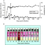
To evaluate the specificity and sensitivity of the ICT for detection of B. gibsoni infection, sera from dogs experimentally infected with B. gibsoni, closely related parasites, and sera from SPF dogs were used. All 30 sera from SPF dogs were negative, whereas all 10 sera from experimentally infected dogs were clearly positive. In addition, all sera from dogs experimentally infected with closely related parasites, B. canis canis (0/3), B. canis vogeli (0/2), and B. canis rossi (0/3), and Neospora caninum (0/4), and Leishmania infantum (0/5) were found to have no cross-reaction in the ICT (Fig. 2). The sensitivity of the ICT was also evaluated using the sequential sera from an experimentally infected dog and simultaneously compared to the parasitaemia level and ELISA antibody titre (Fig. 3). The specific antibodies to P50t were detectable 6 days p.i. and 8 days p.i. by use of ICT and ELISA, respectively. Whereas the parasites were undetectable microscopically at the late infection phase, strong antibody responses to BgP50 remained both in ICT and ELISA until day 144 p.i., the end of experiment. Similar results were obtained from the other 9 experimentally infected dogs (data not shown).

Fig. 3. Specific antibody responses in a dog experimentally infected with Babesia gibsoni. (A) Detection of the parasitaemia level (%) by microscopical examination of a Giemsa-stained thin blood smear and detection of the antibody (IgG) against P50t by the ELISA. (B) Detection of antibodies (IgG+IgM+IgA) against P50t by the ICT.
Detection of the specific antibodies in sera from domestic dogs
To evaluate the clinical application of the ICT, a total of 54 serum samples collected from domestic dogs that had been diagnosed as having anaemic signs at veterinary hospitals in Japan were tested with the ICT, and the results were compared with that of previously established IFAT (Yamane et al. 1993 a) and ELISA (Verdida et al. 2004). Twenty-eight of the tested samples (51·8%) were positive in IFAT while 24 (44·4%) were positive in both ICT and ELISA. The concordance of ICT with ELISA was 100%, while there was a slight discordance (85·7%) with that of IFAT (Table 1).

DISCUSSION
The ICT continues to be one of the most effective means to diagnose infectious diseases and offers the most benefit with the easiest performance and lowest cost. Recently, ICT has been used to detect and evaluate some protozoan parasite infections, such as Plasmodium falciparum, Leishmania infantum, Cryptosporidium parvum, Toxoplasma gondii, and Babesia equi infections, with sensitivities ranging from 72% to 100% and specificities from 61% to 100% (Mills et al. 1999; Chan et al. 2000; Reithinger et al. 2002; Richardson et al. 2002; Huang et al. 2004 a,b; Mohebali et al. 2004).
Here, we established a practical ICT using recombinant P50 for the diagnosis of B. gibsoni infection in dogs. P50 is known as an immunodominant surface antigen of B. gibsoni (Fukumoto et al. 2001), and an ELISA using recombinant GST-P50t expressed in E. coli could detect specific antibodies from both acutely and chronically B. gibsoni-infected dogs (Verdida et al. 2004). In the present study, the leader protein GST was removed from the GST-P50t fusion protein using thrombin protease in order to reduce the possibility of non-specific reactions with antibodies against the GST. P50t was conjugated with a gold colloid for a conjugate pad, immobilized on NC for a test line, and used as an immunogen to induce the specific antibody for a control line. Based on these reagents, an ICT for the detection of B. gibsoni infection was established, and it was demonstrated that the ICT has high sensitivity and specificity. To our knowledge, this is the first report describing the development of a practical ICT diagnosis of B. gibsoni infection in dogs.
For sequential serum samples from an experimentally infected dog, the specific antibody responses were detectable as early as 6 days p.i. by use of the ICT. This early detection of antibodies by the ICT could be attributed to its ability to detect all classes of immunoglobulins, especially immunoglobulin M (IgM), present in the sera during early infection. On the other hand, strong antibody responses to BgP50 in ICT remained until day 144 p.i., the end of experiment, although the parasites were undetectable at the latent period of infection. This result indicates that the ICT is sensitive enough for the diagnosis of acutely and latently infected dogs.
Cross-reactions between antibodies against B. gibsoni and the closely related protozoan parasites have been reported (Yamane et al. 1993 a,b; Birkenheuer et al. 2003 b). Therefore, it is important to discriminate B. gibsoni infection from other related protozoan parasite infections. The ICT using recombinant P50t detected no cross-reactivity with sera from dogs experimentally infected with B. canis canis, B. canis vogeli, and B. canis rossi, or with N. caninum, and L. infantum.
Since canine anaemic signs can be caused not only by Babesia infections but also by other diseases, such as autoimmune haemolytic anaemia (AIHA), and the individual disease has different treatment means, it is important to distinguish babesiosis from other diseases with anaemic signs. Of 54 serum samples collected from domestic dogs, which had clinical anaemic signs, 24 samples (44·4%) were positive for B. gibsoni infection by the ICT and ELISA while 28 (51·8%) samples were positive by the IFAT. All 24 ICT-positive serum samples were IFAT positive, but 4 ICT-negative serum samples were IFAT positive. It is not known whether the IFAT+/ICT- 4 samples are real positive or false positive for B. gibsoni infection. The remaining 30 serum samples, however, might have been from dogs that had other unknown disease(s) with anaemic signs. Previous workers have reported that blood smear test-positive or PCR-positive dogs sometimes lack the detectable antibodies by IFAT, and IFAT-positive dogs sometimes lack the detectable parasites (Birkenheuer et al. 2003 b). To confirm whether all ICT-positive dogs are infected with B. gibsoni, larger samples from dogs diagnosed by blood smear test or PCR are needed to validate the sensitivity and specificity of the ICT.
Cost is an important factor in the evaluation of a newly developed diagnostic method. In the present study, the final yield of highly purified recombinant P50t was 25 mg per litre culture of E. coli, which is sufficient to make approximately 10000 ICT strips. Therefore, the total cost of the ICT can be reduced to as little as US$ 0.1 per strip.
The ‘gold standard’ diagnostic method for B. gibsoni infection in dogs should have: (i) high sensitivity for detecting both acute and latent infections; (ii) high specificity for discriminating the disease from other closely related diseases; (iii) easy to perform and cheap (Bruning, 1996). The ICT meets the gold standard. While ICT and ELISA are similar with regard to the first two advantages because both are considered highly sensitive and specific diagnostic tools, the ICT has the advantage of being simple, economical, rapid, and easy to manipulate even in laboratory or field conditions.
Recent reports have suggested that there are at least 3 distinct subtypes for small canine piroplasms: the classic Asian-type B. gibsoni identified in Asia; a small organism identified as California-genotype B. gibsoni; and Theileria annae, identified in northern Spain (Kjemtrup et al. 2000; Zahler et al. 2000 a,b; Camacho et al. 2001; Jefferies et al. 2003). Since the P50t gene is cloned from the Asian-type B. gibsoni, work is needed to evaluate whether the ICT with P50t can be used for detecting antibodies to T. annae or the California-genotype small piroplasm in dogs in Europe or North America.
This study was supported by a grant from The 21st Century COE Program (A-1), Ministry of Education, Culture, Sports, Science, and Technology, Japan, and Grants-in-Aid for Scientific Research from the Japan Society for the Promotion of Science. Sera from experimentally infected dogs with B. canis subspecies, and L. infantum were kindly obtained from Dr Th. Schetters, Intervet International BV, Boxmeer, The Netherlands. B. canis vogeli parasite was kindly obtained from Dr Namikawa, Department of Infectious Disease, School of Veterinary Medicine, Azabu University, Kanagawa, Japan.