A 19-year-old man presented in the emergency room with acute chest pain. At 7 weeks of age, aortic coarctation had been repaired using inferior translocation of the origin of the subclavian artery to enlarge the coarcted segment. At that time, he was also noted to have a mildly stenotic aortic valve with 2 leaflets, and mild mitral valvar stenosis. Regular clinical and echocardiographic follow-up from about 3 years of age documented development of a subaortic obstructive fibrous shelf, but no systemic hypertension, recoarctation, or aneurysmal formation. He had no signs of connective tissue disorder, nor was there any family history of aortic pathology. Subsequent genetic testing was negative for Marfan’s and Ehlers-Danlos syndromes.
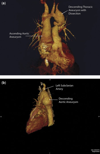
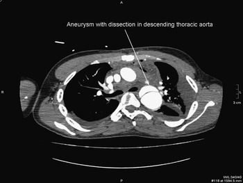
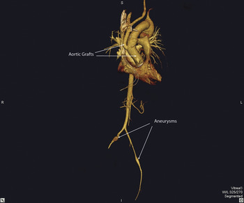
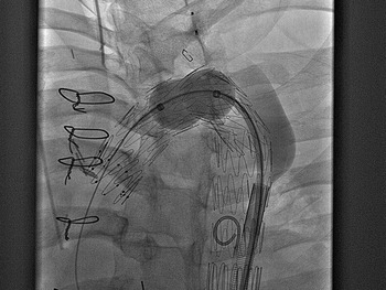
Computed tomography, reconstructed from 5 millimetre slice datasets (Fig. 1), revealed an ascending aorta with a diameter of 4.5 centimetres, a hypoplastic transverse arch, and an aneurysm of the descending thoracic aorta extending from the origin of the left subclavian artery to the level of the 7th thoracic vertebra. The aneurysm (Fig. 2) was associated at its apex with a localized flap of 2.4 centimetres originating on the inner curvature of the aorta in an area of mural calcification. Additional small aneurysms were seen in the femoral and iliac arteries (Fig. 3). Taking note of the new and progressive ascending aortic dilation, in combination with obstructive valvar and subvalvar lesions in the left ventricular outflow tract, as well as the need for a safe proximal landing zone for any interventional repair of the distal aneurysm, we elected to replace the aortic valve, the ascending aorta, and the transverse aortic arch with a 23 millimetre Carbomedics valve and conduit. Arch debranching was accomplished using a bifurcated Hemashield graft to the right brachiocephalic and left common carotid arteries (Fig. 3). After a period of 10 days following this operation, a further episode of chest pain heralded pseudoaneurysmal formation at the distal anastomosis. We deployed a tapered Zenith TXS TAA endovascular graft across both the pseudoaneurysm and the initial site of dissection in the descending aorta. The endovascular leak noted on the initial angiogram (Fig. 4) cleared after embolization of the left subclavian artery with a 12 millimetre Amplatzer plug. The left vertebral artery was preserved, and the proximal site was dilated using a chorda balloon. His subsequent recovery was uneventful. Histology of the ascending aorta showed only adventitial haemorrhage, while mild myxomatous changes were noted in the aortic valvar leaflets. Although infrequent, occasional and increasing reports of aortic disease in patients treated in infancy for a variety of congenital cardiac malformations raises the question whether this is a chance occurrence, the consequence of previous interventions and/or residual pathology, or a manifestation of an underlying genetic predisposition. Our experience illustrates how management can be successful when using a combination of surgical and interventional procedures, and emphasizes the need for careful, long-term follow-up.
Figure 1

Figure 2

Figure 3

Figure 4