Introduction
Synovial sarcoma is a rare, aggressive neoplasm. Eighty per cent of cases occur in the peri-articular areas of the lower extremities in adolescents and young adults.Reference Kawai, Woodruff, Healey, Brennan, Antonescu and Ladanyi1 The head and neck region is the second most common site, accounting for 5–10 per cent of cases. Specific head and neck sites include the tonsil, tongue, retropharyngeal and parapharyngeal regions, hard palate, carotid bifurcation, floor of mouth, retromolar trigone area, maxillary sinus, buccal mucosa, ethmoid sinus, and parotid gland.Reference Weiss, Goldblum and Enzinger2–Reference Mostafapour and Futran12
Synovial sarcomas are so called on account of their morphological resemblance to synovial cells; they usually do not involve a synovial structure. Synovial sarcoma is now believed to arise from undifferentiated pluripotent mesenchymal cells.Reference Szendroi and Deodhar13
Pathologically, synovial sarcoma has three main histological subtypes: monophasic, biphasic and poorly differentiated. The biphasic subtype exhibits both spindle cells and epithelioid cells arranged in gland-like or papillary configurations, whereas the monophasic subtype is composed solely of spindle cells.Reference Amble, Olsen, Nascimento and Foote11
Over 90 per cent of synovial sarcomas have the characteristic chromosomal translocation t(X;18)(p11;q11). Cloning studies of the translocation breakpoints have revealed the fusion of two novel genes: SYT and SSX. Further studies have shown that biphasic synovial sarcoma manifests mainly the SYT–SSX1 translocation, whereas the monophasic subtype demonstrates SYT–SSX2 translocation.Reference Kawai, Woodruff, Healey, Brennan, Antonescu and Ladanyi1 The translocation is found in the poorly differentiated variant.
We report here a case of primary cervical tracheal monophasic synovial sarcoma. The diagnosis was confirmed by observing SYT–SSX gene rearrangement following interphase fluorescence in situ hybridisation. We could find only one previously reported case of synovial sarcoma arising in the trachea.Reference Sykes, Rokkas, Kajdacsy-Balla and Haasler14 Furthermore, our patient represents the first reported case of tracheal synovial sarcoma confirmed by molecular analysis.
Case
A 48-year-old woman suffered several episodes of haemoptysis and progressively worsening dyspnoea in the three months prior to admission.
Evaluation included bronchoscopy, which showed a large mass obstructing the tracheal lumen, requiring a tracheostomy. Histological examination of biopsy tissue demonstrated a spindle cell neoplasm. Further evaluation of this biopsy material included immunohistochemical staining; the spindle cells were not immunoreactive against cytokeratins, S100 protein or muscle actin. Interphase fluorescence in situ hybridisation was performed, and demonstrated a SYT–SSX1 translocation, thus confirming synovial sarcoma of the trachea.
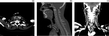
A computed tomography (CT) scan demonstrated a 3 cm intra-tracheal mass extending from 1 cm below the inferior border of the cricoid cartilage to just above the tracheostomy tube opening (Figure 1). The mass appeared to arise from the right lateral wall, and occluded the tracheal lumen. No pathologically enlarged lymph nodes were identified radiologically.
(a) Axial computed tomography (CT) scan showing a tracheal mass nearly occluding the lumen. (b) Sagittal CT scan showing the tracheal mass just above the tracheostomy tube entrance. (c) Coronal CT scan showing the tracheal mass.

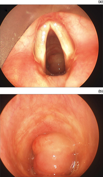
The patient underwent direct laryngoscopy to better visualise the extent of the tumour. A large, lobulated mass was found emanating from the left side of the fourth tracheal ring and the posterior wall, and occluding the tracheal lumen (Figure 2). The tumour was debulked endoscopically and a 0° endoscope was successfully passed. Distal to the mass, the airway was clear. The mass itself terminated above the tracheostomy tube opening.
Direct laryngoscopy views. (a) View just above the vocal folds, demonstrating a large tracheal mass just beneath the subglottic area. (b) The same tracheal mass viewed with the rigid scope passed below the vocal folds.

The patient then underwent en bloc cervical tracheal resection, including the tracheostomy site from the second to the sixth tracheal ring, via an anterior neck approach. The mass did not extend beyond the tracheal lumen. The thyroid gland and oesophagus were not involved by the tumour, and the trachea separated easily from the anterior oesophagus. The surgical margins were uninvolved by tumour. A suprahyoid laryngeal release was performed to provide a tension-free primary anastomosis. At the conclusion of the procedure, fibre-optic laryngoscopy was performed; both vocal folds were mobile.
The patient was observed in the intensive care unit overnight, and discharged home on post-operative day four. There were no post-operative complications.
The patient was followed up by serial examinations and combined positron emission tomography CT scanning. One year post-operatively, she showed no evidence of recurrent disease.
Histopathology
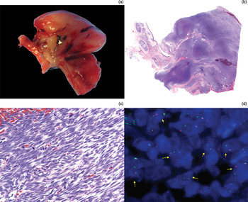
Tracheal resection revealed a polypoid luminal mass measuring 3.2 × 2 × 1 cm (Figure 3). The cut surface revealed a firm, homogeneous mass without necrosis or haemorrhage.
(a) Macroscopic view of resected tracheal specimen, showing a polypoid, tan mass attached to tracheal rings. (b) Low power photomicrograph showing the relationship of the synovial sarcoma to the trachea; the cellular tumour is composed of nodules and sheets of spindle cells with a thin overlying squamous mucosal layer (H&E; ×12.5). (c) High power photomicrograph showing spindle cells and numerous mitotic figures (H&E; ×400). (d) Fluorescence in situ hybridisation analysis performed on the initial incisional biopsy specimen, showing separation of the 5′ and 3′ SYT signals in many of the tumour cell nuclei (arrows).

Microscopic sections showed a malignant spindle cell lesion composed of uniform cells with ovoid nuclei, prominent nucleoli and scant cytoplasm. Numerous mitotic figures were distributed throughout the neoplasm.
Immunohistochemical analysis of the initial biopsy tissue showed that the spindle cells were negative for S100, desmin, pan-cytokeratin, myogenin, cluster of differentiation 34 glycoprotein, high molecular weight cytokeratin 5 and 6, and showed equivocal staining for epithelial membrane antigen (EMA).
Interphase fluorescence in situ hybridisation using a dual-colour, break-apart SYT probe was performed on paraffin-embedded tissue, and demonstrated numerous separation signals (Figure 3). This confirmed the diagnosis of synovial sarcoma.
Discussion
In 1954, Jernstrom was the first to report a case of synovial sarcoma of the pharynx.Reference Jernstrom15 There have been many subsequent reports of synovial sarcoma in the head and neck region, with sites including the tonsil, tongue, retropharyngeal and parapharyngeal regions, hard palate, carotid bifurcation, floor of the mouth, retromolar trigone area, maxillary sinus, buccal mucosa, ethmoid sinus, and parotid gland. Even so, synovial sarcomas of the head and neck are exceptionally rare, with less than 200 cases reported since Jernstrom's initial description.
• Synovial sarcomas are rare, and occurrences in the head and neck are exceptionally rare; this region accounts for 5–10 per cent of cases
• Synovial sarcoma has three main histological subtypes: monophasic, biphasic and poorly differentiated
• Over 90 per cent of synovial sarcomas have the t(X;18)(p11;q11) chromosomal translocation; cloning studies of translocation breakpoints have revealed the fusion of two novel genes, SYT and SSX
• If the immunohistochemical profile from the initial biopsy is equivocal, interphase fluorescence in situ hybridisation using a dual-colour, break-apart SYT probe can confirm synovial sarcoma
• En bloc surgical excision is the treatment of choice
If the literature on synovial sarcoma of the head and neck is sparse, reports of tracheal synovial sarcoma are even more limited. To our knowledge, our patient represents the second reported case of primary cervical synovial sarcoma of the trachea, and the first case to have the diagnosis confirmed by interphase fluorescence in situ hybridisation, detecting SYT–SSX gene translocation.