Lactobacillus delbrueckii is a lactic acid bacterium (LAB) of great technological relevance. Lb. delbrueckii subsp. bulgaricus and Lb. delbrueckii subsp. lactis are part of commercial yogurt cultures and they are also present in whey cultures used for the production of Argentinean (Reinheimer et al. Reference Reinheimer, Suárez, Bailo and Zalazar1995, Reference Reinheimer, Quiberoni, Tailliez, Binetti and Suárez1996) and Italian (Neviani & Carini, Reference Neviani and Carini1994; Giraffa et al. Reference Giraffa, De Vecchi and Reinheimer1997; Giraffa & Rosetti, Reference Giraffa and Rosetti2004) hard cheeses.
Bacteriophage inhibition during dairy fermentations is an ever-present threat for industry. To minimize phage dissemination in dairy plants, various strategies are applied, such as direct vat-inoculation of starters, optimized sanitation, and rotation of phage-resistant starter cultures (Moineau et al. Reference Moineau, Tremblay and Labrie2002). Unfortunately, with the constant use of LAB strains carrying phage defense mechanisms, new phages resistant to these systems have emerged, but only a few of them have been characterized (Labrie & Moineau, Reference Labrie and Moineau2007).
Within host-related, phages defense mechanisms, restriction/modification (R/M) systems are one of the most commonly spread in LAB. Restriction/modification systems have been mainly studied in Lactococcus genus (Schouler et al. Reference Schouler, Clier, Lerayer, Ehrlich and Chopin1998), but they have also been demonstrated for Lactobacillus helveticus (de los Reyes-Gavilán et al. Reference de los Reyes-Gavilán, Linsowtin, Séchaud, Veaux and Accolas1990), Streptococcus thermophilus (Allison & Klaenhammer, Reference Allison and Klaenhammer1998), and Lb. delbrueckii (Auad et al. Reference Auad, Ascárate, Ruiz Holgado and Raya1998; Bourniquel et al. Reference Bourniquel, Casey, Mollet and Pridmore2002). In response to R/M systems, phages have evolved several anti-restriction measures to ensure their survival, such as modification of the phage genome, transient occlusion of restriction sites, subversion of host R/M activities and direct inhibition of restriction enzymes (Tock & Dryden, Reference Tock and Dryden2005).
The present study demonstrates the ability of R/M systems present in a commercial Lb. delbrueckii strain to modify a phage originally derived from another Lb. delbrueckii commercial strain, which was also lysogenic. A Type I R/M system was molecularly characterized and the hsdR and hsdM genes were sequenced after PCR amplification.
Materials and Methods
Strains and culture conditions
The source and identification of the strains used in this work are listed in Table 1. Stocks were maintained at −80°C, in MRS broth (Biokar, Beauvois, France) with the addition of 15% (v/v) glycerol as a cryoprotective agent, and in reconstituted (10% w/v) skimmed milk (RSM), and routinely cultured overnight at 42°C in MRS broth.
Source and Lactobacillus delbrueckii strains used in this work

Bacteriophages
The bacteriophages Cb1/204 and Cb1/342 were obtained by induction with mitomycin C (0·5 mg/ml) (MC, Sigma Chemical Co., St. Louis, MO, USA) of the commercial strain Lb. delbrueckii subsp. lactis Cb1 (Suárez et al. Reference Suárez, Zago, Quiberoni, Carminati, Giraffa and Reinheimer2008). After centrifugation at 12 000 g (10 min at room temperature), the supernatant was filtered (Millipore, Săo Paulo, Brazil) with a pore diameter 0·45 μm, and propagated on the strains Lb. delbrueckii subsp. lactis 204 (Lb.l 204) and Lb. delbrueckii subsp. bulgaricus 342 (Lb.b 342), respectively.
MRS broth or agar, which had been supplemented with 10 mm-CaCl2 (MRS-Ca when specified), was routinely used to propagate and count phages (Svensson & Christiansson, Reference Svensson and Christiansson1991). Phage stocks were maintained at −80°C with 15% (v/v) glycerol added.
Cross sensitivity and adsorption rates
The sensitivity of Lb.l 204 and Lb.b 342 against the bacteriophages Cb1/204 and Cb1/342 was investigated by the turbidity test (Svensson & Christiansson, Reference Svensson and Christiansson1991). Briefly, 5 ml MRS-Ca broth were inoculated with an overnight culture of each Lb. delbrueckii strain and each phage at a multiplicity of infection (m.o.i.) of 0·5. Tubes inoculated only with the strains were used as controls. A total of three subcultures were made to increase the concentration of phages. Tubes were incubated at 42°C and the turbidity was compared with the control tubes.
Phage adsorption rates on bacterial cells were performed at 30 min in MRS-Ca broth as described by Séchaud et al. (Reference Séchaud, Callegari, Rousseau, Muller and Accolas1989). Results were expressed as percentages of the initial phage counts.
Detection of R/M systems in Lactobacillus delbrueckii subsp. lactis 204
The presence of R/M systems was investigated as proposed by de los Reyes-Gavilán et al. (Reference de los Reyes-Gavilán, Linsowtin, Séchaud, Veaux and Accolas1990). The Cb1/342 bacteriophage was propagated on the strain Lb.l 204 and the presumptive modified phage was named Cb1/342m. This one was propagated on strain Lb.b 342, obtaining another phage named Cb1/342m-d. For all propagated phages, the EOP (Efficiency of Plaquing) values were calculated by using the double-layer plaque titration method (Svensson & Christiansson, Reference Svensson and Christiansson1991).
DNA manipulation and analysis
Phages were propagated in a volume of 100 ml MRS-Ca broth and phage particles were concentrated overnight (Yamamoto et al. Reference Yamamoto, Alberts, Benzinger, Lawhorne and Treiber1970). Phage DNAs were obtained by three phenol-chloroform-isoamyl alcohol extractions and concentrated by ethanol precipitation (Quiberoni et al. Reference Quiberoni, Guglielmotti, Binetti and Reinheimer2004). Their quantification and visualization was performed according to Sambrook & Russell (Reference Sambrook and Russell2001). One-Kbp DNA ladder was used as molecular weight marker (Amersham Biosciences UK limited, UK).
For restriction analysis, phage DNA aliquots were cleaved with BglII, HindIII, EcoRI, EcoRV and SalI (Amersham Biosciences UK Limited, UK) and restriction fragments were then treated for 10 min at 70°C (Quiberoni et al. Reference Quiberoni, Guglielmotti, Binetti and Reinheimer2004). After heat treatment, gel electrophoresis in agarose (0·8% w/v) was performed (Sambrook & Russell, Reference Sambrook and Russell2001) to resolve DNA fragments.
The total DNA of the strain Lb. delbrueckii subsp. lactis 204 was obtained by phenol-chloroform extractions as was previously described (Quiberoni et al. Reference Quiberoni, Guglielmotti, Binetti and Reinheimer2004). The alkaline lysis method was used for plasmid isolation from the strain Lb. delbrueckii subsp. lactis 204 (Quiberoni et al. Reference Quiberoni, Reinheimer and Tailliez1998).
Primer design and PCR conditions
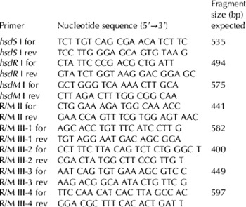
Pairs of primers were designed to characterize the R/M systems of Lb.l 204 (DNAMAN Sequence Analysis Package Program Version 5.2.9, Lynnon Corporation, Copyright 1994–2001). Primer sequences are detailed in Table 2. The primers to amplify the restriction and modification subunits (hsdR and hsdM, respectively) for Type I R/M system were designed on the basis of the complete genome sequence of Lb. delbrueckii subsp. bulgaricus ATCC 11842 (GenBank, accession number NC_008054). Type II R/M system, specifically Type II restriction enzyme NgoFVII, harbored in Lb. delbrueckii subsp. bulgaricus ATCC BAA-365 (GenBank, accession number NC_008529) was used to design another pair of primers. Finally, the methyltransferase enzyme gene sequence of Lb. delbrueckii subsp. bulgaricus ATCC 11842 (GenBank, accession number NC_008054) was used to design the four pairs of primers corresponding to Type III R/M system.
Sequences of primers used in this study

The PCR reactions were performed in 25 μl containing 200 μm-deoxynucleoside triphosphate, 0·5 μm of each primer, 1·25 U Taq DNA polymerase (Invitrogen), Taq buffer (20 mm-Tris-HCl, pH 8·4, 1·5 mm-magnesium chloride, 50 mm-potassium chloride) and 1 μl of total bacterial DNA. The PCR conditions were as follows: 5 min at 94°C followed by 30 cycles (30 s at 94°C, 30 s at 53°C, 1 min at 72°C) and a final step of 7 min at 72°C. The PCR products were separated on a 0·8% (w/v) agarose gel, stained with ethidium bromide, and visualized under UV light.
DNA sequencing
DNA sequencing of the PCR-amplified, infragenic fragment of the hsdR and hsdM genes was performed. Before PCR extension, amplified products were excised with the NucleoSpin Extract (Macherey Nagel, Germany). After a second amplification, the amplified products were purified with the Exo-SAP-IT (GE Healthcare, Italy) according to the manufacturer's recommendations. The PCR extension reactions were performed in 20 μl volumes with 3·2 pm of each primer (BioTez, Berlin, Germany), 4 μl of sequencing mix (Applera, Italia), and 7 μl of purified, amplified product.
PCR extension consisted of 25 cycles of 96°C for 10 s, 50°C for 5 s, and 60°C for 4 min. Rapid thermal ramps (1 deg C/s) between steps were performed. Prior to sequencing, extended products were purified with Big-Dye X-Terminator (Applera, Italia) according to the supplier's indications. Nucleic acid sequences were obtained using an ABI PRISM 310 automated DNA sequencer (Applied Biosystems, Foster City, CA). Sequence alignments and analyses were performed with the Sequence Navigator software (Applied Biosystems). Consensus sequences obtained were also compared with hsdR and hsdM gene sequences of Lb. delbrueckii subsp. lactis strain NCC82 (GenBank, accession number AJ938156) and Lb. delbrueckii subsp. lactis strain NCC88 (GenBank, accession number AJ938155) (DNAMAN Sequence Analysis Package Program).
Results
Cross sensitivity and adsorption rates
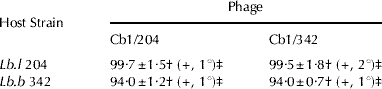
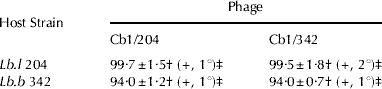
Both phages, Cb1/204 and Cb1/342, were able to produce lysis of both Lb. delbrueckii strains, Lb.l 204 and Lb.b 342. However, the lysis of Lb.l 204 strain with phage Cb1/342 was slightly delayed (i.e. in the second subculture) with respect to that observed on its host strain Lb.b 342, which was observed in the first subculture (Table 3).
Adsorption rates (%) and cross sensitivity in broth (MRS-Ca) for phages Cb1/204 and Cb1/342, on Lactobacillus delbrueckii host strains
Values are the means±SD for three replicate trials
† Adsorption rate (30 min – 42°C)
‡ +: Lysis in broth, subculture number when lysis was observed
The adsorption rates (%) of both bacteriophages to Lb.l 204 and Lb.b 342 were similar, with values ranging between 94·0±0·7% (both phages on Lb.b 342) and 99·7±1·5% (both phages on Lb.l 204) (Table 3). Specifically for strain Lb.l 204 with phage Cb1/342, the adsorption rate was higher than 99%, but the lysis in MRS broth was not observed until the second subculture. This fact may indicate the presence of a R/M system in the Lb.l 204 strain, active against phage Cb1/342.
Detection of R/M systems in Lactobacillus delbrueckii subsp. lactis 204
The EOP values for phages Cb1/342, Cb1/342m and Cb1/342m-d on the restrictive strain Lb.l 204 are shown in Table 4. A restrictive development of bacteriophage Cb1/342 after propagation on the original host was observed when it was subsequently propagated on the Lb.l 204 strain. After a second passage on the restrictive strain, the progeny of the bacteriophage (named Cb1/342m) grew on this strain as well as on the host strain (Lb.b 342). When this new phage was propagated back through its original host, a drop of the EOP value on Lb.l 204 was observed again. These data could indicate the presence of R/M systems in the strain Lb.l 204, active on phage Cb1/342.
EOP (Efficiency of Plaquing) of phage Cb1/342 and the corresponding propagated isolates on the Lb.b 342 host strain and the Lb.l 204 restrictive strain
Values are the means±SD for three replicate trials

† Strain succession of phage propagation
‡ EOP (phage count on the restrictive strain/phage count on the host strain)
Phage DNA analysis
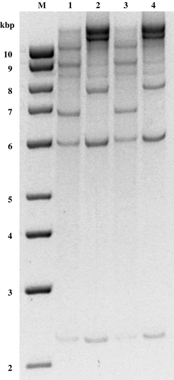
The DNA restriction profile obtained with BglII on the bacteriophage Cb1/342m (presumptively modified by Lb.l 204) was identical to that found for the phage Cb1/204, originally isolated as temperate phage from the commercial strain Lb. delbrueckii subsp. lactis Cb1 (Fig. 1). More specifically, phage Cb1/204 was obtained by propagation of an induced prophage from Lb.l Cb1 on Lb. delbrueckii subsp. lactis 204. The DNA restriction profile of phage Cb1/342m-d (propagated again on the original strain Lb.b 342) reverted to that exhibited by the originally isolated phage (Cb1/342). These results might indicate the existence of an operative Type II R/M system in Lb.l 204 strain.
Agarose gel electrophoresis of DNA fragments obtained from cleavage with the BglII restriction enzyme of Cb1/342 (line 1), Cb1/204 (line 2), Cb1/342m-d (line 3) and Cb1/342m (line 4) phages. Lines M: 1 kbp molecular weight marker.

The profiles obtained with the rest of the restriction enzymes did not show any differences among the four bacteriophages (data not shown).
Characterization of R/M systems
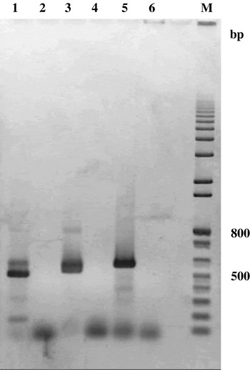
A Type I R/M system was amplified from total DNA of strain Lb.l 204 (Fig. 2). Fragments with the sizes expected for the hsdS (535 bp), hsdR (494 bp) and hsdM (575 bp) subunits were obtained. No amplification products were observed with primers designed for Type II and III R/M systems (data not shown).
Agarose gel electrophoresis of amplified DNA fragments from hsdR (line 1), hsdS (line 3) and hsdsM (line 5) genes. Lines 2, 4 and 6: negative controls. Line M: 1 kbp molecular weight marker.

DNA sequencing
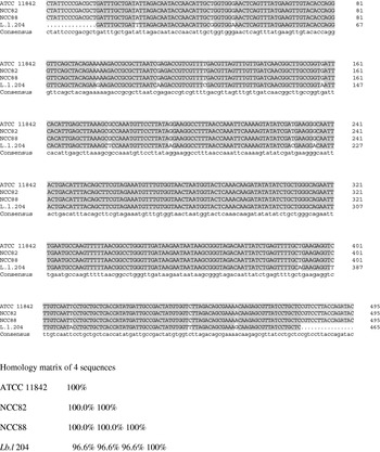
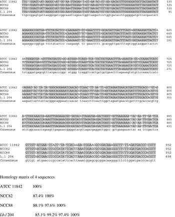
The two sequences obtained from the amplified fragments of the putative Lb.l 204 hsdR (494 bp) and hsdM (575 bp) genes showed similarities higher than 90% with other hsdR and hsdM Lb. delbrueckii genes reported in GenBank (Fig. 3 & 4). The alignments of the sequenced hsdR gene fragment obtained from Lb.l 204 with published sequences of the same gene present in other Lb. delbrueckii strains gave a consensus of 96·6% (Lb. delbrueckii subsp. bulgaricus ATCC 11842, Lb. delbrueckii subsp. lactis NCC82 and Lb. delbrueckii subsp. lactis NCC88). On the other hand, the similarity percentages of the sequenced hsdM gene fragment obtained from Lb.l 204 with the other published Lb. delbrueckii hsdM genes were highest with Lb. delbrueckii subsp. lactis NCC82 (99·2%) and Lb. delbrueckii subsp. lactis NCC88 (97·4%) and the lowest with Lb. delbrueckii subsp. bulgaricus ATCC 11842 (85·1%).
Sequence alignments of the hsdR gene for Lactobacillus delbrueckii susbsp. bulgaricus ATCC 11842, Lb. delbrueckii subsp. lactis NCC82 and Lb. delbrueckii subsp. lactis NCC88, with the corresponding amplicon sequence obtained from Lb. delbrueckii subsp. lactis 204.

Sequence alignments of the hsdM gene for Lactobacillus delbrueckii susbsp. bulgaricus ATCC 11842, Lb. delbrueckii subsp. lactis NCC82 and Lb. delbrueckii subsp. lactis NCC88, with the corresponding amplicon sequence obtained from Lb. delbrueckii subsp. lactis 204.

Discussion
Lysogeny is widespread in lactococci and lactobacilli (Séchaud et al. Reference Séchaud, Cluzel, Rousseau, Baumgartner and Accolas1988; Davidson et al. Reference Davidson, Powell and Hillier1990; Carminati & Giraffa, Reference Carminati and Giraffa1992; Séchaud et al. Reference Séchaud, Rousseau, Fayard, Callegari, Quénée and Accolas1992; Klaenhammer & Fitzgerald, Reference Klaenhammer, Fitzgerald, Gasson and de Vos1994; Carminati et al. Reference Carminati, Mazzucotelli, Giraffa and Neviani1997; Josephsen & Neve, Reference Josephsen, Neve, Salminen and Wright1998) and, specifically, a high frequency of lysogenic strains has been demonstrated for Lb. delbrueckii (Suárez et al. Reference Suárez, Zago, Quiberoni, Carminati, Giraffa and Reinheimer2008).
Temperate phages can disturb the normal process of fermentations when they infect sensitive strains in starter cultures (Davidson et al. Reference Davidson, Powell and Hillier1990). The source for the bacteriophages used in this study is the commercial strain Lb. delbrueckii subsp. lactis Cb1 (Suárez et al. Reference Suárez, Zago, Quiberoni, Carminati, Giraffa and Reinheimer2008). The problem of phage release from lysogenic strains in the dairy industry environment and its consequences are clearly demonstrated in this work.
One effective way to control phages in dairy processes is through the use, in rotation, of strains harboring phage defense mechanisms. Based on their general modes of action, these mechanisms are classified as follows: inhibition of phage adsorption, DNA ejection blocking, restriction/modification systems and abortive infection mechanisms (Abi) (Labrie & Moineau, Reference Labrie and Moineau2007). More recently, CRISPRs (Clustered regularly interspaced short palindromic repeats) have been demonstrated as the latest defense mechanism revealed in prokaryotes (Barrangou et al. Reference Barrangou, Fremaux, Deveau, Richards, Boyaval, Moineau, Romero and Horvath2007; Deveau et al. Reference Deveau, Barrangou, Garneau, Labonté, Fremaux, Boyaval, Romero, Horvath and Moineau2008).
Restriction/modification systems are found in many bacteria where they act to protect the cell from foreign DNA such as bacteriophage DNA. In this work, the lysis of Cb1/342 phage on Lb.l 204 strain was slower than on its host strain Lb.b 342. This fact, in addition to a high adsorption level in both cases, suggested that Lb.l 204 strain had intracellular, chromosome-coded (since no plasmids were detected in the strain) phage resistance mechanisms. The activity of R/M mechanisms in Lb.l 204 was proven by obtaining the modified and unmodified Cb1/342 bacteriophages.
Restriction/modification systems of LAB collectively fall into Types I, II and III systems (Coffey & Ross, Reference Coffey and Ross2002). In Type I R/M systems, the active enzyme comprises three subunits encoded by three closely linked genes, hsdR (restriction), hsdM (methylation), and hsdS (specificity) (Giraffa et al. Reference Giraffa, De Vecchi and Reinheimer1997; Bourniquel, Reference Bourniquel2000). In Lb. delbrueckii, Type I R/M is widespread but a few complete genes were sequenced (Bourniquel, Reference Bourniquel2000; Van de Guchte et al. Reference Van de Guchte, Penaud, Grimaldi, Barbe, Bryson, Nicolas, Robert, Oztas, Mangenot, Couloux, Loux, Dervyn, Bossy, Bolotin, Batto, Walunas, Gibrat, Bessières, Weissenbach, Ehrlich and Maguin2006). The amplicons obtained by PCR on Lb.l 204 DNA using the primers designed for the three R/M systems (I, II and II) demonstrated the presence of a Type I system. The results obtained after sequence alignments suggested that all systems belong to the same family, because the hsdR and hsdM subunits are much conserved into the Type I R/M families (Murray, Reference Murray2000). Conversely, the identical BglII-restriction profiles shown for Cb1/204 – Cb1/342m and Cb1/342 – Cb1/342m-d allowed us to hypothesize that a Type II R/M system is also present in Lb.l 204. These data seem to indicate that both Type I and Type II R/M systems may be present in Lb.l 204, with the latter being operative within the cell. Further research is needed to better clarify the type(s) of restriction/modification systems actively involved in Lb.l 204 phage resistance.
In this work we proved that R/M systems of Lb.l 204 generate a modified phage progeny from the original phage Cb1/342, which continue the infectious process. This fact becomes even more critical when considering that the original bacteriophage is a prophage isolated from another Lb. delbrueckii commercial strain. These facts contribute strongly to diversify the phage population in a factory environment.
This work was supported by the Universidad Nacional del Litoral (Santa Fe, Argentina) (Programación CAI+D 2006, Secretaría de Ciencia y Técnica, Proyecto N° 37-200), the Consejo Nacional de Investigaciones Científicas y Técnicas (CONICET, Argentina) (Project PIP 2004 N° 5321) and the Agencia Nacional de Promoción Científica y Técnológica (ANCyT, Argentina) (Project PICT 2004 N° 20358).