Introduction
Radical neck dissection was first described by George Crile in 1906.Reference Crile 1 It involved the removal of three key structures of the neck: the internal jugular vein (IJV), the sternocleidomastoid muscle and the spinal accessory nerve. This method provided oncological clearance, but often left the patient with severely decreased mobility of the shoulder and impacted upon quality of life. Since Crile's first description, many surgeons have modified the technique. In 1945, Dargent and Papillon were the first to propose the preservation of the spinal accessory nerve.Reference Dargent and Papillon 2 Then, in 1963, Suarez was the first to describe selective neck dissection,Reference Suarez 3 and his work was later greatly enhanced by Bocca and colleagues.Reference Bocca, Pignataro, Oldini and Cappa 4 , Reference Bocca, Pignataro and Sasaki 5 The final result of these pioneering works is today's modified neck dissection, which has been conclusively shown to have equal oncological efficiency.Reference Brandenburg and Lee 6 – Reference Teymoortash, Hoch, Eivazi and Werner 12
An independent review of the literature regarding spinal accessory nerve anatomy was conducted using Medline, PubMed and Q Read databases (for papers published up to August 2015). This involved a search of combinations of the following terms: ‘spinal accessory nerve’, ‘anatomy’, ‘surgical anatomy’, ‘anatomical variant’, ‘cranial nerve XI’ and ‘shoulder syndrome’. We also manually searched the reference lists of articles and used institutional access to the South Australia Health Library Service to identify further relevant papers. A case report is used to demonstrate an example of an interesting but rare anatomical variant of the spinal accessory nerve and IJV.
‘Shoulder syndrome’ and iatrogenic injury
A major complication of neck dissection or indeed any surgery involving the posterior triangle of the neck is severe shoulder disability from accessory nerve damage.Reference Aramrattana, Sittitrai and Harnsiriwattanagit 13 Damage to this nerve in radical neck dissection was first described by Ewing and Martin in 1952,Reference Ewing and Martin 14 and occurred in 50–70 per cent of patients.Reference Osgaard, Eskesen and Rosenorn 15 , Reference van Wilgen, Dijkstra, van der Laan, Plukker and Roodenburg 16 In 1961, Nahum et al. went on to describe the ‘shoulder syndrome’.Reference Nahum, Mullally and Marmor 17 It comprises a multitude of symptoms: shoulder pain, impaired abduction of the shoulder, drooping of the affected shoulder, winging of the scapula, atrophy of the sternocleidomastoid and trapezius, and compensatory hypertrophy of the other muscles of the affected shoulder.Reference Canella, Demondion, Abreu, Marchiori, Cotten and Cotten 18 , Reference Celik, Coskun, Kumas, Irdesel, Zarifoglu and Erisen 19
Iatrogenic injury of the spinal accessory nerve has not been completely eradicated by the advancement of nerve sparing neck dissections. Van Wilgen et al. found that shoulder complaints occurred in 33.3 per cent of patients after modified radical neck dissection.Reference van Wilgen, Dijkstra, van der Laan, Plukker and Roodenburg 16 Birinci et al. found that shoulder dysfunction occurs less significantly in those patients who have minor changes on intra-operative neuromonitoring of the spinal accessory nerve, compared with those patients with larger changes on neuromonitoring.Reference Birinci, Genc, Ecevit, Erdag, Guneri and Oztura 20 Other surgical procedures of the neck can also cause injury to the nerve. Iatrogenic spinal accessory nerve injury still occurs in about 3–6 per cent of patients undergoing cervical lymph node biopsies,Reference Cesmebasi and Spinner 21 and iatrogenic damage to the nerve is a major source of malpractice litigation.Reference Morris, Ziff and DeLacure 22
Anatomy review
Although the spinal accessory nerve has been widely discussed in the literature, its function has not been conclusively agreed upon. It is generally accepted as being a motor nerve, supplying the sternocleidomastoid and the trapezius muscles. Emerging works have postulated a nociceptive role of the spinal accessory nerve as well.Reference Bremner-Smith, Unwin and Williams 23 – Reference Tubbs, Sorenson, Watanabe, Loukas, Hattab and Cohen-Gadol 25 The nerve is frequently encountered in operations involving both the anterior and posterior triangles of the neck. Accurate, timely and consistent identification of the spinal accessory nerve is crucial for preserving this nerve, and this requires extensive anatomical knowledge.
The XIth cranial nerve is classically described as having a spinal and a cranial root. The spinal root of the spinal accessory nerve arises from the spinal nucleus of the lateral grey matter of cervical vertebrae C1 to C5.Reference Salgarelli, Landini, Bellini, Multinu, Consolo and Collini 26 Filaments from these segments form a trunk before ascending through the foramen magnum into the posterior fossa. This spinal root joins briefly with the cranial root, which originates from the dorsolateral surface of the medulla oblongata,Reference Ryan, Blyth, Duggan, Wild and Al-Ali 27 and together they exit the skull as the common trunk.Reference Restrepo, Tubbs and Spinner 24
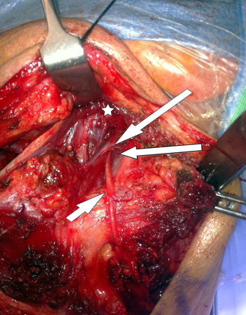
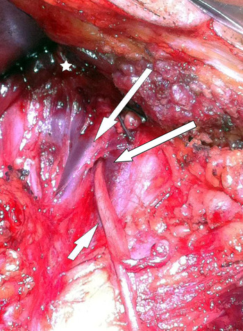
The common trunk exits the skull via the middle compartment of the jugular foramen, lateral to the vagus nerve and anterior to the IJV.Reference Aramrattana, Sittitrai and Harnsiriwattanagit 13 , Reference Lloyd 28 From here it enters the neck, lying compactly between the internal carotid artery and the IJV.Reference Canella, Demondion, Abreu, Marchiori, Cotten and Cotten 18 The common trunk terminates in the retrostyloid space,Reference Salgarelli, Landini, Bellini, Multinu, Consolo and Collini 26 and again separates into cranial and spinal roots. The cranial root joins the superior ganglion of the vagus nerve, and supplies muscles of the palate, pharynx and larynx.Reference Restrepo, Tubbs and Spinner 24 The spinal root continues on and passes most commonly laterally (anterior) to the IJV, although incidences vary widely.Reference Canella, Demondion, Abreu, Marchiori, Cotten and Cotten 18 , Reference Brennan, St J Blythe, Alam, Green and Parry 29 – Reference Taylor, Boone, Schmalbach and Miller 32 The largest study of 207 necks found a lateral relationship in 96 per cent of cases,Reference Taylor, Boone, Schmalbach and Miller 32 whilst another study of 84 necks found it in 67 per cent.Reference Saman, Etebari, Pakdaman and Urken 33 Less commonly, as shown in our case study described below, the spinal accessory nerve can pass through the IJV. Incidence of this variant ranges from 0.48 to 3.3 per cent, although only nine cases have been reported.Reference Lee, Lee, Jin, Kim, Park and Chu 31 , Reference Taylor, Boone, Schmalbach and Miller 32 , Reference Hashimoto, Otsuki, Morimoto, Saito and Nibu 34 A fourth variation has been described, where the spinal accessory nerve itself divides and passes both medial and lateral to the IJV.Reference Taylor, Boone, Schmalbach and Miller 32
After crossing the IJV, the spinal accessory nerve progresses anterior to the transverse process of the atlas,Reference Singh 35 although this is another site of anatomical variance. Durazzo et al. found that in 77.5 per cent of cadavers, the spinal accessory nerve lies anteriorly to the transverse process of the atlas, in 20 per cent it lies in the lateral position and in 2.5 per cent it lies medial.Reference Durazzo, Furlan, Teixeira, Friguglietti, Kulcsar and Magalhães 30 The spinal accessory nerve descends medial to the styloid process and stylohyoid and digastric muscles. It then passes into (70–80 per cent) or under (20–30 per cent) the sternocleidomastoid,Reference Lloyd 28 in close proximity to the sternocleidomastoid branch of the occipital artery.Reference Hill and Olson 36
After taking a tortuous course through the sternocleidomastoid muscle, it exits the posterior border at a point 7–9 cm above the clavicle.Reference Kierner, Zelenka, Heller and Burian 37 It passes 1 cm superior to Erb's point (where the bundle of sensory nerves from the cervical plexus emerges from the posterior border of the sternocleidomastoid muscle).Reference Durazzo, Furlan, Teixeira, Friguglietti, Kulcsar and Magalhães 30 It crosses the posterior triangle of the neck in an inferolateral direction, superficial to the levator scapulae, separated from it by the pre-vertebral layer of deep cervical fascia and adipose tissue.Reference Canella, Demondion, Abreu, Marchiori, Cotten and Cotten 18 , Reference Salgarelli, Landini, Bellini, Multinu, Consolo and Collini 26 , Reference Durazzo, Furlan, Teixeira, Friguglietti, Kulcsar and Magalhães 30 It then pierces the trapezius muscle, most commonly at a point 2–4 cm above the clavicle.Reference Soo, Hamlyn, Pegington and Westbury 38
Anatomical landmarks
Several anatomical landmarks have been described in the literature as useful in the identification of the spinal accessory nerve within the posterior triangle of the neck. They include: (1) the distance between the clavicle and the point at which the spinal accessory nerve passes under or pierces the anterior trapezius;Reference Aramrattana, Sittitrai and Harnsiriwattanagit 13 , Reference Tatla, Kanagalingam, Majithia and Clark 39 (2) Erb's point – the point at which the bundle of sensory nerves from the cervical plexus emerges from the posterior border of the sternocleidomastoid muscle;Reference Salgarelli, Landini, Bellini, Multinu, Consolo and Collini 26 , Reference Durazzo, Furlan, Teixeira, Friguglietti, Kulcsar and Magalhães 30 (3) the superficial cervical vein at the point where it vascularises the anterior border of the trapezius muscle;Reference Shiozaki, Abe, Agematsu, Mitarashi, Sakiyama and Hashimoto 40 and (4) the sternocleidomastoid branch of the occipital artery.Reference Tatla, Kanagalingam, Majithia and Clark 39
Within the anterior triangle of the neck, the anatomical landmarks most commonly reported in the literature are the transverse process of the atlasReference Durazzo, Furlan, Teixeira, Friguglietti, Kulcsar and Magalhães 30 , Reference Singh 35 and the IJV.Reference Canella, Demondion, Abreu, Marchiori, Cotten and Cotten 18 , Reference Salgarelli, Landini, Bellini, Multinu, Consolo and Collini 26 , Reference Lloyd 28 , Reference Lee, Lee, Jin, Kim, Park and Chu 31 , Reference Taylor, Boone, Schmalbach and Miller 32 , Reference Hashimoto, Otsuki, Morimoto, Saito and Nibu 34
Case report
We present the case of a selective neck dissection in a 48-year-old male non-smoker, who had squamous cell carcinoma of the left tonsil with a tumour–node–metastasis grading of T2N1M0. The neck dissection was undertaken following a transoral oropharyngectomy. The intra-operative photograph demonstrates the spinal accessory nerve entering the IJV (Figures 1 and 2).
Intra-operative photograph taken during a left selective neck dissection, demonstrating the accessory nerve entering the internal jugular vein (IJV). (The short white arrow points to the accessory nerve, the long white arrows indicate the IJV either side and the star overlies the posterior belly of digastric muscle.)

Magnified (3×) view of Figure 1, showing the accessory nerve running through the internal jugular vein (IJV). (The short white arrow points to the accessory nerve, the long white arrows indicate the IJV either side and the star overlies the posterior belly of digastric muscle.)

Discussion
Preservation of the spinal accessory nerve in modified radical neck dissection results in significantly increased quality of life for the patient. The literature demonstrates that there is a great deal of variance in the route of the spinal accessory nerve from the skull base through the neck to the anterior border of the trapezius. Many structures in both the anterior and posterior triangles of the neck have been postulated to be useful anatomical landmarks. However, variation clearly exists in the relationship of the XIth cranial nerve to these structures. It is imperative that the head and neck surgeon is mindful of the anatomical variability of the spinal accessory nerve and IJV relationship when dissecting the accessory nerve, in order to avoid accidental injury to either structure at the skull base.
-
• Spinal accessory nerve anatomy has been widely discussed in the literature
-
• The nerve is generally considered to have a motor-related function, although emerging evidence indicates a nociceptive role
-
• The spinal accessory nerve is frequently encountered in surgery involving the anterior and posterior triangle of the neck
-
• The nerve is vulnerable to surgery from the skull base to point of entry into trapezius
-
• This article highlights significant anatomical variations at certain points of the nerve's route, particularly at the internal jugular vein
-
• Awareness of variation is important to prevent significant morbidity associated with nerve injury