Introduction
Hereditary haemorrhagic telangiectasia is an autosomal dominant disorder characterised by diffuse telangiectasia and arteriovenous malformations on both cutaneous and mucosal surfaces.Reference AAssar, Friedman and White1
The clinical course of the illness is dominated by chronic, recurrent epistaxis which can be difficult to manage.Reference Haitjema, Westermann, Overtoom, Timmer, Disch and Mauser2 Many techniques have been used to control epistaxis in such patients, including hormonal manipulation,Reference Sadick, Riedel, Oulmi, Hörmann and Bergler3, Reference Jameson and Cave4 various lasers,Reference Harvey, Kanagalingam and Lund5–Reference Jovancević and Mitrović8 microdebriding,Reference Bublik, Sargi and Casiano9 chemical and electrical cautery,Reference Ghaheri, Fong and Hwang10 septodermoplasty,Reference Saunders11 and nasal closure (Young's procedure).Reference Gluckman and Portugal12
The ideal technique for hereditary haemorrhagic telangiectasia would be effective, long-lasting and minimally invasive while preserving mucociliary function.
The KTP laser can arguably claim to be the current ‘gold standard’ for hereditary haemorrhagic telangiectasia management.Reference Harvey, Kanagalingam and Lund5 However, it is an expensive piece of equipment and can be difficult to use in a bloody field.
Microdebriders have been successfully used to remove telangiectasia, but also require bipolar diathermy to seal the feeding vessels.
Radiofrequency coblation is a relatively new technique which is being increasingly used in ENT surgery. This technique destroys tissue by a process of radiofrequency energy application to a conductive medium (e.g. normal saline), which produces a localised plasma field that breaks molecules into inert, low molecular weight gases at low temperature (classically 60–70°C). This contrasts with conventional electrosurgery, in which direct electric energy applied to tissue causes temperatures of over 400°C. Coblation has been demonstrated to promote good healing and to preserve surrounding normal tissue.Reference Grobler and Carney13 Despite low temperatures, small blood vessels are sealed by this process. Additional, prospective studies have demonstrated that radiofrequency coblation significantly decreases blood loss during endoscopic tumour removal,Reference Kostrzewa, Sunde, Riley and Woodworth14 and is a fast, effective method for reducing encephalocoeles.Reference Smith, Kostrzewa, Riley and Woodworth15
Radiofrequency coblation can therefore theoretically achieve both ablation and haemostasis of telangiectatic and arteriovenous malformations, using the same instrument. The use of coblation in patients with hereditary haemorrhagic telangiectasia has not previously been formally reported. However, informal discussion has indicated that several surgeons worldwide have now begun to use coblation to treat hereditary haemorrhagic telangiectasia.
We describe a multicentre series of five patients with hereditary haemorrhagic telangiectasia, in whom radiofrequency coblation was used for disease management.
Case reports
We present the experience of two surgeons from different centres, both of whom have used coblation to manage epistaxis related to hereditary haemorrhagic telangiectasia. Cases one and two were treated at the Division of Otolaryngology, Department of Surgery, University of Alabama at Birmingham, USA, while cases three to five were treated at Flinders Medical Centre, Adelaide, Australia.
The operating time for all procedures ranged from 8 to 40 minutes (Table I). All procedures were performed under general anaesthesia using the Coblation PROcise EZ View Sinus Wand (Arthrocare ENT, TX, USA).
Overview of cases with regards to interventions and outcomes

Min = minutes; F = female; M = male
Case one
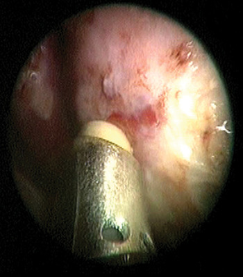
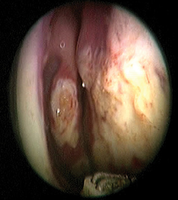
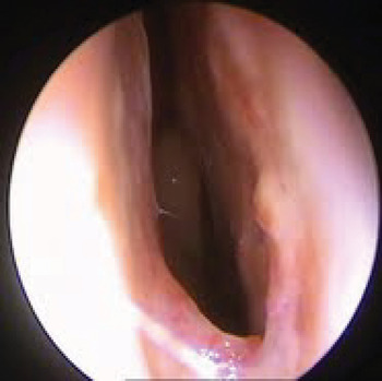
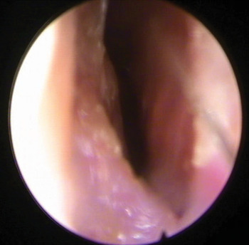
A 45-year-old woman with hereditary haemorrhagic telangiectasia presented with recurrent episodes of epistaxis for two years, despite previous septodermoplasties. Bleeding was from hereditary haemorrhagic telangiectatic lesions involving the lateral nasal wall (Figure 1). She received several four-weekly coblation treatments (Figure 2), after which she remained symptom-free.
Case one: telangiectasia involving the nasal septum.

Case one: excision of the lesion and haemostasis achieved with coblation.

Case two
A 70-year-old woman was diagnosed with hereditary haemorrhagic telangiectasia four months before presentation. She was referred after several attempts at embolisation had failed to improve her recurrent, bilateral epistaxis.
The patient received two coblation treatments six weeks apart, resulting in excellent epistaxis control.
Case three
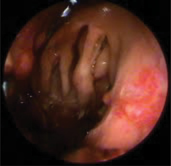
A 54-year-old man with a long history of hereditary haemorrhagic telangiectasia and secondary pulmonary hypertension presented with life-threatening epistaxis. Previous treatments included septodermoplasty, septectomy, ND:YAG laser and KTP laser (Figure 3).
Despite an extensive coblation procedure, the wand failed to clear blood clots, and the patient required nasal packing and subsequent embolisation. A second coblation procedure gained control for a further six months, but another episode of life-threatening epistaxis necessitated further embolisation and a Young's procedure. The patient then developed telangiectasia on the skin over his Young's closure, but these were easily controlled with coblation. He remained well following this procedure.
Case three: recurrence of telangiectasia despite extensive surgery.

Case four
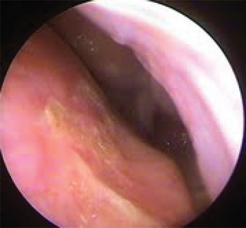
A 61-year-old woman presented with a two-year history of hereditary haemorrhagic telangiectasia. Previous lesions had been treated with silver nitrate cautery. A single, quick coblation procedure ablated all lesions (Figure 4), and the patient remained well eight months post-operatively.
Case four: minimal post-operative crusting following coblation.

Case five
A 61-year-old man presented with recently diagnosed hereditary haemorrhagic telangiectasia. Coblation was used to ablate all hereditary haemorrhagic telangiectatic lesions.
However, the patient presented one year later with a large arteriovenous malformation on the floor of the right nasal cavity (Figure 5). Coblation ablated this arteriovenous malformation with ease. Ten months later, the patient remained symptom-free (Figure 6).
Case five: telangiectasia on the floor of the nasal cavity.

Case five: a good result following coblation of telangiectasia.

Discussion
Hereditary haemorrhagic telangiectasia usually presents with recurrent epistaxis from the anterior third of the nasal septum or the anterior ends of the turbinates.Reference Stecker and Lake16
A myriad of surgical modalities have been described to manage epistaxis in patients with this condition.Reference Harvey, Kanagalingam and Lund5–Reference Gluckman and Portugal12
As regards laser treatment of hereditary haemorrhagic telangiectasia, there is controversy over which type of laser is best. Carbon dioxide,Reference Haitjema, Westermann, Overtoom, Timmer, Disch and Mauser2 ND:YAG,Reference Sadick, Riedel, Oulmi, Hörmann and Bergler3 pulsed dye laser, diodeReference Jameson and Cave4 and KTPReference Harvey, Kanagalingam and Lund5 lasers have all been used for this purpose, with the KTP laser now gaining consensus as the most popular device. It has been suggested that laser can easily ablate the periphery of larger lesions to reduce central blood flow, but that direct laser focussed on the centre of a lesion causes extensive bleeding and makes further treatment difficult.Reference IlIum and Bjerring6 In one study, the majority of hereditary haemorrhagic telangiectasia patients with moderate to severe bleeding reported no change in bleeding severity and no improvement in quality of life following laser ablation of telangiectasias.Reference Lennox, Hitchings, Lund and Howard17 In fact, the use of carbon dioxide laser has been found to provoke lesions on the mucosal surface,Reference Kardos, Holt and Ferguson18, Reference Shapshay, Rebeiz, Bohigian and Hybels19 in contrast with other laser types which target submucosal tissues.Reference Byahatti, Rebeiz and Shapshay20 Whilst in the USA and UK most teaching centres will have access to a wide spectrum of lasers for treatment, this is not the case in Australian hospitals, nor in the private clinic. Hereditary haemorrhagic telangiectasia remains a rare condition, and it is difficult to justify the cost of a US$100 000 (or more) laser which may only be used a few times a year. Unlike the laser, coblation works at low temperatures (classically less than 60°C), thereby causing less thermal injury to adjacent tissues, and theoretically reducing the amount of crusting and scarring.Reference Grobler and Carney13
Microdebriders have also been trialled and found to be beneficial for the removal of telangiectasias on nasal mucous membranes; however, bipolar cautery is also needed to achieve haemostasis after lesion removal.Reference Bublik, Sargi and Casiano9 There is a risk of septal perforation if both sides of the nose are treated simultaneously. In contrast, the use of coblation enables both lesion removal and haemostasis, with the same instrument. The low temperatures minimise the risk of septal perforation. As well as traditional coblation probes (e.g. the Evac 70; Arthrocare ENT, TX, USA), newer probes designed for the nasal wall are now available (e.g. the PROcise EZ View Sinus; Arthrocare ENT, TX, USA).
Bipolar cauteryReference Ghaheri, Fong and Hwang10 remains an option for hereditary haemorrhagic telangiectasia treatment. However, it produces thermal injury, with secondary crusting, mucosal damage and an inevitable reduction in mucociliary function. As such, it has fallen from the list of recommended techniques for the management of hereditary haemorrhagic telangiectasia.
• Hereditary haemorrhagic telangiectasia can present with chronic, recurrent epistaxis
• There is currently no single standard treatment
• Radiofrequency coblation is an effective, logical and well tolerated treatment for mild to moderate cases of epistaxis due to hereditary haemorrhagic telangiectasia
• In severe epistaxis cases, coblation may struggle to achieve control (as may other treatment modalities)
SeptodermoplastyReference Saunders11 involves removal of nasal mucosa from the anterior part of the nasal cavity and replacement by a split skin graft. This technique has been found to have good initial outcomes in patients with hereditary hemorrhagic telangiectasia patients, which unfortunately decline over time due to contraction and revascularisation of the graft. The technique has also been found to be associated with nasal crusting and halitosis.Reference Lund and Howard21
Young's procedureReference Taylor and Young22 is a radical technique involving closure of the nasal vestibule. Although it provides long term relief in patients with moderate to severe epistaxis secondary to hereditary haemorrhagic telangiectasia,Reference Gluckman and Portugal12 the disadvantages (dry mouth, loss of smell and complete nasal obstruction) are often not tolerated by patients. Upon reviewing the procedure, Young himself found that many patients had to have the procedure reversed because of these problems. Young's procedure is best reserved for cases of hereditary haemorrhagic telangiectasia unresponsive to other treatment modalities. One patient in the current series failed coblation management and required a Young's procedure to control his disease.
The use of coblation for hereditary haemorrhagic telangiectasia epistaxis is a much more conservative procedure, which can be safely repeated without significant complications.
One limitation of coblation for hereditary haemorrhagic telangiectasia related epistaxis is the difficulty experienced when manoeuvring the instrument inside the nasal cavity, along with the endoscope, due to its width and angulation. Smaller coblation wands are being developed which will hopefully enable easier manipulation. We have also found that severe bleeding can block the suction port of the coblation wand. This potentially limits its use in severe cases of hereditary haemorrhagic telangiectasia. However, in the current series coblation was successful in managing a patient with a large arteriovenous malformation. We must concede that more experience with coblation is required in order to clarify the ideal candidates for, and limitations of, its use in hereditary haemorrhagic telangiectasia therapy.