Introduction
Rhinorrhoea is a frequent symptom reported amongst patients with allergic and vasomotor rhinitis. Most of these patients usually respond well to medical treatment. Indications for surgical treatment are warranted only when medical treatment fails or a patient wants a permanent solution.
In 1961, Golding-WoodReference Golding-Wood1 first described vidian neurectomy for the treatment of allergic and vasomotor rhinitis. There was a high incidence of post-operative complications, such as disturbed lacrimal secretion and numbness of the cheek and gums. In 2007, KikawadaReference Kikawada2 reported an endoscopic technique involving resection of the posterior nasal nerve near the sphenopalatine artery. With this technique, any intra-operative bleeding can be controlled under direct vision. In 2008, Ikeda et al.Reference Ikeda, Yokoi, Saito, Kawano, Yao and Furukawa3 described submucosal reduction of the inferior turbinate and resection of the posterior nasal nerve. This resulted in significant improvements in nasal symptoms for patients with resistant chronic rhinitis (rhinorrhoea).
The posterior nasal nerve is a peripheral branch of the sphenopalatine ganglion. It enters the nasal cavity through a separate foramen, 4–5 mm below the sphenopalatine foramen, after bifurcation of the nerve into the lacrimal nerve. The posterior superior nasal nerves innervate the superior and middle turbinates, and the superior and middle meatus. Other parasympathetic nerve fibres of the nose branches off and joins the greater palatine nerve and enters the nasal cavity through the canaliculi in the perpendicular plate of the palatine bone as the posterior inferior nasal nerves. These nerves innervate the inferior turbinate and the inferior meatus.Reference Kobayashi, Hyodo, Nakamura, Komobuchi and Honda4
Operative procedure
Anaesthesia
This procedure can be conducted either under general anaesthesia or local anaesthesia. Local anaesthesia is preferred at our centre, for a clear bloodless field, which enables better visualisation of the slender nerve fibres.
Positioning
The patient is placed in a supine posture in a reverse Trendelenburg position. The head end is elevated to 30 degrees, to decrease venous return. A 0-degree 4 mm rigid endoscope with a high-definition camera is used. Pre-medication with fentanyl and dexmedetomidine is administered intravenously as per body weight.
Infiltration and nerve block
A dose of 1–2 ml ropivacaine (0.5 per cent) or lignocaine with adrenaline (1 per cent) at a dilution of 1:40,000 solution is administered as a sphenopalatine block. The solution is injected inferior to the posterior attachment of the middle turbinate, just behind the posterior fontanelle, or through the greater palatine foramen (transoral) if there is a gross septal deviation. A 23 gauge spinal needle is used, with about 1.5 cm of the tip of the needle bent to 45 degrees.
Incision
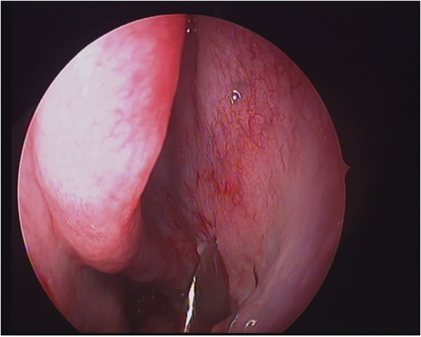
A vertical incision is made (Figure 1) behind the posterior fontanelle. The posterior end of the posterior fontanelle is identified by palpation with an elevator. Just behind this, a vertical incision is made on the lateral nasal wall, starting opposite to the posterior end of the middle turbinate and running all the way down until it reaches the attachment of the inferior turbinate. The incision is made with an angled Cottle elevator. However, one could instead use a needle-tipped electrocautery device or a Colorado needle, to prevent bleeding from the incision site, if the procedure is performed under general anaesthesia.
Left side nasal cavity: incision site.

Posterior nasal nerve identification
The mucoperiosteum is raised gently using a Cottle elevator or a suction freer elevator, after making the initial incision. Alternatively, if middle meatal antrostomy has been performed, the mucoperiosteum is elevated from the posterior edge of the MMA. Care must be taken not to injure the sphenopalatine vessel during flap elevation.
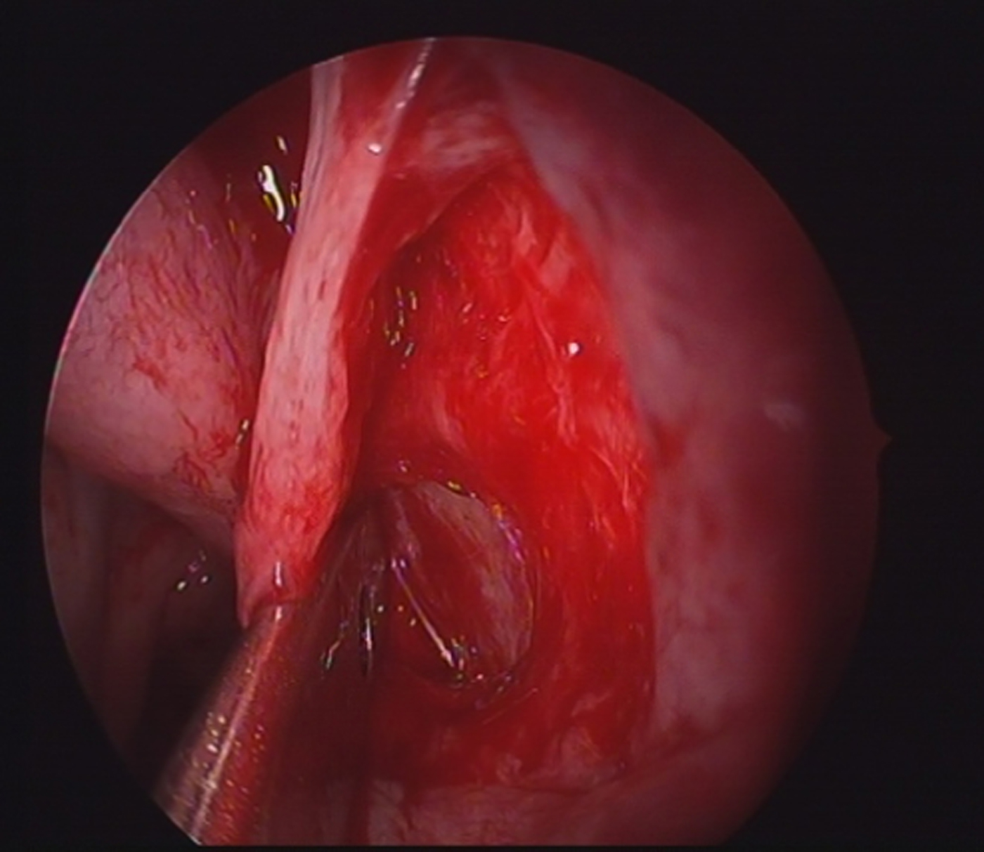
The peripheral part of the posterior nasal nerve can usually be identified just behind the incision, about 4–5 mm inferior to the sphenopalatine artery or crista ethmoidalis (Figure 2). It is always better to identify the main trunk or the proximal part of the posterior nasal nerve below the sphenopalatine foramen area, where the nerve lies inferior to the vessel. The nerve may divide into several branches at its exit into the nasal cavity, each through its foramen. The surgeon may miss a branch if the peripheral part of the nerve is targeted instead of the proximal one near the foramen.
Left side nasal cavity: flap elevated, with posterior nasal nerve identified.

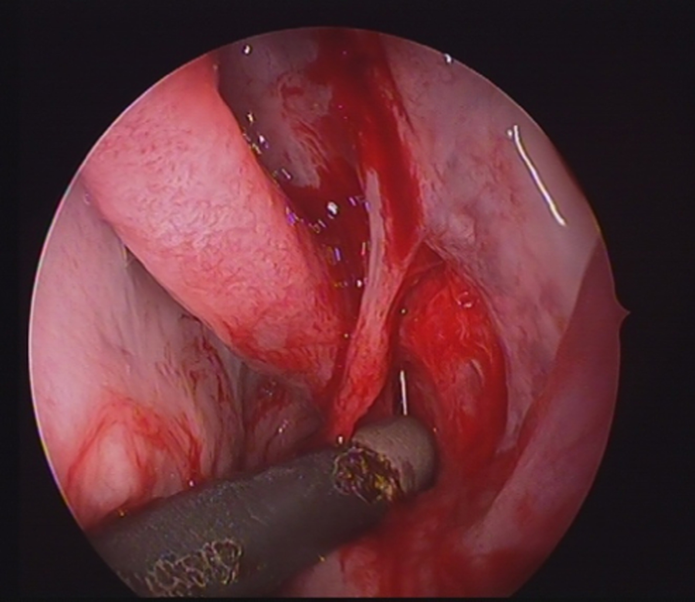
After identifying the nerve fibres, it is cauterised using monopolar suction cautery (Figure 3) or cut using micro-scissors. It is essential to carry out this procedure on both sides for effective results.
Left side nasal cavity: suction cautery of posterior nasal nerve.

Closure
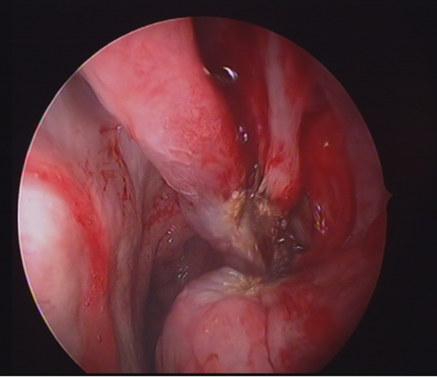
The mucoperiosteal flaps are repositioned (Figure 4). No nasal packing is required. Patients are discharged on the same day.
Left side nasal cavity: repositioning of mucoperiosteal flap.

Complications
In our series of 210 cases operated on between 2012 and 2017, we did encounter 1 case of sphenopalatine artery bleeding, which was coagulated with monopolar suction cautery.
Outcomes
The patients were evaluated 2 weeks before surgery, after surgery, and at 1, 2, 6 and 12 months post-operatively. Subjective evaluation was performed using the 22-item Sino-Nasal Outcome Test (SNOT-22), for which each symptom is scored on a scale from 0 to 5 (0 = no problem, 1 = very mild problem, 2 = mild problem, 3 = moderate problem, 4 = severe problem and 5 = problem as bad as it can be).
Statistical analysis data were obtained and entered into a master chart, and assessed using SPSS® version 16 statistical software. The variables were presented as mean ± standard deviation (SD). Post-operative improvements in symptom scores were evaluated with the Wilcoxon signed-rank test. A p-value of less than 0.0001 was considered statistically significant.
Results
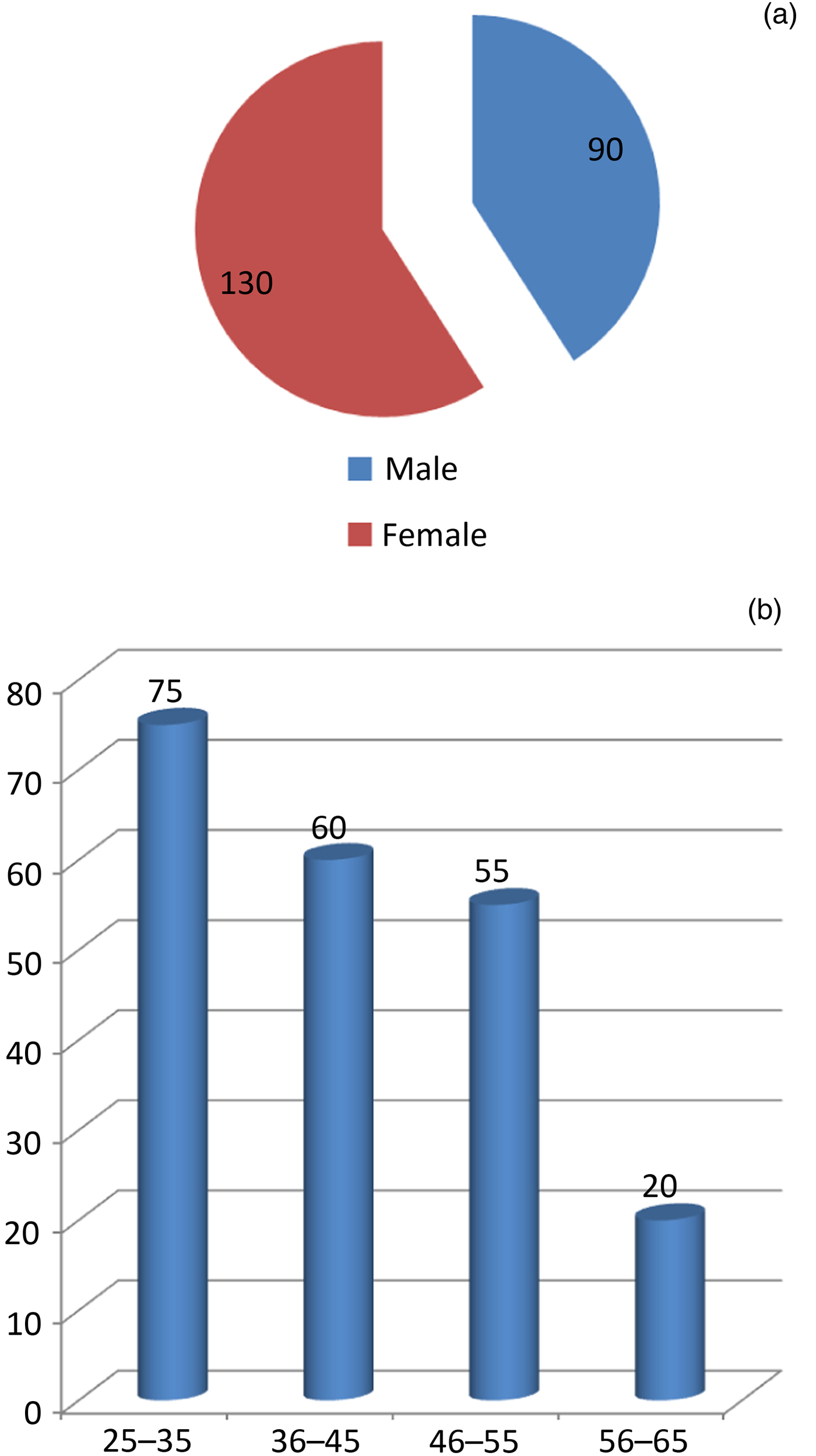
During our study period, from January 2012 to February 2017, 212 patients were enrolled. Two patients were lost to follow up and were hence excluded. There were 90 male patients (42.85 per cent) and 130 female patients (57.14 per cent), with an age range of 27 to 52 years (mean ± SD = 36.24 ± 7.93 years) (Figure 5).
(a) Sex and (b) age distribution of the study population.

We conducted a retrospective review of the patients’ clinical records. Regarding clinical effectiveness, most of the patients reported subjectively excellent or good results. The subjective nasal symptoms (sneezing, rhinorrhoea and obstruction) were recorded and scored using the SNOT-22.
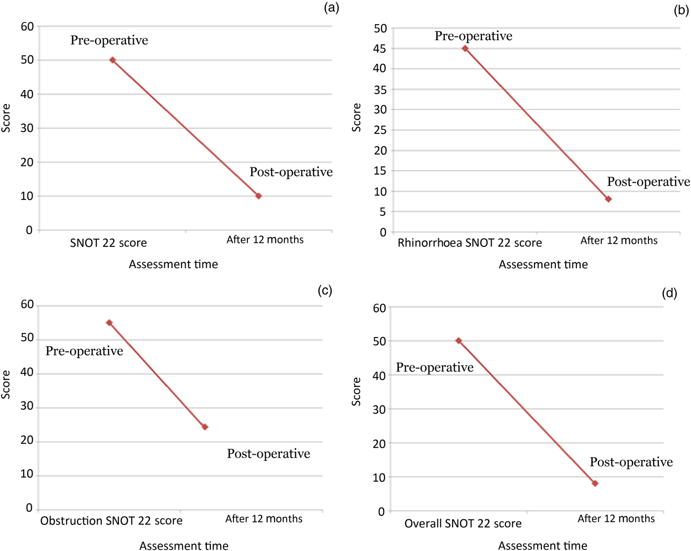
The SNOT-22 score was calculated by adding the scores of the individual nasal symptoms. The mean symptom scores for sneezing, rhinorrhoea and nasal obstruction were all significantly decreased at 12 months compared to the pre-operative baseline scores (Figure 6). The SNOT-22 score data were stratified according to symptom severity, with symptoms defined: as mild (scores of 8–20), moderate (scores of more than 20 to 50) or severe (scores of more than 50).Reference Toma and Hopkins5
Pre- and 12-month post-operative changes in (a) sneezing, (b) rhinorrhoea and (c) nasal obstruction nasal symptom scores, and (d) Sino-Nasal Outcome Test-22 scores (n = 212) (all p < 0.0001, Wilcoxon signed-rank test).

The mean SNOT-22 score decreased from 50 pre-operatively to 8 at 12 months post-operatively. In addition, 39.6 per cent of the patients (84 out of 212) had remained almost free of all symptoms, without need of medication at 12 months. The p-values for these changes were statistically significant (all p < 0.01). We also observed a significant improvement in patients’ quality of life when assessed at the end of the 12th month post-operatively.
Discussion
Resection of the posterior nasal nerve is especially effective for severe rhinorrhoea patients because the interruption of parasympathetic nerve fibres suppresses nasal secretion. The posterior nasal nerve contains afferent sensory fibres supplying the upper two-thirds of the lateral wall of mucosa in the nasal cavity. This procedure can thereby reduce sneezing, making it superior to vidian neurectomy.Reference Ogawa, Takeno, Ishino and Hirakawa6
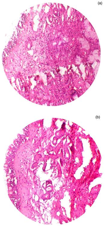
MalcomsonReference Malcomson7 showed conclusively that stimulation of the parasympathetic nerve supply or interruption of the sympathetic nerve supply to the nasal mucous membrane causes vasodilatation, hypersecretion and sneezing. Hence, it is reasonable to assume that under normal conditions there exists a balance between the two systems. The effectiveness of this technique is accompanied by a decrease in local secretary mucous glands and basement membrane thickening, even though the allergy mediator cell eosinophils remained the same, as determined by comparison of pre-operative and post-operative histopathology findings (Figure 7).
(a) Pre- and (b) post-operative histopathological changes in the nasal mucosa. (H&E; ×4)

Conclusion
Endoscopic posterior nasal neurectomy is easy to perform and a less invasive procedure; it can reduce sneezing as well as rhinorrhoea.Reference Toma and Hopkins5 The surgeon can have clear, direct endoscopic visualisation of all the posterior superior nasal nerve fibres and the sphenopalatine artery during the procedure, with appropriate visualisation of anatomical landmarks. This makes it a safe, reliable procedure for patients with resistant chronic rhinitis (rhinorrhoea), providing better results than vidian neurectomy.Reference Malcomson7
Acknowledgement
We would like to thank all patients who underwent this procedure and attended follow-up appointments; this enabled us to share our technique and results via publication in a scientific research journal.
Competing interests
None declared