Percutaneous pulmonary valve implantation is nowadays approved to treat right ventricle outflow tract dysfunction in patients with surgical conduits between the right ventricle and pulmonary artery.Reference Qureshi and Prieto 1 It is increasingly adopted also in patients with “native” right ventricle outflow tract.Reference Boshoff, Cools and Heying 2 , Reference Esmaeili, Bollmann and Khalil 3 In the latter, pre-stenting of the right ventricle outflow tract is more important before implanting the valve.Reference Malekzadeh-Milania, Ladouceur, Cohen, Iserin and Boudjemline 4
Here we report on a tricky situation that occurred during pre-stenting, and we describe how we successfully overcame it.
Case report
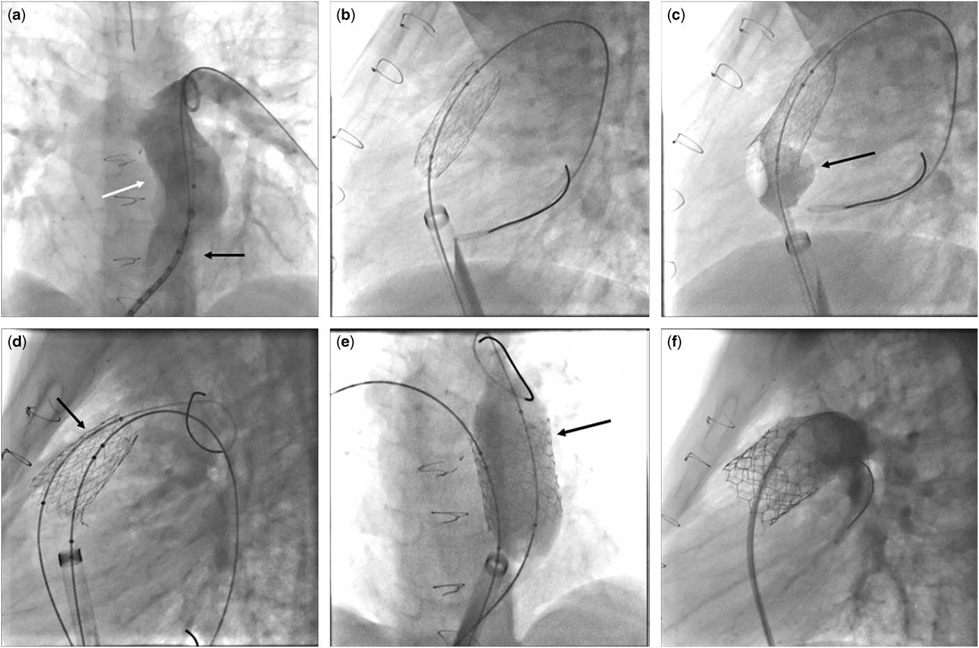
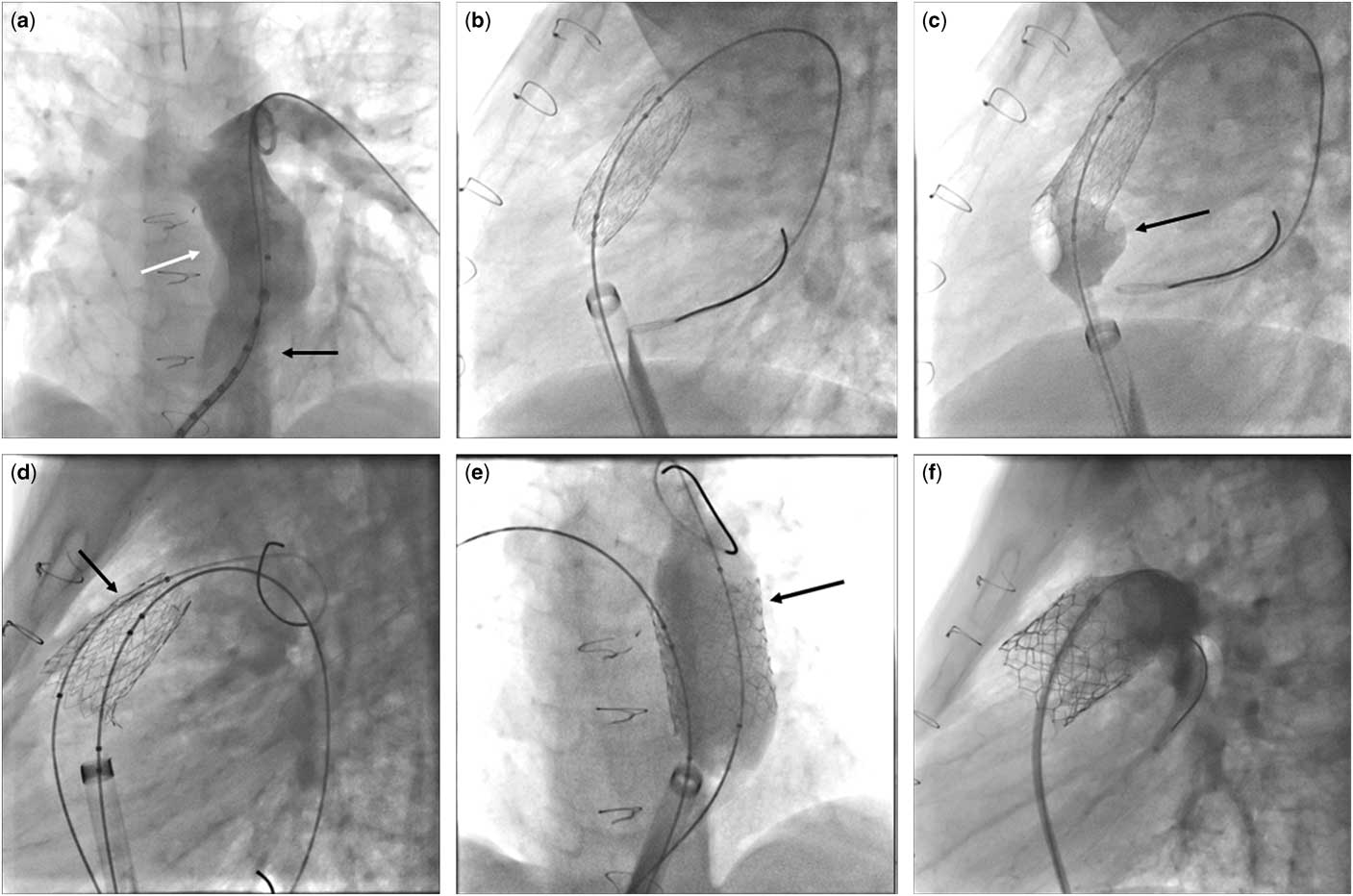
A 13-year-old boy with double-outlet right ventricle, subaortic ventricular septal defect, and pulmonary valvular and subvalvular stenosis underwent surgical palliation with right modified Blalock–Taussig shunt at 5 months of age. When he was 13 months old, he underwent surgical correction with ventricular septal defect closure and right ventricle outflow tract reconstruction with a monocusp patch, taking down the modified Blalock–Taussig shunt. He came to our attention because of progressively poor effort tolerance. Magnetic resonance demonstrated a severe pulmonary valve regurgitation determining severe right ventricle dilation (indexed volume 144 ml/m2), associated with a mild right mid-ventricular stenosis and a slight residual ventricular septal defect. Therefore, he underwent cardiac catheterisation via right common femoral artery access, which confirmed the severe pulmonary valve regurgitation (Fig 1a, black arrow) with a dilated pulmonary trunk, which was 25 mm in the narrower region (Fig 1a, white arrow), associated with a wide sisto-diastolic excursion, the absence of significant interventricular shunt (Qp/Qs=1), and the mild right mid-ventricular stenosis (maximum gradient 10 mmHg). Sizing of the right ventricle outflow tract was performed using a BIB balloon 28×40 mm and 30×40 mm (NuMED Inc., Hopkinton, New York, United States of America) advanced over a Lunderquist guidewire (Cook Medical, Bloomington, Indiana, United States of America), and when completely inflated an angiogram was performed by an 18-Fr long Sheath Mullins (Cook Medical) showing a complete right ventricle outflow tract occlusion.
( a ). Anteroposterior pulmonary angiography showing a severe pulmonary valve regurgitation (black arrow) with a dilated pulmonary trunk (white arrow). ( b ) An Andra Stent XXL 43 mm mounted on a BIB balloon 30×40 mm with the inner balloon completely swelled. ( c ) Expansion of only the proximal part of the outer balloon (black arrow), suggesting a distal small rupture of the outer balloon itself. ( d ) BIB balloon left in place with a Lunderquist guidewire that crosses the stent parallel to the balloon (black arrow). ( e ) Z-MED balloon inflated, after BIB balloon deflation, with a complete stent expansion (black arrow). ( f ) A 29-mm Edward Sapien 3 implantation.
The coronary angiography and the aortography performed with the balloon inflated showed no signs of coronary compression or aortic root distortion.
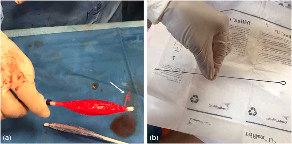
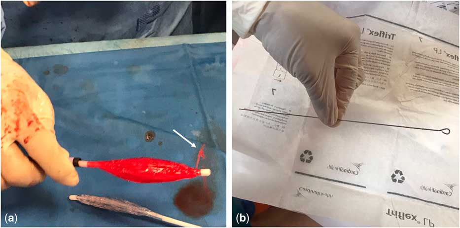
An Andra Stent XXL 43 mm (Andramed, Reutlingen, DE, Germany) mounted on a BIB balloon 30×40 mm was chosen to pre-stent the right ventricle outflow tract to create an adequate landing zone for the subsequent percutaneous pulmonary valve implantation. The inner balloon completely swelled (Fig 1b), whereas only the proximal part of the outer balloon expanded when it was inflated (Fig 1c, black arrow), suggesting a distal small rupture of the outer balloon itself. The BIB balloon was left in place in order to avoid stent embolisation, which was the main risk. An adjunctive venous access was gained through the left common femoral vein by inserting a 12-Fr Introducer Sheath. A guidewire 0.014” and a 4-Fr Judkins right catheter were used to cross the stent parallel to the balloon. Then, a Lunderquist guidewire was changed with the 0.014 wire (Fig 1d, black arrow) and a Z-MED balloon 30×40 mm (NuMED Inc.) was advanced. The BIB balloon was completely deflated, and the Z-MED balloon was inflated up to 2.5 ATM (Fig 1e, black arrow), with a complete stent expansion without sign of dissection at the control angiography. Both balloons were then drawn back and a 29-mm Edward Sapien 3 valve was implanted, with a good final result (Fig 1f). The following inspection of the ruptured BIB balloon revealed a small hole (Fig 2a, white arrow), probably caused by a senior operator while re-inserting the spindle into the balloon catheter to give a higher support to the balloon when mounting the stent (Fig 2b).
( a ). A small hole evidenced when inflating the BIB balloon outside the patient (white arrow). ( b ) The spindle that caused the rupture.
Discussion
Pre-stenting is a required measure before percutaneous pulmonary valve implantation in “native” right ventricle outflow tract.Reference Malekzadeh-Milania, Ladouceur, Cohen, Iserin and Boudjemline 4 This increases the number of procedural steps and the consequent likelihood of possible pitfalls. Here we report on a tricky case of an accidentally dysfunctional balloon, successfully overcome adopting a technique not previously described in this setting.
Once rupture of the stent balloon occurs, the stent usually expands incompletely, and the balloon cannot be easily withdrawn. Moreover, withdrawal may be complicated by displacement of the stent off the balloonReference Keelan, Nunez, Berger, Holmes and Garratt 5 and/or damage of the tricuspid valve on the way down to the inferior caval vein. Albeit rarely, pinhole rupture of a balloon has been described to occur during catheter treatment of highly calcified coronary artery lesions, causing incomplete stent expansion. A case report delineated how this problem was overcome inflating the balloon with a higher concentration of contrast medium.Reference Tsunoda, Shirota, Inoue, Ishii, Sugihara and Mimura 6 In our case, the balloon defect was possibly caused by re-inserting the spindle into the balloon catheter to give a higher support to the balloon when mounting the stent or by stent edge while being crimped.
To our knowledge, this is the first case reported in the literature illustrating the double-balloon technique to complete stent expansion after rupture of the balloon on which the stent was mounted. This method has been previously anecdotally described to prevent recurrent balloon rupture during dilation of heavily calcified conduits before percutaneous pulmonary valve implantation, without implanting a stent.Reference Abu Hazeem, Aldoss and Fagan 7
This case illustrates a useful approach in the case of incomplete stent expansion in the right ventricle outflow tract owing to balloon rupture. It has allowed both to fully expand the stent and to safely withdraw the ruptured balloon.
Conclusion
Pre-stenting is a mandatory step when performing percutaneous pulmonary valve implantation in native right ventricle outflow tract. Stent balloon rupture can occur accidentally when mounting the stent over the balloon and causes incomplete stent expansion. A double-balloon technique can be adopted to both fully expand the stent and safely withdraw the balloon.
Acknowledgements
None.
Financial Support
This research received no specific grant from any funding agency or from any commercial or not-for-profit sectors.
Conflicts of Interest
None.
Ethical Standards
The authors assert that all procedures contributing to this work comply with the ethical standards of the relevant national guidelines on human experimentation and with the Helsinki Declaration of 1975, as revised in 2008, and has been approved by the institutional committees.