Introduction
Chagas disease is a vector-borne neglected disease widely distributed in Latin America. As reported, about 6–7 million people worldwide, mostly in Latin America, are estimated to be infected with Trypanosoma cruzi, the parasite that causes Chagas disease. In the last decades, Chagas disease has spread to non-endemic countries including USA, Canada, and many European and some African, Eastern Mediterranean and Western Pacific countries, due to migration phenomena of the infected population from endemic countries (WHO, 2020). This parasitic disease presents two phases: acute and chronic, the latter characterized by cardiomyopathy, megaoesophagus, megacolon and alterations of the peripheral nervous system that can cause the death. Despite the high number of people infected with T. cruzi, <1% have been diagnosed and treated, so far (Kratz et al., Reference Kratz, Garcia Bournissen, Forsyth and Sosa-Estani2018). Discovered more than 40 years ago, benznidazole (BNZ) and nifurtimox (NFX) are the only approved drugs to treat this parasitic infection (Apt, Reference Apt2010; Sguassero et al., Reference Sguassero, Cuesta, Roberts, Hicks, Comandé, Ciapponi and Sosa-Estani2015). In adults, BNZ is commonly prescribed in two daily doses of 5 mg kg−1 day−1 for 60 days while NFX is recommended in three daily doses of 8–10 mg kg−1 day−1 for 60 days (Meymandi et al., Reference Meymandi, Hernandez, Park, Sanchez and Forsyth2018). BNZ is the drug of choice in several countries of Latin America, USA and Spain being prescribed to treat the acute infection (Cardoso et al., Reference Cardoso, Ribeiro, Oliveira, Oliveira, Ferreira, Bierrenbach, Silva, Colosimo, Ferreira, Lee, Busch, Reingold and Sabino2018). However, its efficacy to treat the chronic stage of the disease is still controversial (Pérez-Molina et al., Reference Pérez-Molina, Pérez-Ayala, Moreno, Fernández-González, Zamora and López-Velez2009). Despite this, clinical studies showed that BNZ is still used for acute and chronic phases in children (Sosa Estani et al., Reference Sosa Estani, Segura, Ruiz, Velazquez, Porcel and Yampotis1998) and adult patients (Viotti et al., Reference Viotti, Vigliano, Lococo, Alvarez, Petti, Bertocchi and Armenti2009). Recently, the BENEFIT study has shown a trypanocidal effect of BNZ, with decreased parasitic loads in patients with severe chronic disease, but no association with clinical outcomes (Morillo et al., Reference Morillo, Marin-Neto, Avezum, Sosa-Estani, Rassi, Rosas, Villena, Quiroz, Bonilla, Britto, Guhl, Velazquez, Bonilla, Meeks, Rao-Melacini, Pogue, Mattos, Lazdins, Rassi, Connolly and Yusuf2015). In addition, it is important to note that BNZ treatment in children showed promising results in terms of efficacy (Sosa Estani et al., Reference Sosa Estani, Segura, Ruiz, Velazquez, Porcel and Yampotis1998; Altcheh et al., Reference Altcheh, Moscatelli, Moroni, Garcia-Bournissen and Freilij2011). However, the appearance of adverse effects, including hepatic and renal damage, anorexia, dermatitis, nervous and digestive problems, immunosuppression and teratogenicity after BNZ administration, is still a serious concern (Maya et al., Reference Maya, Cassels, Iturriaga-Vásquez, Ferreira, Faúndez, Galanti, Ferreira and Morello2007; Salomon, Reference Salomon2012; Bonney, Reference Bonney2014). As described, between 20 and 40% of patients must discontinue treatment due to such undesirable effects (Viotti et al., Reference Viotti, Vigliano, Lococo, Alvarez, Petti, Bertocchi and Armenti2009; Vinuesa et al., Reference Vinuesa, Herráez, Oliver, Elizondo, Acarregui, Esquisabel, Pedraz, Ventosa, Veciana and Viñas2017; Crespillo-Andújar et al., Reference Crespillo-Andújar, Chamorro-Tojeiro, Norman, Monge-Maillo, López-Vélez and Pérez-Molina2018). Taking all these considerations into account, it is imperative to develop chemotherapeutic alternatives to optimize the biological response and minimize the adverse effects, as strongly recommended by other researchers (Kratz et al., Reference Kratz, Garcia Bournissen, Forsyth and Sosa-Estani2018; Ribeiro et al., Reference Ribeiro, Dias, Paiva, Hagström-Bex, Nitz, Pratesi and Hecht2020). BNZ is a poorly water-soluble compound (0.4 mg mL−1) and, therefore, high doses are needed to reach therapeutic concentrations in blood following oral administration (Quijia Quezada et al., Reference Quijia Quezada, Azevedo, Charneau, Santana, Chorilli, Carneiro and Bastos2019). Thus, reduction of the particle size constitutes a useful strategy to enhance the aqueous solubility/dissolution and improve the bioavailability after oral administration (Maximiano et al., Reference Maximiano, de Paula, Figueiredo, de Andrade, Talvani, Sá-Barreto, Bahia and Cunha-Filho2011; Ferraz et al., Reference Ferraz, Alves, Nascimento, Amariz, Ferreira, Costa, Rolim, Lima and Rolim Neto2018). Generally, micronization methodologies, including spray-drying devices, lead to the development of drug-loading microparticulate systems with desired functionalities (Piccirilli et al., Reference Piccirilli, García, Leonardi, Mamprin, Bolmaro, Salomón and Lamas2014; Al-Khattawi et al., Reference Al-Khattawi, Bayly, Phillips and Wilson2018). Particularly, spray-drying technique transforms liquid products into free-flowing dry powders and such methodology can be scaled up from the laboratory to the industrial production (Poozesh and Bilgili, Reference Poozesh and Bilgili2019). Keeping in mind the needs of novel oral formulations for Chagas disease, the aim of this study was to formulate BNZ-loaded microparticles (BNZ-MP) by spray-drying using Eudragit® RL PO (RL) and Eudragit® RS PO (RS) to evaluate their efficacy in an experimental model of acute infection with the virulent T. cruzi Nicaragua (TcN) isolate (Grosso et al., Reference Grosso, Bua, Perrone, Gonzalez, Bustos, Postan and Fichera2010). Dissolution efficiency (DE) and storage stability assays were performed to characterize the formulations. In addition, in vitro studies in cell culture and haemolytic assays of the BNZ-MP were carried out. Then, the levels of T. cruzi-specific antibodies, antiparasitic efficacy and histopathological damages, after oral administration of BNZ-MP, were investigated.
Materials and methods
Materials
BNZ (lot 9978 A, 99% purity; Laboratorios Elea, Buenos Aires, Argentina, was provided by the National Institute of Parasitology ‘Dr. Mario Fatala Chaben’, National Ministry of Health, INP-ANLIS, Buenos Aires, Argentina). Fetal bovine serum (FBS) was purchased from Natocor (Córdoba, Argentina) and horse serum was obtained from Internegocios SA (Córdoba, Argentina). African green monkey kidney epithelial cells (Vero cells) were obtained from Asociación Banco Argentino de Células (ABAC, Pergamino, Argentina). Eudragit® RL PO and RS PO (copolymers of ethylacrylate, methylmethacrylate and methacrylic acid esterified with quaternary ammonium groups) and Sipernat® (speciality silica) were kindly donated by Evonik (Buenos Aires, Argentina). All other reagents and chemicals used for analytical purposes were of chromatography grade.
Formulation of BNZ-MP
BNZ-MP were obtained by spray-drying method (Seremeta et al., Reference Seremeta, Arrúa, Okulik and Salomon2019). On the one hand, 6.0 g of polymer (RL or RS) and 2.4 g of BNZ were dissolved in absolute ethanol (60 mL) under magnetic stirring (IKA® C-MAG HS 4 digital, Staufen, Germany) overnight (200 rpm). On the other hand, Sipernat® was dispersed in distilled water (540 mL, 7.5% w/v). Then, both phases were mixed, magnetically stirred (1 h) and homogenized (5 min, 17 500 rpm) by using T18 Ultra-Turrax (IKA®-Werke GmbH & Co.KG, Staufen, Germany). This suspension was fed into a Mini Spray Dryer Büchi B-290 (Büchi Labortechnik AG, Flawil, Switzerland) with the co-current flow and open-loop mode. The inner diameter of the spray nozzle tip was 0.7 mm. It should be noted that the sample was maintained under magnetic stirring at 200 rpm throughout the process to ensure homogeneity. The operating parameters were previously adjusted (Seremeta et al., Reference Seremeta, Arrúa, Okulik and Salomon2019). The resulting air outlet temperature was of 65°C. The BNZ-MP were recovered from the glass collection vessel and stored in caramel colour glass vials sealed with chlorobutyl rubber septa (Labco Limited, Lampeter, UK) and protected from light and moisture, in laboratory cabinets, until further use. The samples were obtained for triplicate using RL and RS. These were namely BNZ-MP-RL and BNZ-MP-RS, respectively. Size and size distribution of the BNZ-MP were estimated by scanning electron microscopy (SEM). An image analysis system (Image Pro-plus® software 6.0) was used to analyse the images of BNZ-MP with 20× magnification and to obtain the average size value.
In vitro BNZ dissolution
Dissolution studies were performed using a method previously reported with slight modifications (Leonardi et al., Reference Leonardi, Salomón, Lamas and Olivieri2009; Lima et al., Reference Lima, Soares-Sobrinho, Silva, Hernandes, Rolim and Rolim-Neto2011). The USP paddle method (apparatus 2) in USP Standard Dissolution Apparatus Hanson Research SR6 Plus (Chatsworth, CA, USA) was used. The dissolution medium was 900 mL of HCl 0.1 N maintained at 37°C and the stirring speed was set at 75 ± 2 rpm. The following samples were tested in triplicate: (i) BNZ-MP-RL equivalent to 50 mg of BNZ, (ii) BNZ-MP-RS equivalent to 50 mg of BNZ and (iii) free BNZ (50 mg). Sink conditions were used to ensure complete dissolution of the drug in the volume of dissolution medium. The volume of the medium used was not <3 times that required to form a saturated solution of the drug substance, according to the United States Pharmacopeia 30 Edition and the National Formulary 25 (USP 30-NF 25). The samples were placed into the flasks containing dissolution medium and the time counter was set to zero. At regular time intervals (10, 15, 20, 30, 45, 60, 90 and 120 min), samples of medium (4 mL) were taken using a filter and replaced by the same amount of fresh medium to keep a constant volume. The samples were diluted with 0.1 N HCl and the concentration of dissolved BNZ was determined by UV-visible spectrophotometry (UV-1800 Shimadzu UV-VIS Spectrophotometer, Kyoto, Japan) at 324 nm and 25°C using a calibration curve of the drug in absolute ethanol/distilled water (97/3 v/v) (5–40 μg mL−1, R 2 > 0.9997). The dissolution profiles were compared using the DE at 15, 30 and 90 min (DE15, DE30 and DE90, respectively). Determinations were carried out for triplicate and results were expressed as mean ± s.d. Statistical analysis of the DE was performed using a one-way analysis of variance (ANOVA) followed by a least significant difference.
BNZ-MP stability assay
The samples were placed in caramel colour glass vials sealed with chlorobutyl rubber septa (Labco Limited) and stored at room temperature (25 ± 2°C) in laboratory cabinets for 6, 9, 12, 24 and 36 months. The stability was assessed by measuring the drug content of the BNZ-MP-RL and BNZ-MP-RS before (zero time) and after storage. Thus, each sample of BNZ-MP (0.01 g) was dissolved in 5 mL mixture of absolute ethanol/distilled water (97/3 v/v) and left under moderate magnetic stirring for 30 min. Then, 200 μL of the sample were taken and diluted up to 5 mL with the same solvent mixture. Finally, the BNZ concentration in each sample was determined by UV-visible spectrophotometry (see above). Determinations were carried out for triplicate according to equation (1) and results were expressed as mean ± s.d.:

where W BNZ is the weight of the drug in the BNZ-MP and W MP is the total weight of BNZ-MP obtained.
Encapsulation efficiency of BNZ-MP
The encapsulation efficiency (%EE) of the BNZ-MP was determined according to equation (2):

where DCE and DCT are the experimental and theoretical drug content of the BNZ-MP, respectively. Determinations were carried out for triplicate.
Cell toxicity assay
The viability of the kidney epithelial cells from African green monkey (Vero line) was carried out by reduction of yellow tetrazolium salt 3-[4,5-dimethylthiazol-2-yl]-2,5-diphenyltetrazolium bromide to a blue crystalline formazan product (MTT). A 96-well microplate with 1 × 10⁵ Vero cells per well were incubated with increasing concentrations of different BNZ-MP (0, 10, 25 and 50 μg mL−1) suspended in Roswell Park Memorial Institute medium (RPMI) overnight at 37°C. Then, MTT [in phosphate-buffered saline (PBS), 5 mg mL−1] was added and, after 2–4 h, a solution of dimethyl formamide (DMF) and sodium dodecyl sulphate (SDS) (50% of DMF + 10% of SDS) was added to dissolve the MTT. The colour was measured in a microplate reader (Bio-Rad, model 3550, Buenos Aires, Argentina) at 540 nm. Assays were performed in triplicate.
Haemolytic effect
To determine the possible haemolytic effect of the formulated BNZ particles, increasing concentrations of different BNZ-MP (10, 25, 50 and 100 μg mL−1) were added to a 4% suspension of fresh defibrinated human blood prepared in sterile 5% glucose solution and left in contact for 24 h at 37°C. Then, samples were centrifuged and the supernatant absorbance was determined at 540 nm. The absorbance values indicate the haemolysis per cent. Untreated BNZ and Triton X-100 (10%) were used as the haemolytic reference drug and positive control, respectively.
Parasites
Cultured-derived trypomastigotes of the TcN isolate (Grosso et al., Reference Grosso, Bua, Perrone, Gonzalez, Bustos, Postan and Fichera2010) were maintained in culture using RPMI medium supplemented with 10% of FBS obtained by serial passage through Vero cells (ABAC).
Animal model
The animals were obtained from the National Institute of Parasitology ‘Dr. Mario Fatala Chaben’ bioterium (Buenos Aires, Argentina), under specific pathogen-free conditions. Mice were in a controlled room with water and food ad libitum, a circadian light cycle and a temperature of 22°C. Animals were randomly selected prior to infection and assignment to the treatment groups. Physical adverse events that were monitored twice a day included inability to move, reduced appetite, extreme pallor, ruffled hair and body weight loss over 16%. Five groups (n = 4–7) of 1-month-old female C3H/HeN mice were inoculated intraperitoneally with 1000 culture-derived trypomastigotes of the TcN isolate. Raw BNZ and BNZ-MP were dispersed in the corresponding volume of olive oil immediately before administration. Formulations were administered to mice through oral gavage, at 2 days post-infection (d.p.i.). The following groups of mice were systematically compared: (1) infected mice and without treatment, (2) infected mice and treated with BNZ for 30 days with daily doses of 50 mg kg−1 day−1 (BNZ 50), (3) infected mice and treated with BNZ-MP-RL for 30 days with daily doses of 50 mg kg−1 day−1 (RL 50), (4) infected mice and treated with BNZ-MP-RS for 30 days with daily doses of 50 mg kg−1 day−1 (RS 50) and (5) mice without infection (Fig. 1). For the untreated group, a larger number of mice (n = 38) was infected to obtain an appropriate survival.
Treatment schedule in the experimental murine model of acute TcN infection. TcN-infected mice treated at 2 d.p.i. with BNZ or BNZ-MP at 50 mg kg−1 day−1 (RL 50 or RS 50) for 30 days.

Parasitaemia detected by DNA amplification
First, one volume of blood was collected from euthanized infected and uninfected mice at 3 months post-infection (p.i.) after completing the treatments (n = 4–7 samples per treatment). Then, each sample of blood was mixed with an equal volume of guanidine-HCl 6 M, EDTA 0.1 M, pH 8 and kept for 1 week at room temperature and after at 4°C until further use. Thus, DNA was isolated by means of a quantitative polymerase chain reaction (qPCR) (High Pure PCR Template Preparation Kit, Roche, Mannheim, Germany) from 0.2 mL of guanidine-EDTA buffer B mixture and eluted in 0.2 mL, following the manufacturer's protocol. A bacterial commercial plasmid, pQE (Qiagen, MD, USA) was used as an internal standard of DNA extraction. A T. cruzi satellite DNA flanked by the Sat Fw and Sat Rv oligonucleotides highly conserved in the parasite genome was amplified by ABI 7500 thermocycler (Applied Biosystems, Carlsbad, CA, USA) (Duffy et al., Reference Duffy, Bisio, Altcheh, Burgos, Diez, Levin, Favaloro, Freilij and Schijman2009). A commercial kit (SYBR® GreenER® qPCR SuperMix Universal, Invitrogen, Life Technologies, Waltham, MA, USA) was used to run duplicated samples as previously described (Bua et al., Reference Bua, Volta, Perrone, Scollo, Velázquez, Ruiz, De Rissio and Cardoni2013). Epimastigotes of the TcN isolate, DTU TcI were used as a standard in artificially spiked mouse blood. In each determination, the parasite curve, negative samples and non-template DNA were included (Grosso et al., Reference Grosso, Alarcon, Bua, Laucella, Riarte and Fichera2013). The cut-off value was determined at 0.14 Eq parasites mL−1.
Specific anti-T. cruzi humoral response
A sample of blood (500 μL) from uninfected mice, treated infected mice and untreated infected mice was collected from the orbital venous sinus at 3 months p.i. Then, the serum of the collected samples was analysed for IgG antibody levels by the enzyme-linked immunosorbent assay (ELISA). As a source of antigen, a lysate preparation derived from epimastigotes of the strain, Tul2 stock (20 μg mL−1) was used. Briefly, antigen diluted in carbonate buffer pH = 9.6 was used to coat flat-bottomed (96-well, 50 μL well−1) plates at 4°C overnight. Then, 5% skimmed milk in PBS was used to block the plates (100 μL well−1) at room temperature for 1 h. Plates were washed three times with PBS-0.05% Tween 20 (PBS-T) and incubated with sera samples (50 μL well−1, 1:50–1:400 dilution) at 37°C for 30 min. Then, plates were washed with PBS-T and 50 μL well−1 of horseradish peroxidase-labelled goat anti-mouse IgG (Jackson ImmunoResearch Inc, West Grove, PA, USA) were added at room temperature for 30 min. O-phenylenediamine dihydrochloride (50 μL well−1) and 2 N sulfuric acid were used to develop and stop the reaction, respectively. Optical density (OD) was read with an ELISA microplate reader (Dynatech Laboratories, Alexandria, VA, USA) at 490 nm. The mean minus two standard deviations of OD obtained from sera of untreated infected control mice was set up as a cut-off value for significant decreases in antibody levels.
Histopathological studies
Histopathological analysis was carried out with heart tissues removed from treated and untreated infected mice. These tissues were fixed in 10% formaldehyde solution and embedded in paraffin. Then, tissue sections (5 μm) were stained with Haematoxylin and Eosin (H&E) stain and observed by light microscopy. Finally, according to the extension of inflammation, tissues from eight different areas of the heart (left and right atria, upper and lower halves of each ventricular wall and upper and lower septum) were scored. Different numbers were assigned to each section according to the degree of inflammation: (0) absent/none, (1) focal or mild myocarditis with one foci, (2) moderate with two inflammatory foci, (3) extensive with generalized coalescing of inflammatory foci or disseminated inflammation with minimal necrosis and retention of tissue integrity, and (4) severe with diffused inflammation, interstitial oedema and loss of tissue integrity (Gupta and Garg, Reference Gupta and Garg2010). An estimate of the degree of cardiac tissue inflammation was obtained from an average of the values found in the eight sections of the heart.
Statistical methods
The normality of the variable distribution was assessed by using the Shapiro–Wilk normality test. Differences among groups were evaluated by ANOVA or Kruskal–Wallis test as appropriately followed by Bonferroni or Dunn's multiple comparison tests. Statistical significance was considered at P < 0.05 (two-tailed). Data analyses and graphs were performed with GraphPad Prism 7.0 software.
Results
Formulation of BNZ-MP
The corresponding BNZ formulations were obtained in nearly 70% yield with a media size of 0.87–1.08 μm. The %EE was of 95.41% (2.31) and 96.21% (6.11) for BNZ-MP-RL and BNZ-MP-RS, respectively. It is important to mention that BNZ-MP were obtained as free-flow powders and no aggregation phenomena were observed. In contrast, as reported by Cortesi et al. (Reference Cortesi, Lahm Ajanji, Sivieri, Manservigi, Fundueanu, Menegatti and Esposito2007), the preparation of acyclovir-containing MP by spray-drying with Eudragit® RS lead to the formation of aggregates, mainly.
In vitro BNZ dissolution
Dissolution studies showed that both BNZ microparticulate polymeric formulations exhibited faster dissolution rates than the raw drug. DE at 15 min (DE15) indicated a 3.80 and 3.12-fold increase in dissolution rate for BNZ-MP-RL and BNZ-MP-RS, respectively. Furthermore, DE at 30 min (DE30) was 2.39 and 2.07-fold greater for BNZ-MP-RL and BNZ-MP-RS, respectively, to the raw drug (Table 1). This indicates that the DE of the drug from the microparticulate systems was greater in the first minutes and decreasing with the passage of time with respect to untreated BNZ.
In vitro BNZ dissolution efficiency and stability of BNZ-MP stored at room temperature up to 36 months

a BNZ-MP-RL: microparticles of Eudragit® RL PO loaded with benznidazole.
b BNZ-MP-RS: microparticles of Eudragit® RS PO loaded with benznidazole.
c BNZ: raw benznidazole.
Difference is statistically significant with respect to BNZ: *P < 0.05; **P < 0.01; ***P < 0.001; ****P < 0.0001.
BNZ-MP stability assay
The stability assay of BNZ-MP was carried out by measuring the drug content at zero time (i.e. immediately after their fabrication) and after storage at room temperature for 6, 9, 12, 24 and 36 months. To provide long shelf-life, it is necessary to avoid the leakage of the drug from the MP for a long period. In this case, the results showed no significant changes (P > 0.05) in drug content values under storage (Table 1). After 36 months, the drug content values slightly decreased to 95.46 and 93.72% of initial values for BNZ-MP-RL and BNZ-MP-RS, respectively (Table 1).
Cell toxicity assay
Due to the clinical interest of BNZ for treatment of a neglected disease as Chagas disease, the biocompatibility and cell toxicity of the novel formulations should be evaluated. Thus, a cell viability study was carried out by MTT assay to evaluate the possible toxicity associated with the BNZ-MP. After incubation of Vero cell culture with the BNZ-MP, there were no visible morphological changes or cell membrane destabilization. The OD values showed no significant differences after the addition of such formulations with respect to untreated cells (Supplementary Table 1 1). As seen, BNZ-MP-RL 50, 25 and 10 showed a toxicity percentage of 7.7, 8.4 and 27%, respectively. In a similar fashion, BNZ-MP-RS 50, 25 and 10 exhibited 3.7, 9.6 and 26%, respectively.
Haemolytic effect
Due to the potential interest in clinical research, an in vitro haemolytic assay was performed to evaluate whether BNZ-MP might damage the red blood cells. BNZ was used as the haemolytic reference drug and Triton X-100 (10%) as the positive control. Low percentage of haemolysis (4.6–8%) was observed in defibrinated human blood incubated with BNZ or different concentrations of BNZ-MP (10, 25, 50 and 100 μg mL−1). Thus, such formulations did not promote any alteration or degradation in erythrocyte membranes suggesting that RL and RS may be used as carriers for BNZ microencapsulation. As expected, Triton X-100 produced complete lysis of the erythrocytes (Supplementary Table 1 2).
Course of infection
An assay in the acute phase of TcN-infected mice was carried out with different BNZ-MP to evaluate the effectiveness of this novel treatment for Chagas disease. All infected mice (100%) treated with 50 mg kg−1 day−1 of BNZ-MP (RL 50 or RS 50) for 30 days survived as seen in Fig. 2. This acute infection shows a peak of parasitaemia 30–35 d.p.i. in untreated C3H/HeN mice inoculated with 1000 TcN trypomastigotes, with a survival rate of 15% (Grosso et al., Reference Grosso, Alarcon, Bua, Laucella, Riarte and Fichera2013). Additionally, untreated infected mice and infected mice treated with BNZ-MP 50 mg kg−1 day−1 showed the same appearance, behaviour and weight (data not shown).
Survival rate curve of TcN-infected C3H/HeN mice. Mice were infected with 1000 trypomastigotes of TcN and the survival rates were checked daily up to 90 days. Each line indicates the survival rate of untreated infected mice and mice treated with 30 oral doses of BNZ-MP or BNZ at 50 mg kg−1 day−1 (RL 50, RS 50 or BNZ 50, respectively). To assess differences among survival curves, a log-rank test of Kaplan–Meier was performed.

Specific anti-T. cruzi humoral response
Trypanosoma cruzi-specific IgG antibody levels were measured in uninfected mice, untreated infected mice and infected mice treated with BNZ-MP or raw BNZ. As shown in Fig. 3, treatment with BNZ-MP (50 mg kg−1 day−1) led to a decrease in T. cruzi-specific antibodies of all mice compared to untreated infected control mice. No antibodies were detected in all animals treated with BNZ-MP. These treatments were significantly more effective than those with the same doses of raw BNZ.
Trypanosoma cruzi-specific antibody levels in serum samples from TcN-infected untreated and treated mice after 3 months of follow-up. Serum samples from TcN-infected untreated mice and treated with BNZ-MP or BNZ at 50 mg kg−1 day−1 (RL 50, RS 50 or BNZ 50, respectively) were analysed for IgG antibody levels specific for T. cruzi antigens by ELISA. The cut-off value for negative antibody levels, as described in the Materials and methods section, is represented by the horizontal dotted line. Each dot represents an individual mouse. *P < 0.05; ***P < 0.001 as determined by the Kruskal–Wallis test.

Parasitaemia detected by DNA amplification
As seen in Fig. 4, the results of this assay indicated that no detectable parasitaemia was observed in mice treated with BNZ-MP (RL 50 or RS 50) at 3 months p.i. In contrast, raw BNZ-treated mice reduced their parasite load by 50%.
Parasitaemia by qPCR. Quantitative PCR amplification of the T. cruzi satellite DNA from the blood of TcN-infected untreated mice and treated with BNZ-MP or BNZ at 50 mg kg−1 day−1 (RL 50, RS 50 or BNZ 50, respectively). Each symbol represents the parasite equivalents mL−1 (Eq mL−1) at 92 d.p.i. measured by qPCR in each mouse as described in the Materials and methods section. The dotted line represents the limit of detection of the technique.

Histopathology
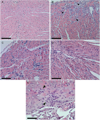
The results of this study showed that infected mice treated with raw BNZ exhibited similar heart tissue inflammatory damage to that of the untreated infected group. In contrast, it is important to note that a significant decrease of inflammatory cells in the heart tissue was observed after treatment with BNZ-MP (RL 50 and RS 50) (Fig. 5). In Fig. 6A, it is possible to see the cardiac tissue of uninfected mice without any sign of inflammation. On the other hand, the cardiac tissues of both untreated infected mice (Fig. 6B) and infected mice treated with the raw drug (Fig. 6E) showed amastigotes nests, extensive and multiple inflammatory foci of mononuclear cell infiltrates and some necrotic areas with structural alterations and fibrotic foci (Grosso et al., Reference Grosso, Alarcon, Bua, Laucella, Riarte and Fichera2013). In contrast, the cardiac tissues of mice treated with BNZ-MP-RL (Fig. 6C) and BNZ-MP-RS (Fig. 6D) were not damaged and only minor inflammatory foci with respect to untreated infected animals were detected (Fig. 6B).
Evaluation of inflammation in the heart of acute T. cruzi-infected mice treated with BNZ or BNZ-MP. Animals were treated at 2 d.p.i. with BNZ-MP or BNZ at 50 mg kg−1 day−1 (RL 50, RS 50 or BNZ 50, respectively) for 30 days. Data represent the morphometric quantification of inflammatory cells in heart tissue stained with Haematoxylin and Eosin (H&E). *P < 0.05; **P < 0.01, analysed by the Kruskal–Wallis test, bars represent mean + s.d.

Haematoxylin and Eosin (H&E) staining of heart tissues from mice infected with T. cruzi and treated with BNZ-MP at 3 months post-infection. (A) Uninfected mice, (B) untreated infected mice, arrows show amastigotes nests, (C) infected mice treated with RL 50, (D) infected mice treated with RS 50 and (E) infected mice treated with BNZ 50. (A–D) Magnification: 20× and (E) 40×. Scale bars: 25 μ m. This figure appears in colour in the online version and in black and white in the printed version.

Discussion
BNZ, like many other benzimidazole derivatives, is poorly water-soluble and exhibits a low dissolution rate which may require the administration of high doses to achieve therapeutic effects (Buckley et al., Reference Buckley, Frank, Fricker and Brandl2013). Thus, BNZ belongs to Class II of the Biopharmaceutical Classification System (BCS) (Del Moral Sanchez et al., Reference Del Moral Sanchez, Gonzalez-Alvarez, Cerda-Revert, Gonzalez-Alvarez, Navarro-Ruiz, Almidon and Bermejo2018; Ferraz et al., Reference Ferraz, Alves, Nascimento, Amariz, Ferreira, Costa, Rolim, Lima and Rolim Neto2018). However, according to the experimental data provided by Maximiano et al. (Reference Maximiano, de Paula, Figueiredo, de Andrade, Talvani, Sá-Barreto, Bahia and Cunha-Filho2011), it is classified as Class IV, which indicate a reduced solubility and permeability with a Log P in distilled water, gastric fluid and enteric fluid of 0.77, 0.78 and 0.76, respectively (Maximiano et al., Reference Maximiano, de Paula, Figueiredo, de Andrade, Talvani, Sá-Barreto, Bahia and Cunha-Filho2011). In this regard, the reduction of particle size and increase of the surface area of such lipophilic compounds through the micronization process is one of the most effective methods to overcome such drawbacks (Singh and Van den Mooter, Reference Singh and Van den Mooter2016; Abuzar et al., Reference Abuzar, Hyun, Kim, Park, Kim, Park and Hwang2018). Although there are several technological tools to produce MP (Paulo and Santos, Reference Paulo and Santos2017; Barrera et al., Reference Barrera, Tejada, Leonardi, Lamas and Salomon2020) the spray-drying technique is a versatile and efficient method to produce particles <10 μm when spray-dryers with two-fluid nozzles are used (Kemp et al., Reference Kemp, Hartwig, Herdman, Hamilton, Bisten and Bermingham2016; Davis and Walker, Reference Davis and Walker2018). It is important to note that MP prepared by spray-drying are usually organic solvent-free, in contrast to other methodologies that use toxic organic solvents and/or toxic reagents (Li et al., Reference Li, Rouaud and Poncelet2008; Shim and Sah, Reference Shim and Sah2020). Furthermore, the selection of a suitable carrier is also a key step during the development of final products (Lengyel et al., Reference Lengyel, Kállai-Szabó, Antal, Laki and Antal2019). In general, most of the synthetic polymers are dissolved in volatile hazardous chlorinated organic solvents immiscible with water. Thus, the solvents are removed by either evaporation at elevated temperatures or extraction in a large amount of water, resulting in the formation of MP. However, the traces of such hazardous solvents are serious concerns during the development of pharmaceutical, cosmetic and food products. Additionally, the rate and temperature of solvent elimination are reported to affect the morphology of the product and/or the stability of the drug (Dixit et al., Reference Dixit, Athawale and Singh2015; Haque et al., Reference Haque, Boyd, McIntosh, Pouton, Kaminskas and Whittaker2018). Taking all these considerations into account, methacrylate copolymers (Eudragit®) were selected as polymeric carriers to formulate BNZ-MP, because of their solubility and harmless properties and ability to encapsulate hydrophobic substances (Thakral et al., Reference Thakral, Thakral and Majumdar2013; Higashi et al., Reference Higashi, Hayashi, Yamamoto and Moribe2015). Because these carriers are easily dissolved in ethanol, a Generally Recognized As Safe (GRAS) solvent, it is not necessary to use hazardous solvents to solubilize the carriers. In particular, Eudragit® derivatives (RL and RS) were proved to be able to encapsulate BNZ in a very effective manner, providing interesting mechanical and stability characteristics to the final products (Seremeta et al., Reference Seremeta, Arrúa, Okulik and Salomon2019). However, there are many other polymers with good capability as carriers for hydrophobic drugs. Therefore, in the future, other carriers will be used in our research group in order to encapsulate BNZ and compare them with Eudragit® RS and RL. It is important to note that the combination of the methodology and the selected carriers allowed an excellent drug encapsulation efficiency (95–96%). As can be seen in Table 1, there was a significant difference in the dissolution of BNZ from both polymeric MP compared to the raw drug during the first 60 min of the assay. It could be due to the reduced size of the BNZ-MP (<2 μm) after the process of spray-drying and the increased surface area which lead to a fast drug dissolution. These results are consistent with those using chitosan as a polymeric carrier to encapsulate BNZ by the coacervation methodology (Leonardi et al., Reference Leonardi, Salomón, Lamas and Olivieri2009; Barrera et al., Reference Barrera, Tejada, Leonardi, Lamas and Salomon2020). In this regard, it should be mentioned that the dissolution rate of BNZ was also increased using solid dispersions (SDs). Lima et al. (Reference Lima, Soares-Sobrinho, Silva, Hernandes, Rolim and Rolim-Neto2011) obtained SDs of BNZ with hydrophilic polymers. Dissolution study showed a significant increase up to 8-fold in dissolution rate of s.d. vs standard BNZ at 30 min. In addition, complexation with supramolecular hosts, such as cyclodextrins (CDs), was also applied for this aim. Leonardi et al. (Reference Leonardi, Bombardiere and Salomon2013) prepared BNZ-CDs complexes able to improve the dissolution rate of BNZ up to 4.3-fold in comparison with the untreated drug. Another approach to improved BNZ solubility and dissolution was proposed Maximiano et al. (Reference Maximiano, de Paula, Figueiredo, de Andrade, Talvani, Sá-Barreto, Bahia and Cunha-Filho2011) who increased the drug performance by preparing BNZ microcrystals using different hydrophilic stabilizing agents. Taking into account such results, in this work, BNZ loaded into RL and RS-MP lead to a faster drug release as compared to the raw drug. It was found that RL formulation released a higher amount of drug than RS formulation, particularly in the first 10 min of the assay. It could be because RL is more soluble in water and more permeable than RS, which can facilitate the drug release (Apu et al., Reference Apu, Pathan, Shrestha, Kibria and Jalil2009). Stability of pharmaceutical products is one of the critical aspects of ensuring the safety and efficacy of these products (Wu et al., Reference Wu, Zhang and Watanabe2011; Sosnik and Seremeta, Reference Sosnik and Seremeta2015). In particular, the problems of instability of liquid formulations upon storage could be avoided by converting them to solid products (Sanchez-Vazquez et al., Reference Sanchez-Vazquez, Lee, Strimaite, Buanz, Bailey, Gershkovich, Pasparakisa and Williams2019). As reported, several drugs are more stable in the solid state than the liquid state at ambient temperature (Broeckx et al., Reference Broeckx, Vandenheuvel, Henkens, Kiekens, van den Broek, Lebeer and Kiekens2017; Emami et al., Reference Emami, Vatanara, Park and Na2018; Ziaee et al., Reference Ziaee, Albadarin, Padrela, Femmer, O'Reilly and Walker2019). In this study, stability assay showed no significant difference (P > 0.05) in the drug content of BNZ-MP for 3 years. This finding is in agreement with a report of Cilurzo et al. (Reference Cilurzo, Selmina, Minghettia, Gennaria, Demartin and Montanaria2008), who found no variation of nifedipine content after 3 months storage of fast-dissolving MP. These results confirm that spray-drying process is a suitable method to keep the drug content in MP for a long period of time. Due to the potential importance in clinical research, in vitro haemolytic and cytotoxicity assay was performed to evaluate whether BNZ-MP might damage cells. The results showed intact red blood cells and no toxic effects as no morphological change of the mammalian cell membrane, as occurs with other nanoparticles evaluated as carriers for cancer therapy (Liu et al., Reference Liu, Park, Zhang, Conwell, Liu, Bathula and Huang2010), indicating that such BNZ-MP would present desirable properties in terms of cell viability. In addition, it was not observed in any BNZ-MP-treated mice physical alterations nor any change in their behaviour during the course of our experiments. We used as standard 50 mg kg−1 day−1 of BNZ with the aim to compare the same amount of drug used in the BNZ-MP formulation, based on the effects observed with BNZ 50 mg kg−1 day−1 on the parasitaemia and parasite in hearts, in the same mice model (Grosso et al., Reference Grosso, Alarcon, Bua, Laucella, Riarte and Fichera2013) and other models (Strauss et al., Reference Strauss, Lo Presti, Bazán, Baez, Fauro, Esteves, Sanchez Negrete, Cremonezzi, Paglini-Oliva and Rivarola2013; Rial et al., Reference Rial, Scalise, López Alarcón, Esteva, Búa, Benatar, Prado, Riarte and Fichera2019). Oral administration of drugs to the animal of experimentation is usually performed using an aqueous solution with different excipients (cellulose derivatives or surfactants) (Piccirilli et al., Reference Piccirilli, García, Leonardi, Mamprin, Bolmaro, Salomón and Lamas2014). However, when BNZ-MP were dispersed in water, a small precipitate was observed and, therefore, it was decided to disperse those MP in oil instead of water, as already reported (Rial et al., Reference Rial, Scalise, Arrúa, Esteva, Salomon and Fichera2017). In agreement with the results of qPCR assay, mice treated with BNZ-MP did not exhibit parasitic infection in comparison to the mice treated with the raw drug (Fig. 4). This finding could be due to the faster release of BNZ from the Eudragit® formulations (Table 1), which could improve its absorption and further biological activity. A similar finding was reported by Piccirilli et al. who described a faster release and improved pharmacokinetic properties of albendazole from the polymeric microparticles, in comparison with the raw drug (Piccirilli et al., Reference Piccirilli, García, Leonardi, Mamprin, Bolmaro, Salomón and Lamas2014). Thus, the decrease in the levels of specific antibodies after BNZ-MP administration could be directly related to the decrease or absence of the parasite load as has been seen in other BNZ treatments using the same experimental model (Scalise et al., Reference Scalise, Arrúa, Rial, Esteva, Salomon and Fichera2016; Rial et al., Reference Rial, Scalise, Arrúa, Esteva, Salomon and Fichera2017). One of the most important and serious events of Chagas disease is the damage of the cardiac tissue which can lead to myocarditis and pericarditis (Tanowitz et al., Reference Tanowitz, Machado, Spray, Friedman, Weiss, Lora, Nagajyothi, Moraes, Garg, Nunes and Ribeiro2015). Thus, the reduction or elimination of any inflammation of the cardiac tissue produced by T. cruzi infection by novel BNZ formulations would be a valuable result. As seen in Fig. 5, the histopathology result of mice heart tissue treated with BNZ-MP showed a clear decrease in inflammation, compared to those treated with raw BNZ and untreated infected mice, probably due to the capacity of these polymeric formulations to improve the delivery of BNZ to those tissues. Thus, such lower levels of inflammation could prevent a possible progression towards fibrosis in the chronic stage of the infection, avoiding cardio pathological complications. Similar findings were observed in previous studies during the development of nanocrystals of BNZ (Rial et al., Reference Rial, Scalise, Arrúa, Esteva, Salomon and Fichera2017, Reference Rial, Arrua, Natale, Bua, Esteva, Prado, Laucella, Salomon and Fichera2020), confirming the significance of reformulated BNZ, as novel and efficient treatment for Chagas disease. In summary, fast-release BNZ-MP with a mean particle size of <2 μm and long-term storage stability (3 years) were produced by spray-drying methodology using as carriers Eudragit® RL PO and RS PO. Neither detectable morphological change or cell membrane destabilization nor haemolysis in defibrinated human blood was observed after incubation with such BNZ formulations. Preclinical studies in the murine model showed that BNZ-MP exhibited a remarkable effect on the acute T. cruzi infection. Infected mice survived an acute infection with 30 doses of BNZ-MP (50 mg kg−1 day−1). Also, a decrease of T. cruzi-specific antibody levels and absence of heart tissue damages were observed. Therefore, these fast-release BNZ-MP, prepared by spray-drying, are a very convenient approach to successfully treat Chagas disease in an experimental acute T. cruzi infection.
Supplementary material
The supplementary material for this article can be found at https://doi.org/10.1017/S0031182020002310.
Acknowledgements
We thank the animal facility staff of INP, Gabriela Barja, Laura Potenza and Leticia Orellana. We are also very grateful to Claudia Nose for her excellent technical assistance.
Author contributions
L.E.F. and C.J.S. conceived and designed the study. M.S.R., K.P.S., M.I.E. and J.B. performed the studies. M.S.R., K.P.S., L.E.F. and C.J.S. analysed and interpreted the results. C.J.S., K.P.S., M.S.R. and L.E.F. wrote the first draft of the manuscript and supplied all test compounds. All authors read and approved the final manuscript.
Financial support
This work was partially supported by the National Institute of Parasitology ‘Dr. Mario Fatala Chaben’, National Ministry of Health, INP-ANLIS, Buenos Aires, Argentina (Grant FOCANLIS 2017, NRU: 1704 to M.S.R.), by CONICET (Argentina), ASACTEI-SANTA FE (Argentina), National University of Rosario (Argentina), Universidad Nacional del Chaco Austral (Argentina) (PI N°63).
Conflict of interest
None.
Ethical standards
All procedures involving animals were approved by the Bioethics Committee of the National Institute of Parasitology ‘Dr. Mario Fatala Chaben’ (Register RENIS N°: 000028, Buenos Aires, Argentina) and followed the rules of the ethical legislation and regulatory entities established in Argentina. The international recommendations for the use of laboratory animals (World Medical Association in the Declaration of Helsinki) were also followed.