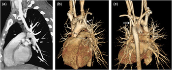
An 11-year-old girl was admitted due to dyspnoea during physical exertion. Transthoracic echocardiography showed a vertical vein draining into the left innominate vein. For further evaluation, the patient underwent multidetector computed tomographic angiography, using a 64-row computed tomographic scanner (Aquilion; Toshiba Medical Systems, Tokyo, Japan). It clearly showed the left upper lobe pulmonary vein draining into the left innominate vein through a vertical vein (Fig 1). The left lower and right pulmonary veins had normal drainage into the left atrium. The left vertebral artery arising directly from the aortic arch between the left common carotid and left subclavian artery was also present. No surgical treatment was indicated due to the stabile haemodynamic situation and absence of associated cardiac malformations.
Sagittal oblique multiplanar reformatted (a), and volume rendered (b and c) images show left-sided partial anomalous pulmonary venous drainage. Left upper lobe pulmonary vein drains into the left innominate vein (LIV) through a vertical vein (long arrow). The left vertebral artery (short arrow) arising directly from the aortic arch between the left common carotid and left subclavian artery is also seen (AO = aorta, SVC = superior vena cava).

Partial anomalous pulmonary venous return is a congenital anomaly in which one or more of the pulmonary veins are connected to a systemic vein or the right atrium. It is very often associated with an atrial septal defect of the sinus venosus type. Isolated partial anomalous pulmonary venous return (namely without an associated atrial septal defect) is a rare anomaly. Today, multidetector computed tomographic angiography has become a principal imaging modality for evaluation of thoracic vascular anomalies because of its advantages of widespread availability, short acquisition times, and high spatial resolution. It allows optimal visualisation of the entire course of the main thoracic vessels, disclosing the presence of vascular anomalies, if any, as well as precise anatomical characteristics. In conclusion, the combination of multidetector computed tomographic angiography with echocardiography provides optimal diagnostic accuracy in patients with partial anomalous pulmonary venous return.Reference Moral, Ortuño and Aboal1
Acknowledgements
There is no conflict of interest (including financial and other relationships).