Introduction
The use of Duddingtonia flagrans for biological control of gastrointestinal nematode parasites in ruminants has been studied extensively in recent years. This fungus produces adhesive three-dimensional net-type traps that capture and destroy the pre-parasitic stages of several nematode species (Barron, Reference Barron1977; Larsen, Reference Larsen2000).
In spite of this research, there is little information about the ultrastructural aspects of the interaction between this fungus species and infective larvae of gastrointestinal nematode parasites in ruminants. The characterization of processes involving fungus–nematode interaction is essential and may influence the selection of isolates for use in biological control programmes (Mendoza-de-Gives et al., Reference Mendoza-de-Gives, Davies, Clark and Behnke1999).
The objective of this study was to investigate interaction processes between the nematode predator fungus D. flagrans and infective larvae of Haemonchus contortus, using scanning electron microscopy.
Material and methods
Infective larvae of Haemonchus contortus
Goat faeces infected with H. contortus were collected using a collecting bag made from cotton sacking. Faecal samples of 20 g each were mixed with 8 g vermiculite and 20 ml distilled water and incubated for 8 days at 25°C in darkness. Larvae were recovered from the culture in tubes used for haemolysis, using a Baermann funnel and water, initially at 42°C, for 12 h.
The larval solution was filtered to eliminate debris and to recover the active larvae, using the methodology described by Barçante et al. (Reference Barçante, Barçante, Dias, Lima and Negrão-Corrêa2003). Larvae were then washed five times in sterile distilled water in a centrifuge at 1000 rpm for 5 min and the supernatant was discarded at the end of each centrifugation. Following this, larvae were resuspended and stored at 4°C in a solution containing 0.05% chloramphenicol and 0.05% streptomycin sulphate. To estimate the total larval number, five 20-μl aliquots of larval solution were counted and total larvae in the suspension were estimated.
Scanning electron microscopy
The Nordbring-Hertz (Reference Nordbring-Hertz1983) technique, with modifications, was used to prepare the material for scanning electron microscopy. Dialysis membrane discs (Sigma, St Louis, Missouri, USA), 6 cm in diameter, were cut and placed in Erlenmeyer flasks containing distilled water. The material was autoclaved at 121°C for 15 min. Discs were removed from flasks using tweezers, and placed on the surface of 2% agar–water in 6-cm diameter Petri dishes, so that membrane edges covered all the agar surface and were raised and adhered to plate edges to prevent larvae passing to the underside of the membrane.
Fragments of 1.7% CMA (Corn Meal Agar, Difco, USA) containing mycelia and spores of D. flagrans isolate CG722 were removed from the culture tubes and replicated in three 9-cm diameter Petri dishes containing 20 ml CMA medium plus 0.5% gentamicin. Petri dishes were incubated in a biochemical oxygen demand (BOD) incubator (FANEM, Brazil) at 25°C in darkness. After 7 days, approximately 4-mm diameter fragments were replicated from the edges of contamination-free colonies on to the surface of dialysis membranes (Nordbring-Hertz, Reference Nordbring-Hertz1983). Petri dishes were then incubated in a BOD at 25°C in darkness.
Seven days after incubation, Petri dishes were removed from the incubator and suspension droplets containing approximately 2000 infective larvae of H. contortus were placed over D. flagrans cultures grown on the surface of dialysis membranes. An equal number of larvae were placed into five Petri dishes containing agar–water without fungus and used as a control of larval viability.
In the first 12 h after nematode inoculation on Petri dishes, these cultures were removed from the incubator (25°C in darkness) and observed hourly under optical microscopy (light field) with 100 × magnification. Predated nematodes in a determined area of Petri dishes were marked on the underneath of the plate with a permanent marker, and each plate was numbered in order to time the fungus–larvae interaction and facilitate the finding of areas with predated nematodes. After predation, observations were made under optical microscopy with 100 × magnification at 6-hourly intervals.
Pieces of dialysis membranes with samples of larvae at stage L3 exposed to capture for 12, 24, 36 and 48 hours were cut with a scalpel and collected with fine pointed tweezers and fixed in 2.5% glutaraldehyde in 0.05 M phosphate buffer, pH 7.4 for 24 h, washed six times in the same buffer and dehydrated by passing the material through an ethanol series (30, 50, 60, 70, 95 and 100%). The material was dried to critical point using carbon dioxide, coated with gold and observed using a LEO scanning electron microscope at 10–15 kV, at the Centre for Electron Microscopy and Microanalysis of the Federal University of Viçosa.
Results and discussion
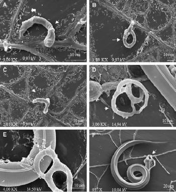
Hyphal ramifications were observed 9 h after inoculating nematodes on to Petri dishes, showing differentiation of predation structures. First, these structures were concentrated in areas of greater nematode concentration and later spread all over the membrane surface. The morphology of predation structures changed gradually and, at 24 h, most presented three-dimensional net formation (fig. 1A–D).
Scanning electron micrographs of trap formation process and interaction of the fungus Duddingtonia flagrans with infective larvae of Haemonchus contortus on dialysis membrane surface. (A) ↑ , Hyphal branches showing the initial phase of trap formation by the fungus;  , rod-type bacterial cells on the dialysis membrane surface. (B) and (C) ↑ , Traps in formation phase. (D) Fungus forming three-dimensional adhesive nets. (E) ↑ , Presence of mucilaginous substance on larval cuticle 12 h after predation. (F) ↑ , H. contortus larva 12 h after predation.

The time necessary for trap formation after adding nematodes to plates recorded in the present experiment differed from the results reported by Nansen et al. (Reference Nansen, Gronvold, Henriksen and Wolstrup1986) who observed trap presence 3–6 h after adding 150 larvae (L1, L2 and L3) of Cooperia oncophora on the surface of 3.2-cm diameter Petri dishes containing Arthrobotrys oligospora grown on the CMA surface. However, factors such as medium type, fungal species and nematode hamper the comparisons.
In predator fungi, the transition from saprophytic growth to parasitic stage, when traps are formed, is influenced by biotic and abiotic factors. In the presence of nematodes, mycelia and spores are induced to form structures that will eventually capture nematodes (Nordbring-Hertz, Reference Nordbring-Hertz1988).
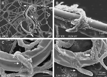
The first predated larvae were observed 11 h after adding larvae to plates. Larvae were found adhering to the surface of the predation structures (fig. 1E and F). Points of larval capture varied, and there were no sites in the cuticle specific for hyphal adhesion, besides, in some cases, larvae were captured by several predation structures at different sites of the cuticle surface (see fig. 2A). An adhesive substance could be seen in areas of contact between traps and nematode cuticle (figs. 1E and 2C).
Scanning electron micrographs showing the interaction between Duddingtonia flagrans with infective larvae of Haemonchus contortus on dialysis membrane surface. (A) , Traps adhering to different surface regions of a stage L3 larva 12 h after predation; ↑ , chlamydospores of D. flagrans. (B) ↑ , Bacterial cells at the point of adherence between traps and nematode cuticle 24 h after predation. (C) ↑ , Presence of mucilaginous substance between net traps and nematode cuticle within 36 h of nematode–fungus interaction. (D) , Mucilaginous substance released from the L3 larva cuticle close to the adherence zone of the trap, 48 h after predation; ↑ , fungal hyphae emerging from the cuticle showing penetration of L3 by the fungus.

Some larvae, even after predation, began to move when exposed to light during examination under optical microscopy. Live larvae were observed up to 24 h after predation, but among larvae predated for 36 h this event was not recorded on any of the Petri dishes. These results are similar to those reported by Nansen et al. (Reference Nansen, Gronvold, Henriksen and Wolstrup1986), who observed that larvae of C. oncophora showed great motility 20 h after predation by A. oligospora.
Larval predation by nematophagous fungi involves several steps. Adhesion between nematodes and fungi starts as a contact between the surfaces of larval cuticle and trap. After capture, hyphae penetrate the cuticle, colonize the interior of the body and emerge again on the surface of the nematode cuticle, producing spores and mycelia (Barron, Reference Barron1977).
In the present study, only observations of fungus–larvae interaction for 48 h showed invasion of larval cuticle by the fungus (fig. 2D), indicating that this may have occurred in the 36–48-h interaction interval. Nevertheless, optical microscopy did not show larval movement 24 h after interaction, which might indicate that larvae may have been penetrated by hyphae within the 24–36-h interval. In contrast, larvae from control plates remained alive and with vigorous movement throughout the assessment period.
Veenhuis et al. (Reference Veenhuis, Nordbting-Hertz and Harder1985) reported that nematode penetration by A. oligospora hyphae occurred 2–4 h after capture. Their findings, however, were based on the action of the fungus on the nematode Panagrellus redivivus, which did not have an external cuticle derived from second-stage larvae. An external cuticle is present on H. contortus infective larvae, which might explain the longer time for hyphal penetration in the present study.
In a differential interference contrast microscopy study, Murray & Wharton (1990) showed that in the penetration of third-stage larvae of Trichsotrongylus colubriformnis by A. oligospora a secondary infection bulb is formed between the sheath and the cuticle, which explains the long time necessary for larval penetration. In the present study, secondary infection pegs were not seen in the micrographs. Ultrastructural studies by transmission electron microscopy or other microscopical techniques may help to clarify the penetration phase of H. contortus larvae by D. flagrans.
Scanning electron microscopy showed the presence of rod-shaped bacteria. These were distributed randomly on membrane surfaces (fig. 1A) and were sometimes associated with the fungus–larvae interaction zone (fig. 2B). Maia et al. (Reference Maia, Santos and Di Mauro2001) reported the association of bacteria with nematophagous fungi, observing a constant presence of rods in fungal cultures of Monacrosporium robustum.
Dupponois et al. (Reference Dupponois, Amadou and Matteile1998) studied the activity of several bacterial strains associated with an isolate of A. oligospora. Interaction between some strains and the fungus enhanced predator activity, radial growth and conidia production. The same study showed that the fungus predator activity, alone, against juveniles of plant parasite nematodes Meloidogyne incognita and M. mayaguanensis was greater than on M. javanica. However, when the association with some bacteria was used, there was no specificity and fungus aggressiveness increased. The authors named these bacteria ‘nematophagous helper bacteria’ and raised the hypothesis of bacteria producing mucilaginous substances that act as molecular bridges between fungi and nematodes.
The presence of bacteria in fungus–larvae interaction zones was not constant in this study. This might indicate that the finding of bacteria was accidental and they would not have relevant functions for D. flagrans activity, or that during the processing of material bacteria were washed off and carried to other areas of the membrane surface. No reports were found in the literature on the association of bacteria with D. flagrans isolates. The isolation of these bacteria and the study of the effect of their association with nematophagous fungus may clarify their real role in the activity of D. flagrans against larvae of animal nematode parasites.
The dialysis membrane technique proposed by Nordbring-Hertz (Reference Nordbring-Hertz1983) was shown to be practical and applicable to the study of fungus–nematode interaction. Some dialysis membranes lost moisture and shrank or wrinkled with time though, causing a number of larvae to move to the under side and preventing the contact with traps. In another study, to prevent this larval movement, liquid agar was added to the membrane edges using a 1 ml syringe (unpublished data).
Previous studies on fungus–nematode interaction have demonstrated that fungi showing earlier production with greater number of traps and greater aggression against several nematode species could be selected for biological control programmes. Molecular and biochemical studies of factors involved in the interaction between D. flagrans and infective larvae of gastrointestinal nematode parasites in ruminants could bring important insights, allowing the identification of substances involved in the interaction, with implications for selection of isolates to be used in nematode biological control programmes.
Acknowledgements
This work was supported financially by Coorden-ação de Aperfeicoamento de Nivel Superior (CAPES), Conseiho Nacionai de Desenvolvimento Cientifico e Tecnoiogico (CNPq) and Fundação de Amparo à Pesquisa do Estado de Minas Gerais (FAPEMIG). The authors would like to thank to Claúdia Vanetti at the Centre for Microscopy and Microanalyses of the Universidade Federal of Viçosa for technical assistance.