Ductal arterial spasm is a very potentially dangerous incidence during percutaneous device closure of patent ductus arteriosus (PDA), which, otherwise, is a very safe catheter intervention. It is essential to notice its occurrence before device sizing and deploying. Without awareness, it can mislead device selection and can result in serious complication.
In our institution, 2 cases of severe ductal spasm were encountered out of 300 procedures of device closure for patent ducts. In this report, we shared our nightmare of ductal spasm during transcatheter closure of PDA in two children which had led to death in one patient.
Case report
We reported two cases of PDA, in which, during percutaneous device closure, the duct spasmed. Both were the children attended to Cardiac Catheterization Laboratory of Yankin Children Hospital for elective percutaneous device closure.
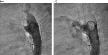
Our case 1 is a 2-year girl, with body weight 10 kg. She had signs and symptoms of heart failure such as poor growth, exercise intolerance, and mild hepatomegaly. Her duct was 5 mm in echo. On the angiogram, PDA was 7 mm at ampulla, minimal luminal diameter at pulmonary artery end was 1.6 mm, length was 8.8 mm, and more or less Krichenko type A in shape [Figure 1 (a)]. Mean PA pressure was 30 mmHg, while mean aortic pressure was 80 mmHg. Qp/Qs (The ratio of total pulmonary blood flow to total systemic blood flow) was not calculated. Amplatzer Duct Occluder II 3/4 was chosen first to close the duct. However, the device was easily slipped out. Then, the device size was increased to Duct Occluder I 8/6 mm and deployed again. However, after releasing device, it was seemed almost dropped into aorta [Figure 1 (b)]. So, it was tried to retrieve. Unfortunately, it then completely embolised to iliac bifurcation. The device was successfully retrieved after a few hours of trying, but the child was expired soon.
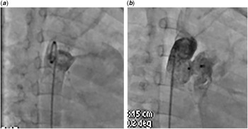
In our second case, the girl is 6 years and 16 kg. She also had clinical signs and symptoms of large duct with heart failure. Although large duct, 6 mm, was seen in echo, it appeared tiny in aortic angiogram. The measurement was as follows: minimal luminal diameter 1.7 mm at pulmonary end, ampulla 9.5 mm, length 9.8 mm, and Krichenko type A [Figure 2 (a)]. When we chose Amplatzer Duct Occluder I 6/4 mm, the recheck angiogram showed noticeable residual shunt around the device. It seemed that the ductal spasm was recovered at that time. On remeasuring, the duct was at least 6.3 mm at pulmonary end. Finally, it was closed with Duct Occluder I 12/10 mm [Figure 2 (b)].
Discussion
According to the literature, there are a number of factors contributing to the ductal spasm.Reference Lozier and Cowley1, Reference Tomita, Fuse and Chiba2, Reference Rohit, Garg and Kumar5 Lozier et al hypothesised direct contact of catheter with pulmonary artery end of ductus as a reason for vasospasm.Reference Lozier and Cowley1 Tomita et al showed catecholamines to be capable of inducing constriction of PDA in some children, especially those with low birth weight and ex-premature infants.Reference Tomita, Fuse and Chiba2 Some author described even oxygen as stimulus for ductal reactivity.Reference Lozier and Cowley1
In our case, catheter induced is less likely as we do not usually perform right heart catheterisation and do not need to enter pulmonary artery before any angiogram. The procedure was usually conducted under general anaesthesia. Other proposed stimuli such as stress-induced catecholamine or oxygen cannot be excluded.
Awareness and high index of suspicion are required to deal with ductal spasm during catheter intervention. Pre-intervention echo diameter is essential to be precise. During procedure, if there is any discrepancy between echo and angiographic measurements, it is wise to wait for a few minutes and take angiogram again.Reference MacDonald, Bhindi, Ormerod and Wilson3, Reference Alkamali and Alasrawi4
The chronological changes in auscultation findings are also essential. In our cases, loud continuous machinery murmur of PDA was detected in both girls preoperatively. However, we had to admit that we missed to recheck heart murmur during the event and, therefore, we were unable to track changes in auscultatory finding.
Conclusion
Ductal spasm, significant and severe enough to cause serious complication during device closure, may be rare. However, once occurred, this can lead to prolong procedure and related complications, even death. High index of suspicion and careful correlation of angiographic measurement with echo finding are highly essential. Owing to the previous bad experience, we were able to avoid serious complications in second case.
(a) Case 1. Very tiny PDA in initial angiogram. (b) Duct occluder 8/6-mm device immediately before embolisation. Note marked residual shunt.

(a) Case 2. Initial angiogram showed minute flow across the duct. (b) Actually large PDA fitted with duct occluder 12/10-mm device.

Acknowledgements
We thank Professor Toshiro Nakanishi for giving advice to write this article. We heartily thank the parents for allowing us to report the events.
Financial Support
The work in this report received no specific grant from any fun ding agency, commercial or not-for-profit sectors.
Conflict of Interest
None.