INTRODUCTION
Encephalitozoon cuniculi is an obligate intracellular single-cell sporoform microsporidian parasite that infects a variety of mammalian hosts, including immunocompromised humans. However, the most commonly infected animals are domestic rabbits (Mathis et al. Reference Mathis, Weber and Deplazes2005). Encephalitozoonosis in laboratory and pet rabbits is of clinical significance worldwide. Before microsporidian-negative rabbit colonies were established, a high prevalence of encephalitozoonosis had been recorded for laboratory rabbits. Nowadays, infections with E. cuniculi are being observed in pet rabbits in which the seroprevalence rates were between 42% and 68% (Halánová et al. Reference Halánová, Cisláková, Valencáková, Bálent, Adam and Trávnicek2003; Dipineto et al. Reference Dipineto, Rinaldi, Santaniello, Sensale, Cuomo, Calabria, Menna and Fioretti2008). On the contrary, low prevalence has been reported in wild rabbits (Thomas et al. Reference Thomas, Finn, Twigg, Deplazes and Thompson1997).
Rabbits are usually infected by ingestion or inhalation of spores passed in the urine. Infected rabbits show a range of clinical signs from latent chronic infections to death. In many cases, the onset of clinical signs is sudden and often occurs after a stressful situation (Künzel and Joachim, Reference Künzel and Joachim2010). Vestibular disease dominates among neurological signs when clinical manifestations of encephalitozoonosis occur (Harcourt-Brown and Holloway, Reference Harcourt-Brown and Holloway2003). Kidney disease is characterized by subclinical chronic renal failure (Cox and Gallichio, Reference Cox and Gallichio1978). Signs of ocular disease include lens rupture, uveitis and cataracts (Wolfer et al. Reference Wolfer, Grahn, Wilcock and Percy1993). Definitive in vivo diagnosis of encephalitozoonosis is difficult. Direct detection of microsporidia in the urine or faeces is not dependable because spore excretion is sporadic (Cox et al. Reference Cox, Hamilton and Attwood1979). For that reason, serological testing is a commonly used method for in vivo diagnosis, even though its value is limited due to the fact that presence of only IgG antibodies indicates exposure to infection, but does not confirm the organism as a causative agent of a clinical disease (Ewringmann and Göbel, Reference Ewringmann and Göbel1999; Csokai et al. Reference Csokai, Joachim, Gruber, Tichy, Pakozdy and Künzel2009). This problem could be solved using determination of specific IgM antibodies reflecting acute, reactivated infection or reinfection of rabbits. But exact kinetics of IgM and IgG during the infection course, including reactivated infection or reinfection confirmed by highly sensitive serological techniques, are not known.
The aim of this study was to characterize antibody and cell responses in rabbits after experimental infection with E. cuniculi using low and high infection doses and 2 different most probable infection routes (oral and ocular).
MATERIALS AND METHODS
E. cuniculi preparation
Spores of a rabbit strain of E. cuniculi (CH-K-2169; kindly provided by Professor P. Deplazes, University of Zurich, Switzerland) were produced on the RK 13 cell line in minimal essential medium with antibiotics (10 U/ml penicillin; 0·1 mg/ml streptomycin and 0·25 μg/ml amphotericin) and 5% fetal bovine serum. The spores were harvested, re-suspended in the culture medium, and stored at 4°C. Spores were purified by density-gradient centrifugation in Percoll (Sigma-Aldrich, USA) using a standard procedure (Visvesvara et al. Reference Visvesvara, Moura, Leitch, Schwartz, Wittner and Weiss1999). For oral and ocular infection, spores were diluted in 1 ml and 0·05 ml of PBS, respectively. The viability of the parasites was established by the inoculation of an aliquot into tissue culture.
Animals and experimental design
Outbred New Zealand White SPF European rabbits, strain Crl:KBL (free of common rabbit pathogens including E. cuniculi) were obtained from the Charles River Laboratories, Munich, Germany. Rabbits were housed individually in wire-mesh cages in the animal care facility with controlled conditions at the Veterinary Research Institute, Brno, Czech Republic. The rooms were maintained at a temperature of 21°C with 40–60% relative humidity, ventilation of approximately 15 air changes per hour and a 12-h light-dark cycle. The rabbits had free access to water and were fed ad libitum with complete pelleted diet (Bonagro, Czech Republic) that had been heated to 120°C for 20 min. A total of 30 male 4-month-old rabbits were randomly divided into 5 equal groups. Uninfected control rabbits were housed in a separate room and all infected rabbits were physically separated from the other groups and all rabbits housed in the facility. The animals were housed and handled with the agreement of the Branch Commission for Animal Welfare of the Ministry of Agriculture of the Czech Republic.
Oral infections were performed by means of a stomach tube, in the case of ocular infection, a drop of E. cuniculi spores in PBS was dropped into the conjunctival sac as follows: group PO/3 – peroral administration of 4×10³ spores, group PO/7 – peroral administration of 4×10⁷ spores, group OC/3 – ocular administration of 4×10³ spores and group OC/7 – ocular administration of 4×10⁷ spores.
After infection, rabbits were monitored daily and rectal temperature was recorded twice a week. Three days after infection and then at 1-week intervals, samples of blood were collected from the central ear artery for whole leukocyte and erythrocyte counts, differential leukocyte count, proliferation assay and detection of specific antibodies in serum. At the same intervals, samples of urine were collected during spontaneous urination. At week 13 post-infection, half of the rabbits in each group were intramuscularly administered 2 mg/kg of dexamethasone sodium phosphate (Dexamed, Medochemie Ltd, Cyprus; DXP) 3 times at 6-h intervals (Jeklova et al. Reference Jeklova, Leva, Jaglic and Faldyna2008), and then another 3 times at 24-h intervals. At week 18 post-infection, cerebrospinal fluid (CSF) was obtained by cerebellomedullary cistern puncture of anaesthetized rabbits (5 mg/kg xylazine and 35 mg/kg ketamine) and the animals were euthanized. Samples of organ tissues (liver, kidney, lung, brain, leptomeninges, medulla oblongata, intestine and eye) were fixed in 10% neutral buffered formalin for histological examination.
Blood analysis
Total RBC and total WBC counts were assessed using the Coulter Counter M4 (Beckman Coulter, USA) cell counter. Differential leukocyte counts were determined by counting 200 cells in blood smears stained with May-Grünwald and Giemsa-Romanowski (Penta, Czech Republic).
Proliferation assay
Detection of proliferation activity of peripheral blood lymphocytes was based on incorporation of [3H]-thymidine as described previously (Jeklova et al. Reference Jeklova, Leva, Jaglic and Faldyna2008). After 5 days of in vitro whole-blood lymphocyte stimulation (37°C, 5% CO2) with E. cuniculi spores (4×10⁵/well), 50 μl of medium with 3H-thymidine (5 μCi/ml) was added for the last 20 h. Incorporation of [3H]-thymidine was analysed by a microplate scintillation and luminescence counter (TopCount NXT™, Packard Bioscience Instrument Company, USA). The results were expressed in terms of stimulation indices (SI), which have been calculated as the ratio of counts per minute (CPM) in stimulated samples versus CPM in non-stimulated ones.
Serological testing
Specific anti-E. cuniculi IgM and IgG were detected using ELISA as described previously (Jeklova et al. Reference Jeklova, Jekl, Kovarcik, Huptman, Koudela, Neumayerova, Knotek and Faldyna2010). For preparation of soluble antigen, spores were mixed with solid glass beads (⩽106 μm; Sigma-Aldrich, USA) and sonicated on ice (30 min, 60 W). For characterization of antibody response, absorbances (iEMS Reader Labsystems, Finland) read at dilutions of 1:30 for IgM and 1:100 for IgG in serum samples were used.
Detection and quantification of spores in urine and CSF
Samples of urine (5 ml) were centrifuged (500 g, 10 min) and the pellets were re-suspended in 0·5 ml of PBS. Calcium carbonate was removed from each sample by 99% acetic acid (Jass et al. Reference Jass, Matiasek, Henke, Küchenhoff, Hartmann and Fischer2008). After washing twice in PBS, the pellet was mixed with 0·2 ml of PBS, frozen immediately and kept at −20°C until used. In order to release DNA, the spore wall was destroyed by mechanical disruption (MagNA Lyser Instrument, Roche, Switzerland) with zirconia silica beads (0·1 mm) and total DNA from 0·2 ml of treated urine or 0·2 ml of CSF was isolated using DNeasy 96 Blood and Tissue kit (Qiagen, USA). Quantitative PCR was performed with the LightCycler LC480 (Roche, Switzerland). Each reaction was carried out in a total volume of 10 μl, which consisted of QuantiTect SYBR Green PCR Kit (Qiagen, USA), 0·1 μ m of each primer (Hester et al. Reference Hester, Varma, Bobst, Ware, Lindquist and Schaefer2002) and 1 μl of template DNA. Cycling conditions were as follows: 15 min of initial denaturation at 95°C was followed by 40 cycles consisting of 95°C for 20 sec, 58°C for 30 sec and 72°C for 30 sec. Analysis of melting temperature of PCR products was performed for each sample.
Histological and immunohistochemical examination
Paraffin sections (6 μm) were stained with haematoxylin and eosin for routine microscopic examination. Severe lesions were defined as extensive pathological changes throughout the tissue, moderate as focal changes, and mild as occasional areas of increased cellularity, or perivascular or periportal cuffing. E. cuniculi spores in kidney, brain and eye were detected immunohistochemically with immune rabbit antiserum (diluted 1:20). The incubation was conducted at room temperature for 60 min. Horse-radish peroxidase-conjugated, affinity-purified chicken anti-rabbit IgG diluted 1:300 (Rockland, USA) and diaminobenzidine (Fluka, Germany) as a chromogen were used for visualization of the immunoreaction complexes. Sections were counterstained with haematoxylin. Staining was performed in parallel with positive and negative controls.
Statistical analysis
Because of relatively small numbers of experimental animals in respective groups, a one-tail Mann-Whitney non-parametric test was used for comparison of results from infected animals from the respective groups with control non-infected rabbits. Differences with the value of P<0·05 were considered statistically significant. All calculations were performed with GraphPad Prism® version 3.03 software (Graph Pad Software, USA).
RESULTS
Clinical signs and blood analyses
Generally, clinical signs were mild. The most striking of these were presented in group OC/7. Three days post-infection (p.i.) unilateral conjunctival hyperaemia, which becomes mild conjunctivitis by 7 days p.i., was observed in all of these rabbits. The signs of conjunctivitis disappeared by 21 days p.i. in all rabbits except 2 (rabbit nos 20 and 23) in which mild follicular conjunctivitis persisted up to 49 days p.i. On the 94th day p.i., 1 rabbit from this group (rabbit no. 24) exhibited episodes of rotation around the longitudinal axis and head tilt during manipulation. Ten days after immunosuppression (from 108 to 126 days p.i.), mild head tilt was repeatedly noticed during neurological examination in all DXP-treated rabbits from OC/7 group while only mild head shaking was observed in 1 non-suppressed animal from the same group. No clinical signs were recorded in group OC/3 during the entire experiment.
In rabbits from group PO/7, no changes in health status were observed until immunosuppression. Twenty-four hours after the last DXP dose, 1 immunosuppressed rabbit from this group suddenly died (rabbit no. 10); in another DXP-treated rabbit (no. 11), apathy and lower intake of food that lasted for 10 days were seen. No clinical signs were recorded in group PO/3 during the entire experiment.
When compared with control rabbits, no changes in rectal temperature were detected in any group of the infected animals during this experiment. Blood analyses did not show any significant changes in haematological parameters in infected rabbits, except for changes related to DXP administration as previously described (Jeklova et al. Reference Jeklova, Leva, Jaglic and Faldyna2008).
Proliferation assay
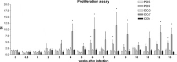
The highest proliferation of lymphocytes after stimulation with E. cuniculi spores was ascertained in rabbits from group OC/7 (Fig. 1). When compared with non-infected rabbits, SI values were significantly increased 4, 6, 8, 9, 12 and 13 weeks post-infection (P<0·05). In group PO/7, a significant cellular response was detected 6, 8 and 13 weeks post-infection (P<0·05). No elevation of proliferation activity was detected in rabbits infected with the lower dose. As a consequence of immunosuppression, a significant decrease in specific proliferation activity of lymphocytes that lasted for 2 weeks occurred in all DXP-treated rabbits from groups OC/7 and PO/7 (data not shown).
Proliferation activity of peripheral blood lymphocytes in rabbits infected with Encephalitozoon cuniculi. Stimulation indices (SI) calculated as the ratio of counts per minute in E. cuniculi-stimulated versus non-stimulated samples from rabbits after peroral infection with low (PO/3, n=6) and high dose (PO/7, n=6), and after ocular infection with low (OC/3, n=6) and high dose (OC/7, n=6) in comparison with control non-infected animals (CON, n=6) at various weeks after infection are shown. The parameters with significant difference (P<0·05) are marked with *.

Detection of antibodies
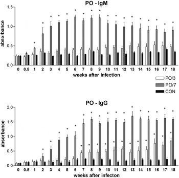
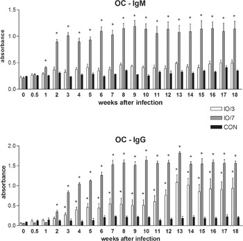
In both groups infected with the higher dose of E. cuniculi (PO/7 and OC/7) strong IgM and IgG antibody responses were detected (Figs 2 and 3). Significant increases in absorbance values of IgM antibodies (P<0·05) were recorded 1 week after infection in rabbits from these groups, as compared with the response of non-infected rabbits. A significant increase in IgG isotype (P<0·05) was detected 1 week later. Specific antibodies of both isotypes persisted up to the 18th week after infection in these groups of rabbits.
Specific antibody response detected by ELISA in rabbits infected with Encephalitozoon cuniculi. Data are shown as mean±s.d. absorbances of IgM or IgG isotype after peroral infection with low (PO/3, n=6) and high dose (PO/7, n=6) of spores in comparison with control non-infected animals (CON, n=6) at various weeks after infection. The parameters with significant difference (P<0·05) are marked with *.

Specific antibody response detected by ELISA in rabbits infected with Encephalitozoon cuniculi. Data are shown as mean±s.d. absorbances of IgM or IgG isotype after ocular infection with low (OC/3, n=6) and high dose (OC/7, n=6) of spores in comparison with control non-infected animals (CON, n=6) at various weeks after infection. The parameters with significant difference (P<0·05) are marked with *.

In rabbits infected with the lower dose (PO/3 and OC/3), significant differences were not detected, even if absorbance values of IgM antibodies tended to be higher than in control animals. Elevation of specific IgG antibodies in group PO/3 was observed from the 7th week post-infection. In contrast, IgG antibodies in group OC/3 were significantly increased (P<0·05) from the 3rd week post-infection.
Changes in antibody response were not detected among DXP-treated and non-treated rabbits in respective groups after immunosuppression.
Detection and quantification of spores in urine and CSF
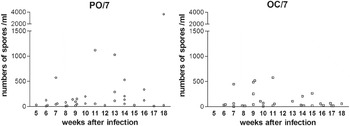
Spores were detected in all rabbits from group PO/7 and OC/7 at least 4 times during the infection course. Excretion of E. cuniculi spores in the urine was first detected 5 weeks post-infection in 1 rabbit from group PO/7 and 1 week later in 2 rabbits from group OC/7 (Fig. 4). Spore excretion tended to decline at 12 weeks post-infection, but from the 13th to the 18th week, spores were found in rabbits from both these groups. Numbers of spores detected in the urine varied from less than 101/ml to 103/ml. Immunosuppression had no influence on spore excretion or their numbers. In rabbits infected with the lower dose, E. cuniculi spores were not detected during the experiment. All samples of CSF, obtained at the 18th week post-infection, were negative for E. cuniculi DNA.
Excretion of spores in the urine of rabbits after peroral (PO/7, n=6) or ocular (OC/7, n=6) infection with a high dose of Encephalitozoon cuniculi. Numbers of rabbits excreting spores at various weeks after infection are indicated. Results of quantitative PCR are expressed as numbers of spores/ml of urine.

Histological and immunohistochemical examination
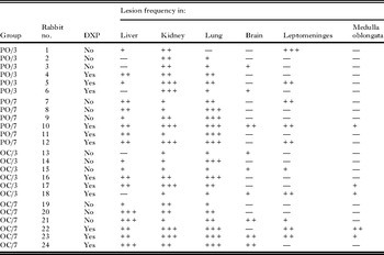
At 18 weeks after infection, mild, moderate or severe microscopic lesions compatible with encephalitozoonosis were seen in the liver, kidney and lung of almost all rabbits (Table 1). There was a positive, dose-dependent increase in the severity of lesions. The DXP-treated rabbits exhibited more severe lesions than untreated animals. Lesions of the brain, leptomeninges and medulla oblongata were detected less often. Control rabbits had no lesions.
Frequency of encephalitozoonosis compatible lesions in rabbits 18 weeks after peroral infection with low (PO/3) and high dose (PO/7), and after ocular infection with low (OC/3) and high dose (OC/7)

Histological examination of the livers from infected rabbits revealed mild to severe periportal and perivascular infiltrations by lymphocytes, plasma cells and eosinophils. Renal lesions were characterized by mild to severe tubular degeneration and interstitial infiltrates of mononuclear cells with lymphocytes and plasma cells. Lung lesions consisted of mild to severe peribronchial and perivascular inflammatory infiltrations of lymphocytes, plasma cells and eosinophils. In all rabbits from the PO/7 group, interstitial pneumonia was diagnosed. Lesions in the brain, leptomeninges and medulla oblongata included mild to moderate tissue and perivascular lymphocyte infiltrates. No lesions that could be attributed to E. cuniculi were seen in the intestine and eyes of any rabbit.
Using immunohistochemistry, a mild presence of proliferative stages or spores of E. cuniculi was detected in the kidneys and brains of all rabbits from groups PO/7 and OC/7. In the rabbit from group PO/7 that died suddenly after immunosuppression (no. 10), a massive presence of parasites in the collecting tubes in the medulla of the kidneys was observed. In rabbits from group PO/3 and OC/3, proliferative stages or spores of E. cuniculi spores were detected occasionally in kidneys and brains. No parasites were found in lens or in other ocular structures of infected rabbits and in kidneys, brains and eyes of non-infected control animals.
DISCUSSION
Encephalitozoonosis is a common infectious disease widespread among rabbits. Due to clinical importance and zoonotic potential of E. cuniculi, a detailed study of the infection course and immunological response of infected rabbits is of interest.
Infection in mammals usually occurs after ingestion or inhalation of spores, but vertical transmission has also been reported (Mathis et al. Reference Mathis, Weber and Deplazes2005). Under experimental conditions, intravenous, oral, intratracheal, intraperitoneal and intracerebral infection routes were also reported (Cox et al. Reference Cox, Hamilton and Attwood1979; Horvath et al. Reference Horvath, Svicky and Sevcikova1998). Despite the fact that ocular transmission is not considered to be a common way of infection, the authors assume it as probable in rabbits, because these animals mark their territories with urine and faeces. Urine marking can take the form of spraying, and both males and females mark (Harcourt-Brown, Reference Harcourt-Brown2002). Due to the fact that spores are excreted via the urine, this behaviour could be a source of ocular and also oral infections for other rabbits and humans. Therefore, immune responses of rabbits after oral or ocular infections with low or high doses of E. cuniculi were investigated in this study.
Severity of clinical signs is dependent on the immunocompetence of the host and virulence of the parasite. The course of experimental encephalitozoonosis in immunocompetent rabbits is usually subclinical (Kunstýr et al. Reference Kunstýr, Lev and Naumannm1986; Horvath et al. Reference Horvath, Svicky and Sevcikova1998). After ocular infection, conjunctivitis with spontaneous resolution within maximally 6 weeks was recorded in group OC/7. This clinical sign is in agreement with the course of microsporidial keratoconjunctivitis in non-immunocompromised humans (Chan et al. Reference Chan, Theng, Li and Tan2003). Before immunosuppression, no clinical signs associated with encephalitozoonosis were present in any other groups of rabbits, except the episode of neurological disorder in 1 rabbit from group OC/7. In contrast to that, the sudden death of 1 rabbit and apathy with a lower food intake in another animal from group PO/7 and mild head tilt were demonstrated in all DXP-treated rabbits from group OC/7 after immunosuppression. These results confirmed that subclinical latent encephalitozoonosis in rabbits could be reactivated after a short-term immunosuppression only.
Cell-mediated immunity is critical for the survival of an E. cuniculi-infected host. In this experiment, cell-mediated immunity was tested by lymphocyte proliferation assay using E. cuniculi spores as an antigen. The cell response was detectable only in groups infected with higher doses. Data from murine models have shown that cytotoxic T lymphocytes are crucial for protection, and their activation does not appear to be dependent upon CD4+ T cells (Khan et al. Reference Khan, Schwartzman, Kasper and Moretto1999). Further studies are necessary to ascertain the cell subsets that are involved in control of this microsporidian parasite in rabbits.
In the present study, strong and rapid humoral responses were detected in both groups of rabbits infected with the higher dose of E. cuniculi. Using ELISA, specific IgM antibodies were detected 1 week after infection, and IgG antibodies followed 1 week later. The antibodies of IgG and unexpectedly also IgM isotype persisted throughout the experiment. Infections with the lower doses led to a delayed and low IgG antibody response, with a reduced and statistically non-significant IgM production. Only in a few studies of experimental encephalitozoonosis, has the detection of IgM antibodies been described. Wicher et al. (Reference Wicher, Baughn, Fuentealba, Shadduck, Abbruscato and Wicher1991) were not able to detect IgM antibodies in intrarectally infected rabbits and Kunstýr et al. (Reference Kunstýr, Lev and Naumannm1986) ascertained IgM antibodies only from the 17th to the 38th day after intravenous infection using an indirect immunofluorescence test. The same results were obtained by Cox et al. (Reference Cox, Hamilton and Attwood1979) in rabbits inoculated intravenously. However, in contrast to that, in 1 orally infected rabbit, the IgM response persisted up to the end of the experiment (98 days p.i.). The commencement of the production of IgG antibodies was in accordance with previous studies (Cox et al. Reference Cox, Hamilton and Attwood1979; Wicher et al. Reference Wicher, Baughn, Fuentealba, Shadduck, Abbruscato and Wicher1991). High titres of IgG isotype antibodies can persist for several years in infected rabbits, even in the absence of apparent clinical signs (Bywater and Kellett, Reference Bywater and Kellett1979). Although the humoral response does not appear to be protective, the antibodies probably contributed to host resistance. Antibodies have been shown to opsonize microsporidia to facilitate killing by macrophages (Schmidt and Shadduck, Reference Schmidt and Shadduck1983). Moreover, serological examination remains the most important tool for ante mortem diagnosis of encephalitozoonosis in rabbits. Simultaneous testing of IgM and IgG specific antibodies give an indication of the infection status. The presence of IgM antibodies is indicative of active infection. The presence of only IgG-specific antibodies denotes chronic/latent infection and animals are at a risk so that stress or another suppressive factor may trigger encephalitozoonosis reactivation. The short-term immunosuppression model used in this study did not allow confirmation of the assumptive presence of IgM antibodies in reactivated infection or reinfection.
The beginning of spore excretion (5 weeks p.i.) and their concentration in the urine (103/ml) is consistent with a previous study by Cox et al. (Reference Cox, Hamilton and Attwood1979). This indicates that infection caused by 1 ml of this urine could lead to subclinical infection that could correspond to infection in groups PO/3 and OC/3. Waller et al. (Reference Waller, Morein and Fabiansson1978) assumed that a single infection dose of spores does not always lead to an infection in experimental studies, and under natural conditions, animals are usually repeatedly exposed to spores. No spores were detected in the urine of rabbits infected with the lower dose even after immunosuppression.
At the 18th week post-infection, all samples of CSF were negative for E. cuniculi DNA. Similar results were obtained by Künzel and Joachim (2010) who tested CSF from 12 pet rabbits with suspected encephalitozoonosis. On the other hand, Jass et al. (Reference Jass, Matiasek, Hartmann, Kuchenhoff and Fisher2006) obtained positive results in 2 of 19 (10·5%) CSF samples from pet rabbits. Further investigations are necessary to evaluate the presence of spores in the CSF at various times after infection and its possible usefulness for intravital diagnosis of encephalitozoonosis.
Histological examination, at 18 weeks p.i., revealed mild to severe lesions. Very severe lesions comprised of multifocal granulomas were not observed in any rabbit. The most affected organs were the lung, kidney and liver. Lesions in the brain, leptomeninges and medulla oblongata were detected less often. There was a positive, dose-dependent increase in the severity of lesions. Furthermore, DXP-treated rabbits showed more severe lesions than untreated animals. These results are comparable with the histological lesions observed by Wicher et al. (Reference Wicher, Baughn, Fuentealba, Shadduck, Abbruscato and Wicher1991). However, according to Cox et al. (Reference Cox, Hamilton and Attwood1979), 60 days p.i., the tissue alterations were seen mainly in the kidney, lungs and liver, while the brain was not changed. After 98 days, the most significant alterations were observed in the kidney and the brain and the extent of changes in the lung and liver was lower. This discrepancy could be caused by different levels of virulence of the pathogen. Even after ocular infection, no histopathological lesions were observed in the eyes and no parasites were found in lens or in other ocular structures. These results support the hypothesis that phacoclastic uveitis caused by microsporidia is a consequence of intrauterine infection (Wolfer et al. Reference Wolfer, Grahn, Wilcock and Percy1993) and localization of the parasite in the lens is less possible in adult rabbits. After experimental ocular infection, only mild unilateral conjunctivitis was observed, and thus authors conclude that E. cuniculi has a higher affinity to another predilection organ than the eye tissue.
Based on data presented here, the authors can conclude that not only oral, but also ocular experimental infection with E. cuniculi resulted in a cellular and antibody response of the infected rabbits. Immunological responses were dose dependent. Subclinical encephalitozoonosis in rabbits could be exacerbated after a short-term immunosuppression. Rabbits could be used as an experimental animal model for the study of ocular microsporidiosis.
ACKNOWLEDGEMENTS
The authors wish to thank Professor MVDr. Bretislav Koudela, CSc. for his assistance with experimental design, and to Professor Dr. Peter Deplazes for kindly providing us with a rabbit strain of E. cuniculi.
FINANCIAL SUPPORT
This study was supported by the Czech Science Foundation (524/08/P568), Ministry of Agriculture of the Czech Republic (MZe 0002716202) and Ministry of Education, Youth and Sports of the Czech Republic (CZ.1.05/2.1.00/01.0006 AdmireVet).