Introduction
Philornis Meinert (Diptera: Muscidae), is a genus of Neotropical muscids represented by about 50 species (Couri Reference Couri1999). While adults are free-living, larvae are associated with nestlings of many altricial or semi-altricial bird (Aves) species (Skidmore Reference Skidmore1985; Teixeira Reference Teixeira1999). Larval feeding behaviour is known for 22 of the described species, most of them (82%) being obligatory subcutaneous parasites (Dudaniec and Kleindorfer Reference Dudaniec and Kleindorfer2006). Subcutaneous larvae burrow into the host integument and develop between the dermis and body musculature, resulting in the formation of individual cysts (Teixeira Reference Teixeira1999; Spalding et al. Reference Spalding, Mertins, Walsh and Morin2002). There, they feed on tissue, blood, and serous fluids (Dudaniec and Kleindorfer Reference Dudaniec and Kleindorfer2006).
Parasitism by subcutaneous Philornis significantly reduces nestling growth (Arendt Reference Arendt1985a; Antoniazzi et al. Reference Antoniazzi, Manzoli, Rohrmann, Saravia, Silvestri and Beldomenico2011; Quiroga and Reboreda Reference Quiroga and Reboreda2012) and survival (Arendt Reference Arendt1985b; Delannoy and Cruz Reference Delannoy and Cruz1991; Antoniazzi et al. Reference Antoniazzi, Manzoli, Rohrmann, Saravia, Silvestri and Beldomenico2011; Segura and Reboreda Reference Segura and Reboreda2011). Over 200 species of birds have been documented as hosts of subcutaneous Philornis (Teixeira Reference Teixeira1999; Salvador and Bodrati Reference Salvador and Bodrati2013), some of which are of conservation concern (Arendt Reference Arendt2000; Guedes et al. Reference Guedes, Werneck, Couri and Araújo2000; Dominguez et al. Reference Dominguez, Reboreda and Mahler2014; Woolaver et al. Reference Woolaver, Nichols, Morton and Stutchbury2015).
Subcutaneous Philornis and their hosts are useful systems to study the ecology of host-parasite interactions in nature (Manzoli et al. Reference Manzoli, Antoniazzi, Saravia, Silvestri, Rorhmann and Beldomenico2013). Nonetheless, a limitation for its use as a model system resides in the paucity of data on their life cycles. This information is also essential to minimise the impact of these parasites on endangered species of birds. The only species in the genus with considerable available data about its life cycle, including information about reproductive biology, is Philornis downsi Dodge and Aitken (Fessl et al. Reference Fessl, Sinclair and Kleindorfer2006; Lincango and Causton Reference Lincango and Causton2008; Dudaniec et al. Reference Dudaniec, Gardner and Kleindorfer2010; O’Connor et al. Reference O’Connor, Robertson and Kleindorfer2010a). This fly was accidentally introduced to the Galapagos Islands, Ecuador (Fessl et al. Reference Fessl, Couri and Tebbich2001) and is considered the main threat for conservation of Darwin finches (Causton et al. Reference Causton, Peck, Sinclair, Roque-Albelo, Hodgson and Landry2006, Reference Causton, Cunninghame and Tapia2013; O’Connor et al. Reference O’Connor, Sulloway, Robertson and Kleindorfer2010b; Kleindorfer et al. Reference Kleindorfer, Peters, Custance, Dudaniec and O’Connor2014; Kleindorfer and Dudaniec Reference Kleindorfer and Dudaniec2016). Contrary to most Philornis species, P. downsi larvae are semi-haematophagous and live freely within nesting material (Dudaniec and Kleindorfer Reference Dudaniec and Kleindorfer2006) making comparisons with subcutaneous species tentative.
Most literature referring to subcutaneous Philornis occurring in southern South America is taxonomical (i.e., Skidmore Reference Skidmore1985; Couri Reference Couri1999; Couri and Carvalho Reference Couri and Carvalho2003; Patitucci et al. Reference Patitucci, Quiroga, Couri and Saravia-Pietropaolo2017) or to interactions between Philornis and their hosts (Nores Reference Nores1995; Rabuffetti and Reboreda Reference Rabuffetti and Reboreda2007; Quiroga and Reboreda Reference Quiroga and Reboreda2012; Manzoli et al. Reference Manzoli, Antoniazzi, Saravia, Silvestri, Rorhmann and Beldomenico2013). The only available information on the life cycle of a subcutaneous Philornis is for P. seguyi García on duration and survival of larval, pupal, and adult stages (Quiroga and Reboreda Reference Quiroga and Reboreda2013).
Unveiling aspects of the life cycle of these parasites is crucial to achieve a better understanding of the ecology of Philornis, which would enhance its use to study host-parasite interactions in nature and would provide crucial information where there is need to predict and minimise its impact. Here we provide data on survival of pupal and adult P. torquans Nielsen and on the duration of the pre-oviposition period.
Materials and methods
Collection of immature stages and development to adults
The study was conducted during the breeding seasons (September to April) of 2013–2014, 2014–2015, and 2015–2016. Third instars of P. torquans were collected from infested nestlings in a native forest from Santa Fe Province, Argentina (60°55'0"W, 31°23'08"S). Molecular studies have shown that P. torquans appears to be the only species present (Monje et al. Reference Monje, Quiroga, Manzoli, Couri, Silvestri and Venzal2013).
Larvae were taken to the laboratory and reared under controlled conditions (temperature: 26±1 °C; humidity: 65±15%). Larvae were kept individually in 25 cm3 plastic containers with pieces of tissue paper until pupation and were examined daily to record adult emergence. Individuals were weighed three to four days before and after pupariation using a precision scale±0.001 g. Mortality was documented for each stage.
In addition, 71 pupae found in the nests of hosts were collected and exposed to the same conditions described above to obtain more adults. Larvae and pupae collected in the 2015–2016 breeding season were subjected to different temperature and humidity regimes and therefore are not included in the analysis of data on the pupal stage.
Adult maintenance and egg collection
Adults obtained during the three breeding seasons were sexed following Couri (Reference Couri1999). Pairs consisting of one female and one male coming from different host broods were placed in 250-mL plastic containers to study their reproductive biology under pre-mentioned room conditions. Adults were fed once a day with a diet consisting of water (17 mL), orange juice (45 mL), white sugar (5.7 g), and hydrolysed protein (2 g) (diet adapted from Lincango and Causton Reference Lincango and Causton2008). We inspected adults daily to record survival and reproductive activity. Dead individuals were preserved in 70% ethanol.
Eggs laid were removed with a moistened fine brush and placed in a 5 cm in diameter glass Petri dishes. Each Petri dish housed all the eggs laid by one female. Dates, number of eggs, and the identification of the female were recorded. Eggs were maintained under the same temperature and humidity used for adults and inspected daily looking for newly hatched larvae. Eggs that did not hatch within 12 days were considered infertile and preserved in 70% ethanol.
After their natural death, a subset of females was dissected and their spermathecae examined under a stereomicroscope (20–40×) to determine their mating status (13 females that laid eggs and 24 females that did not lay eggs). Voucher specimens were deposited in the Laboratorio de Ecología de Enfermedades, Santa Fe, Argentina and in the Museo Argentino de Ciencias Naturales “Bernardino Rivadavia”, Buenos Aires, Argentina.
Developmental time, survival, and reproductive parameters
We examined the fate of each individual to determine percentage pupation for the cohort and, for pupating individuals, the duration of pupariation and pupation. Following Fraenkel and Bhaskaran (Reference Fraenkel and Bhaskaran1973), the pupariation period was estimated as the time elapsed from the retreatment of the larvae and the beginning of cocoon secretion to the darkening of the cuticle (brown colour). The pupation period was estimated as the time elapsed between the darkened puparium and the emergence of the adult. Similarly, adult lifespan was calculated as the time elapsed from emergence from the puparium until death.
We determined survival of each stage by calculating the number of survivors at the end of the stage divided by the number of individuals at the beginning of that stage. Differences in survival between male and females were tested by a non-parametric Wilcoxon rank-sum test.
Sex ratio is the ratio between the total number of females and the total number of males. We use a χ2 test to determine if sex ratio was significantly different from expected (1:1).
The date the first egg was laid and the number of eggs and clutches laid were recorded for each female. The pre-oviposition period was estimated as the number of days elapsed from the emergence of the female and the oviposition of the first egg; the oviposition period was assessed as the number of days during which a female laid eggs. Differences in duration of pre-oviposition period and in total eggs laid were tested by a non-parametric Wilcoxon rank-sum test.
Results
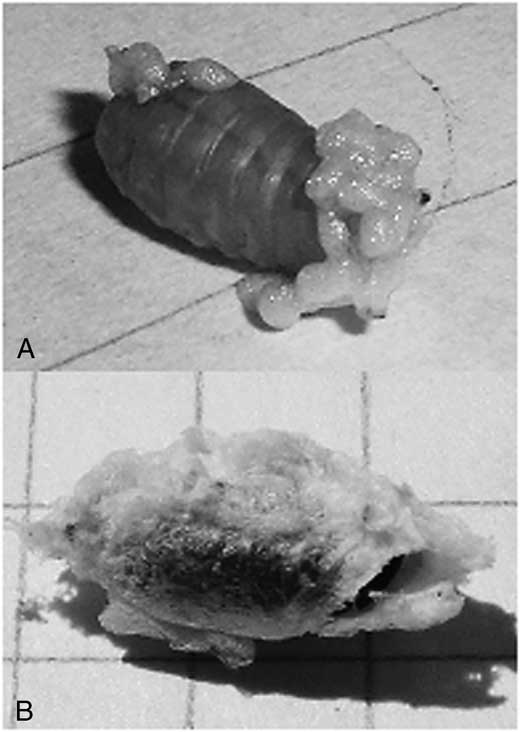
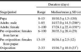
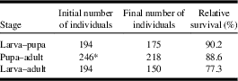
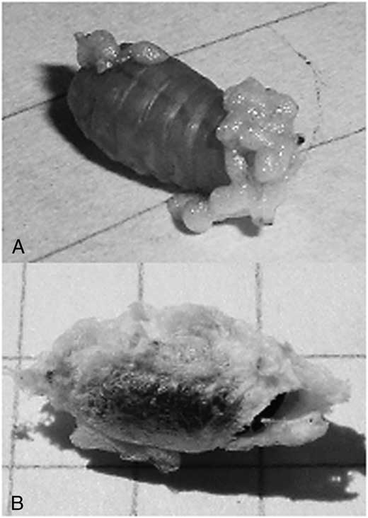
During the breeding seasons of 2013–2014 and 2014–2015, a total of 194 third instars were recovered from infested nestlings. Of these, 90.2% (n=175) pupated within 24–48 hours (pupariation process duration) after recovery, whereas 9.8% died. Pupae were surrounded by a cocoon (white spongy secretion) in 87% of the cases (Fig. 1). A total of 150 (85.7%) pupae successfully produced adults. Out of the 71 individuals collected as pupae from nests, 68 (95.8%) emerged as adults. The duration of pupal and adult stages and the relative survival of each stage are shown in Tables 1 and 2, respectively. Females survived significantly longer than males (W=22944; P=0.002; Table 1).
A, Spongy secretion produced by a third instars of Philornis torquans before pupariation. B, Larva totally enclosed in a cocoon.
Duration of pupal, and adult stages of Philornis torquans, as well as pre-oviposition and oviposition periods under laboratory rearing conditions.

* Total males and females obtained along the three breeding seasons with date of death known (date of death could not be determined for 115 adults).
† Females with more than one clutch.
SD, standard deviation; n, simple size.
Relative survival of each stage of Philornis torquans life cycle.
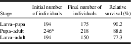
* Pupae obtained from larvae and pupae collected from nest material.
The mean larval and pupal weights were 0.11±0.04 g (n=125) and 0.05±0.02 g (n=125), respectively, equating to a mean weight loss during metamorphosis of 54.5%.
The 512 individuals that develop into adults comprised 259 males (50.6%) and 253 females (49.4%). The sex ratio was not significantly different from the expected 1:1 ratio (χ2=0.07; P=0.79).
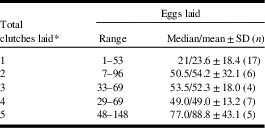
Eggs of P. torquans were obtained for the first time in captivity during the first breeding season (2013–2014). Out of 115 females that were exposed to males, 41 (35.7%) laid eggs. In addition, five females that were not exposed to males laid a total of 112 eggs and oviposited one or two clutches. Those eggs were found on food, on eppendorf tubes where food was provided, and on the walls of plastic containers. Considering all three breeding seasons together, 1843 eggs were collected. The mean number of eggs laid per female was 41.0±31.1 eggs (range=1–148). Females laid 1–8 clutches in their lifetime with one clutch being the mode, occurring in 41.5% (17 of 41) of the cases. Of the remaining females, 14.6% laid two clutches, 9.8% three clutches, and 34.2% four or more clutches (Table 3). There was a positive correlation between number of clutches and median number of eggs laid per clutch (ρ=1, P=0.017). When females laid more than one clutch, the mean duration of the period between first and final clutches was 16.7±10.0 days (Table 1), and the mean interval between clutches was variable: 4.9±3.2 days. Females began to lay eggs at least six days after emerging from the puparium. There was no significant difference in the pre-oviposition period between females that laid only one clutch and females that laid two or more clutches (W=217.5; P=0.73). Nonetheless, females that were collected as pupae had a median pre-oviposition period 53% shorter than females that were collected in the larval stage (Table 1; W=30.5; P<0.001). There were no significant differences in the total number of eggs laid by these females (W=172.5, P=0.98).
Total eggs laid by female Philornis torquans in relation to the final quantity of clutches.

* As we only registered one female each laying seven and eight clutches, they were excluded from the analyses. No females laid six clutches.
SD, standard deviation; n, simple size.
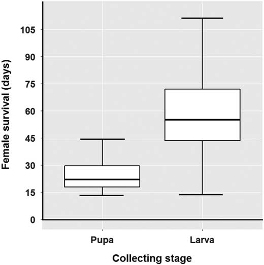
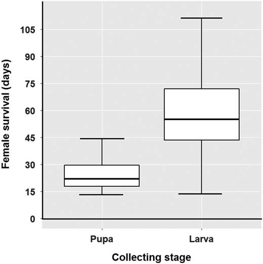
At the time to oviposition, the body size of females collected as pupae was longer and wider (length: range of 7.4–9.0 mm, median 8.0 mm; width: range of 3.0–3.7 mm; median 3.3 mm; n=12) that of females collected as larvae (length: range of 6.4–8.3 mm, median 7.3 mm; width: range of 2.4–3.4 mm, median 2.9 mm; n=29) (length: W=176; P<0.001; width: W=176; P<0.001). Differences in the time of survival of these two groups were also detected (Fig. 2); females collected as pupae survived significantly fewer days than females collected as larvae (W=25; P<0.001).
Survival time of Philornis torquans females obtained under laboratory rearing, by stages of collection (only females that laid eggs were considered). Females collected in pupal stage: n=10; females collected in larval stage, n=27.
Less than 1% (n=15 of 1843) of eggs hatched. Two females laid these eggs and the first instars were detected four days after they were laid. Unfortunately the larvae were already dead when found.
Seven of the 37 females that were dissected contained sperm in their spermathecae. Only two of this seven females laid eggs. A female without sperm in the spermathecae laid eggs that hatched.
Discussion
Studies on Philornis life cycle are crucial to increase the knowledge about these parasitic flies and how they interact with their hosts. We provide data on pupal and adult stages and reproductive parameters of P. torquans under laboratory rearing conditions. Also, these are the first data on the reproductive biology of any subcutaneous species of Philornis.
Information on Philornis life cycle is scarce and mostly restricted to the pupal stage. Previous studies reported duration of pupal stage ranging from 5–18 days, depending on the Philornis species and the environmental conditions (Skidmore Reference Skidmore1985; Spalding et al. Reference Spalding, Mertins, Walsh and Morin2002; Lincango and Causton Reference Lincango and Causton2008). The pupal stage in the life cycle of P. seguyi lasted nine days (Quiroga and Reboreda Reference Quiroga and Reboreda2013), similar values to what we found for P. torquans (10.5 days on average).
The presence of a frothy cocoon in P. torquans was usually observed after the larvae started pupation. This structure is considered of taxonomic importance for Philornis (Couri et al. Reference Couri, Carvalho and Löwenberg-Neto2007), and it has not been previously observed in P. torquans (see Ferrar Reference Ferrar1980; Couri et al. Reference Couri, Antoniazzi, Beldomenico and Quiroga2009). Ferrar (Reference Ferrar1980) and Teixeira (Reference Teixeira1999) suggested that the origin of a cocoon in Muscidae might be related to protection of the pupae against predation and parasitoids. This speculation is also shared by Bennett and Whitworth (Reference Bennett and Whitworth1991) for Protocalliphora Hough species (Diptera: Calliphoridae), which also infest bird nestlings. The pupae of some Philornis that form frothy cocoons (Ferrar Reference Ferrar1980; Couri Reference Couri1999; Couri et al. Reference Couri, Tavares and Stenzel2006) are parasitised by species of Brachymeria Westwood and Conura Spinola (Hymenoptera: Chalcididae) (Couri et al. Reference Couri, Tavares and Stenzel2006; Bulgarella et al. Reference Bulgarella, Quiroga, Brito Vera, Dregni, Cunninghame and Mosquera Muñoz2015; Delvare et al. Reference Delvare, Heimpel, Baur, Chadee, Martinez and Knutie2017). Although this observation supports the hypothesis of predator protection, the origin and function of the cocoon is still unknown for Philornis.
Mortality in pupae reared from larvae collected in the field was almost 15%, similar to reports for P. seguyi (Quiroga and Reboreda Reference Quiroga and Reboreda2013). The mortality of pupae collected as larvae from nestlings was 3.5 times higher than the mortality of those collected as pupae from nests. Although it is possible that larvae were physically damaged during extraction, they were manipulated with the utmost caution to avoid detrimental effects. Because larvae were removed from the host before their natural detachment, they may not have had sufficient nutritional reserves to satisfy their physiological requirements to reach adulthood. Undernourished fly larvae in other studies pupated, but died before metamorphosis (Ullyett Reference Ullyett1950; Kamal Reference Kamal1958). Although Quiroga and Reboreda (Reference Quiroga and Reboreda2013) found pupal mortality similar to ours, they collected P. seguyi larvae after they had naturally detached from nestlings. Further studies considering the stage at which samples are collected and the conditions for successful development in this and other Philornis species are needed to complement our findings.
Recovering individuals as larvae, rather than as pupae, also affected the pre-oviposition period, body size, and longevity of adult females. Females collected as pupae had a shorter pre-oviposition period, were larger and survived for less time than females collected as larvae. Females of species phylogenetically close to Philornis, such as Muscina levida Harris (Diptera: Muscidae) and Synthesiomyia nudiseta Van Der Wulp (Diptera: Muscidae) (Haseyama et al. Reference Haseyama, Wiegmann, Almeida and de Carvalho2015), laid eggs one or two weeks after they emerged from the puparia (Tirone et al. Reference Tirone, Parise and Avancini1996; Aruna Devi et al. Reference Aruna Devi, Abu Hassan, Kumara and Che Salmah2011). Female P. torquans collected as pupae had a pre-oviposition period similar to those in related species, whereas females collected as larvae had longer and more variable pre-oviposition periods.
Pre-oviposition periods provide information on the reproductive maturity rate of females; i.e., the shorter the period, the faster in reaching maturity (Ghoneim et al. Reference Ghoneim, Hamadah, El-Hela, Mohammad and Amer2016). The prolonged pre-oviposition period of smaller female P. torquans collected as larvae may indicate delayed reproductive maturity. Large females of Ceratitis capitata Wiedemann (Diptera: Tephritidae) produce more eggs soon after emerging and tend to copulate earlier than small females (Kaspi et al. Reference Kaspi, Mossinson, Drezner, Kamensky and Yuval2002). The difference in adult size found by Kaspi et al. (Reference Kaspi, Mossinson, Drezner, Kamensky and Yuval2002) was associated with the diet quality at larval stage. Our results reinforce the importance of larval conditions on morphology and reproductive performance in adults.
Experimental studies on trade-offs between reproduction and lifespan in Drosophila melanogaster Meigen (Diptera: Drosophilidae) have found decreased early reproduction with increasing female lifespan (Luckinbill et al. Reference Luckinbill, Arking, Clare, Cirocco and Buck1984; Zwaan et al. Reference Zwaan, Bijlsma and Hoekstra1995; Partridge et al. Reference Partridge, Prowse and Pignatelli1999). Our sample size was relatively small to analyse possible causes of differential lifespan between females collected as larvae and pupae, but it seems likely that early oviposition shortened lifespan in females collected as pupae. In addition, if there is delayed reproductive maturity (and late oviposition) in females collected as larvae this can in part explain the longer lifespan observed. However, further studies analysing gonada development of females at different ages are necessary to confirm these hypotheses.
Despite differences in life histories (subcutaneous versus semi-haematophagous), the adult lifespan observed here was similar to that of P. downsi (Lincango and Causton Reference Lincango and Causton2008). A shorter lifespan was reported for P. seguyi (5.46 days on average: Quiroga and Reboreda Reference Quiroga and Reboreda2013), but rearing conditions (diet composition and size of rearing containers) were different from the ones used here and on P. downsi. Also, we detected sexual differences in adult longevity; P. torquans females lived longer than males, as reported for P. downsi (Lincango and Causton Reference Lincango and Causton2008). In many taxa, including species of Diptera, females live longer than males (Fletcher et al. Reference Fletcher, Axtell and Stinner1990; Davies et al. Reference Davies, Kattel, Bhatia, Petherwick and Chapman2005; Pinilla et al. Reference Pinilla, Patarroyo and Bello2013). Several processes are suggested to explain these differences in lifespan (asymmetric inheritance of sex chromosomes: Tower and Arbeitman Reference Tower and Arbeitman2009; maternal inheritance of mitochondrial DNA: Maklakov and Lummaa Reference Maklakov and Lummaa2013; sex-specific selective pressures: Wolff and Gemmell Reference Wolff and Gemmell2013), which should be investigated for species of Philornis.
Almost half of P. torquans females exposed to males laid eggs. It is difficult to make direct comparisons between our results and those reported by Lincango and Causton (Reference Lincango and Causton2008) for P. downsi, as they did not rear pairs individually. However, these authors did not find any relationship between the number of females per box and the total number of eggs recorded. In other parasitic flies reared under laboratory conditions (Protocalliphora aenea Shannon and Dobroscky (Diptera: Calliphoridae), P. avium Shannon and Dobroscky, P. metallica Townsend, P. sialia Shannon and Dobroscky: Bennett and Whitworth Reference Bennett and Whitworth1991; Dermatobia hominis Linnaeus (Diptera: Oestridae): Ribeiro et al. Reference Ribeiro, Vianna, Costa and Scholl1993), the proportion of females that failed to oviposit was high. During spermathecae examination, we found that the presence of sperm was not a good predictor of egg laying. Surprisingly, the spermathecae of the only female that produced larvae was empty. This suggests that the state of the spermathecae is not informative about mating status in our system. The causes for the failure in reproductive output have not been identified, neither the stage of the reproductive process where this failure occurs. However, this might be related to limited success in recreating the (unknown) conditions required for Philornis reproduction. In order to reach a better understanding of the reproductive process, studies focussing on the constraints of female and male reproductive and sexual maturity should be performed.
The number of eggs laid per female was highly variable, as was the number of clutches. Although most females laid only one clutch, they can oviposit up to eight. The number of clutches appeared to be positively correlated with the number of eggs laid per clutch. This great fecundity highlights the potential of P. torquans to harm bird populations, as one female could parasitise several nestlings and nests in her lifespan.
Lincango and Causton (Reference Lincango and Causton2008) observed that female P. downsi began to oviposit at 12–20 days of age, but they found fully formed eggs when they dissected females six days old. This coincides with the age when P. torquans females began to oviposit, so the time for egg formation could be shorter in P. torquans.
Since less than 1% of the eggs collected hatched, our laboratory conditions probably were not sufficiently favourable for reproduction. First instars were detected four days after eggs were laid. Because they were already dead when found, we could not accurately determine the duration of the egg stage, but at least three days are necessary for eggs of P. downsi to hatch (Lincango and Causton Reference Lincango and Causton2008).
In conclusion, we showed that lifespan and survival in pupae and adults of P. torquans were similar to those reported for P. seguyi and P. downsi. Furthermore, we found that the stage (larvae, pupae) at which they were collected affected survival, body size and pre-oviposition period, but not the fecundity of females. Additional studies are needed to know the optimal requirements for the reproduction of this species under laboratory conditions.
Acknowledgements
The authors are grateful to Pablo Capovilla, Agustín Fasano, Ivana Monsalvo, Lucia Reynoso, Carina Sosa, Daniel Zurvera, and Alejandro Percara for their fieldwork support. This work was funded by Consejo Nacional de Investigaciones Científicas y Técnicas (grant PIP11220100100261). All procedures conducted in this study comply with current national and provincial laws, and were approved by the Bioethics Committee of Universidad Nacional del Litoral.