The successful outcome of percutaneous interventional closure of the atrial septal defect depends on an adequate atrial septal rim (≥5 mm). Reference Amin1 Although a deficient anterior-superior rim and an oversized occluder can increase the risk of long-term complications such as cardiac perforation after transcatheter closure, Reference Mendirichaga, Smairat and Sancassani2–Reference Tchantchaleishvili, Melvin, Ling and Knight5 the lack of an anterior-superior rim is no longer a contraindication for atrial septal defect closure. Reference O’Byrne, Gillespie, Kennedy, Dori, Rome and Glatz6–Reference Li, Li and Yang9 The occlusion of an atrial septal defect with deficient posterior-inferior or inferior vena cava rim is ambiguous, and the complications and long-term outcomes are not clear. Reference Chen, Chen, Cao, Zhang, Chen and Zhang10–Reference Amedro, Bayburt and Assaidi13
At present, most atrial septal defect closures are guided by fluoroscopy and echocardiography. To avoid potential radiation damage and simplify the procedure, some centres only use echocardiography to guide the operation. Reference Schubert, Kainz, Peters, Berger and Ewert14–Reference Ackermann, Quandt and Hagenbuch16 However, these studies did not include patients with deficient posterior-inferior or inferior vena cava rim. We reviewed the data of patients that underwent echocardiography-guided transcatheter atrial septal defect closure at our centre and compared the results and follow-up outcomes between patients with deficient and sufficient posterior-inferior/inferior vena cava rim atrial septal defect. The study is presented in accordance with the STROBE reporting checklist.
Methods
Patient population
The data of patients that underwent non-radiation transcatheter atrial septal defect closure at our centre from March 2017 to March 2020 were retrospectively reviewed. The patients were either admitted after being diagnosed in the outpatient department or were transferred to the cardiothoracic surgery department after deemed unsuitable for atrial occlusion due to deficient posterior-inferior or inferior vena cava rims. The inclusion criteria of patients for transcatheter atrial septal defect closure were as follows: 1) age ≥ 2 years and weight ≥8 kg, 2) atrial defect diameter >6 mm, and 3) echocardiography indicating left to right shunt, and right atrium and right ventricle enlargement. Patients with 1) maximum diameter of atrial defect >38 mm, 2) sinus atrial defect, ostium primum atrial septal defect, or coronary sinus atrial defect, 3) atrial defect combined with other cardiac malformations requiring surgical correction, 4) severe pulmonary hypertension and pulmonary resistance of cardiac catheterisation >7 Wood units, and 5) haemorrhagic diseases and antiplatelet therapy contradiction were excluded. Not all atrial septal defect with deficient posterior-inferior or inferior vena cava rim underwent transcatheter closure, and the operation was conducted on patients with atrial septum shorter than the left disc diameter of the implant. The study was approved by Institutional Ethic Committee of First Affiliated Hospital of Guangxi Medical University [NO.: 2020 (KY-E-102)] and the requirement for patient consent was waived given the retrospective nature of the study.
Pre-operative evaluation
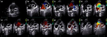
The Philips IE33 ultrasound machine (S5-1 probe, 1˜5 MHz; S8-3 probe, 3 ˜8 MHz, Philips Medical Systems, Cleveland, United States of America) was used for pre-operative assessment. Reference Papa, Gaspardone and Fragasso17 The lengths of the anterior-superior and posterior-inferior rims were measured on the parasternal short-axis view, that of the anterior-inferior and posterior-superior rims on the apical four-chamber view, and the inferior vena cava and superior vena cava rims on the sub-costal view. The size of the defect was recorded in each sector. The four-chamber view was used to measure the overall length of the atrial septum during the systolic circle. The thickness and sturdiness of the rim were evaluated to estimate its stability for device implantation. Rim thickness <0.5 mm was considered unstable, and length <5 mm was defined as deficient. Reference Amin1 GE Vivid E95 (M5SC-D probe, frequency 1.5–4.6 MHz; 6VT probe, frequency 3.0–8.0 MHz; 9T probe, frequency 4.0–10.0 MHz, GE Healthcare, Chicago, United States of America) was used for transthoracic echocardiography or transesophageal echocardiography (Fig 1). Transesophageal echocardiography was performed using various angles in multiple sectors. Based on the observations, the patients were classified into the deficient and sufficient posterior-inferior/inferior vena cava rim groups.
ASD with deficient PI rim was closed under TTE. ( a ) The sheath was advanced into the middle of the right atrium ( b ) and inserted into the left atrium. ( c ) The left disc was ready to be deployed ( d ) and adjusted in a small left atrium under TTE guidance parallel to the atrial septum. ( e ) The right disc was deployed ( f ) with a connected cable. ( g ) The occluder disc clung to the posterior part of the atrial wall. ( h ) TTE showed that the occluder disc clung to the inferior wall of the right atrial wall in a short-axis view. ASD = atrial septal defect; PI = posterior-inferior; TTE = transthoracic echocardiography.

Technique
The Heart R atrial septal occluder consists of a nitinol wire mesh (LifeTech Scientific Co., Shenzhen, China) and a polyester film, and the size ranges from 8 to 40 mm with 2-mm increments. It is a self-expandable device with two discs, and the diameter of the left disc is 2 mm larger than that of the right disc similar to the Amplatzer septal occluder. The delivery sheath size ranges from 7 to 14f.
Deep sedation and local anesthesia were induced in patients older than 12 years with sufficient posterior-inferior or inferior vena cava rims and clear echocardiogram images, and atrial septal defect closure was performed under transthoracic echocardiography guidance. Otherwise, general anesthesia was administered through tracheal intubation, and transthoracic echocardiography or 2D/3D transesophageal echocardiography was used to evaluate the atrial defect and guide the closure. Intraoperative echocardiography was performed using GE E95 Vivid 7. The femoral vein was punctured and a 5f/6f arterial sheath (Cordis Corporation, Miami, United States of America) was inserted, followed by administration of heparin at the dose of 100 IU/kg. The distance from the puncture point to the fourth right rib was preliminarily estimated using the advancing guidewire. Under the transthoracic echocardiography sub-costal view or transesophageal echocardiography bi-caval view, a 260-cm super-stiff straight tip guidewire (Amplatz type, 0.035 in. Cordis Corporation, Miami, United States of America) was advanced through the right femoral vein to the right atrium and the superior vena cava. The delivery sheath was subsequently advanced along the guidewire to the level of the right atrium (Fig 1a). The guidewire was then withdrawn, and the delivery sheath was rotated through the atrial defect under the parasternal short-axis or transesophageal echocardiography bi-caval view (Fig 1b) to prevent the sheath from entering too deep. The occluder was implanted under transesophageal echocardiography or transthoracic echocardiography apical four-chamber or parasternal short-axis view (Fig 1c–f). The size of the device was selected based on the transthoracic echocardiography or 2D/3D transesophageal echocardiography measurements, with the diameter of 0–6 mm larger than the maximum diameter of the atrial defect. A push–pull test was used before releasing the device, and in case the latter was not stable, a larger occluder not exceeding the overall length of the atrial septum was selected. Transthoracic echocardiography/transesophageal echocardiography was performed after detaching the cable, and the procedure was considered successful if there was no occluder dislodgement, moderate or more pericardial effusion, large residual shunt, no new-onset or aggravated mitral regurgitation, no vena cava or pulmonary vein obstruction, and no II°/III° degree atrioventricular block. Cefazolin was given to prevent infection for no more than 24 hours after the procedure, and 3–5 mg/kg oral aspirin was given 6 hours after the operation and continued for 6 months. Patients with G-6PD deficiency were administered Plavix.
Follow-up
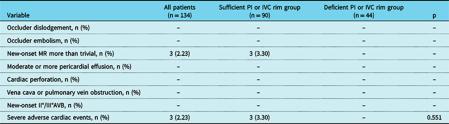
The patients were followed up by transthoracic echocardiography and electrocardiography at our hospital after 1-, 3-, 6- and 12-month post-operation, and annually thereafter. Transthoracic echocardiography was used to detect residual shunt, occluder dislodgement or embolism, new-onset or aggravated mitral regurgitation, pericardial effusion, cardiac perforation, and vena cava or pulmonary venous obstruction. The electrocardiography was used to check for new-onset II°/III° atrioventricular block, new-onset atrial fibrillation or atrial flutter, and other non-sinus rhythms. Occluder dislodgement or embolism, new-onset non-trivial mitral regurgitation, moderate or more pericardial effusion, cardiac perforation, vena cava or pulmonary venous obstruction, and new-onset II°/III° atrioventricular block were defined as severe adverse cardiac events.
Statistical analysis
Continuous variables with normal distribution were represented as the mean ± standard deviation, and those not conforming to normal distribution as median (interquartile range). Discontinuous variables were represented as numbers and percentages. SPSS 23 was used for statistical analysis. The Kolmogorov–Smirnov test was used to test normality. Two groups of continuous variables with normal distribution were compared using the two-tailed Student’s t-test. The Mann–Whitney U test was used to compare two groups of variables that did not conform to normal distribution. The Chi-square test or fisher’s exact test was used for categorical variables. The Kaplan–Meier method was used for survival analysis, and the survival curves were compared by the log-rank test. p < 0.05 was considered statistically significant.
Results
Study population
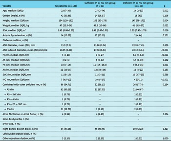
A total of 140 patients underwent echocardiography-guided transcatheter atrial septal defect closure from March 2017 to March 2020, of which 136 patients were included and 4 patients were excluded on account of multiple atrial septal defect closures using multiple devices. The baseline characteristics of the research population are summarised in Table 1. There were 91 patients in the sufficient rim group and 45 patients in the deficient rim group. Patients with deficient rims were younger (p = 0.002) with a smaller body surface area (p = 0.016) and larger atrial septal defect index diameter compared to those with sufficient rims (p < 0.001). The posterior-inferior, inferior vena cava, and posterior-superior rims in the deficient rim group were shorter compared to that in the sufficient rim group [3.5 (0–6.5) versus 9 (5–27) mm, p < 0.001; 4 (0–11) versus 10 (5–27) mm, p < 0.001; 9 (3.6–16) versus 11 (4.5–19.5) mm, p = 0.001, respectively]. There were no significant differences in the incidence of hypertension, type 2 diabetes and pre-operative arrhythmia, or the presence of other deficient rims between both groups.
Basic characteristics of the population

ASD = atrial septal defect; AI = anterior-inferior; AS = anterior-superior; AVB = atrioventricular block; BSA = body surface area; IVC = inferior vena cava; PI = posterior-inferior; PS = posterior-superior; SVC = superior vena cava.
Procedural results
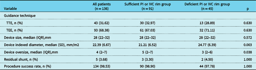
The results of the procedure are shown in Table 2. In the deficient rim group, 13 patients (28.89%) were operated under transthoracic echocardiography guidance and 32 patients (71.11%) under transesophageal echocardiography guidance. In the sufficient rim group, 30 (32.97%) and 61 (67.0%) patients were, respectively, operated under transthoracic echocardiography and transesophageal echocardiography guidance (p = 0.63). Although the size of the occluder was not significantly different between two groups (p = 0.572), the device indexed diameter was greater in the deficient rim group compared to that in the sufficient rim group (24.77 versus 21.21 mm/m2, p = 0.003). Therefore, a less oversize occluder was selected for the deficient rim group compared to the sufficient rim group [3 (2 6) versus 5 (2–7) mm, p = 0.038]. There was no significant difference in the procedure success rate between the deficient rim (97.78%) versus sufficient rim (98.9%) groups (p = 1). All the residual shunts after intervention were no more than moderate in both groups. Reference Boutin, Musewe, Smallhorn, Dyck, Kobayashi and Benson18 One patient in each group suffered device embolism after transcatheter closure. A 42-year-old man with sufficient rim had a large atrial septal defect with a maximum diameter of 38 mm. We therefore used No. 40 occluder and the push–pull test confirmed its stability during the intervention. On the first day after intervention, the patient had dyspnea and transthoracic echocardiography indicated occluder dislodgement to the right ventricle. The occluder was thus removed and atrial defect repair was performed. A 6-year-old boy with deficient rim had a failed atrial septal defect closure. The maximum diameter of atrial septal defect was 25 mm and the posterior inferior rim was 0. Due to the limitation of the overall length of the atrial septum, we selected the size 26 occluder. Electrocardiogram showed frequent pre-mature ventricular contraction in the ICU, and transthoracic echocardiography detected the occluder in the right ventricle. The patient underwent surgical occluder removal and atrial defect repair. Both patients were discharged uneventfully.
Procedure results

IVC = inferior vena cava; PI = posterior-inferior; TEE = transesophageal echocardiography; TTE = transthoracic echocardiography.
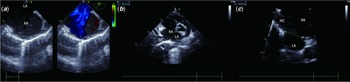
TEE and TTE images of deficient PI or IVC rim. ( a ) Incomplete image of PI rim shown by TEE. ( b ) TTE short-axis view showed deficient PI rim and its relationship with IVC. ( c ) Deficient IVC rim was assessed by TTE in the sub-costal view. TEE = transesophageal echocardiography; TTE = transthoracic echocardiography; PI = posterior-inferior; IVC = inferior vena cava.

Clinical outcomes
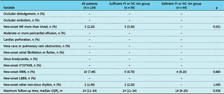
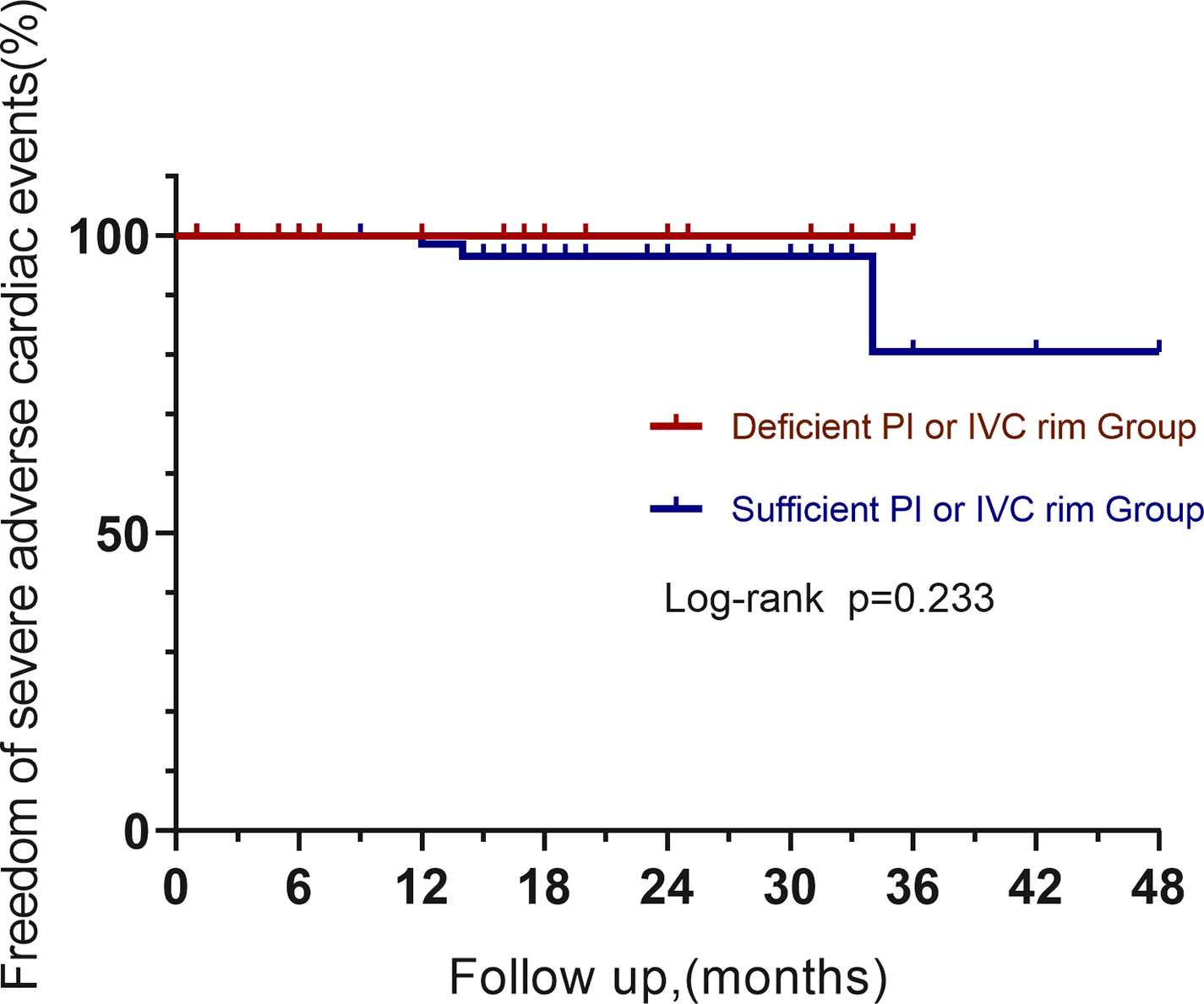
A total of 134 patients were followed up and their results are shown in Table 3. The median follow-up time was 14(IQR11–24) months. Transthoracic echocardiography did not detect any occluder dislodgement or embolisation, greater than mild pericardial effusion, cardiac perforation, vena cava, or pulmonary obstruction. Three adult patients with sufficient rim suffered new-onset mild mitral regurgitation after 6–8 months of the operation. The occluder did not restrict mitral valve movement as per transthoracic echocardiography, and all three patients are currently graded as class I NYHA. No new-onset mitral regurgitation was observed in the deficient rim group. The electrocardiography of two asymptomatic patients with sufficient rim indicated occasional pre-mature ventricular contraction. The adverse cardiac events-free survival was similar in both groups (p = 0.178, Table 4, Fig 3).
Follow-up outcomes

AVB = atrioventricular block; IRBB = incomplete right bundle branch block; LBBB = left bundle branch block; MR = mitral regurgitation.
Results of severe adverse cardiac events during follow-up

AVB = atrioventricular block; MR = mitral regurgitation.
Adverse cardiac events-free survival.

Discussion
Transcatheter closure for atrial septal defect is usually performed under fluoroscopy combined with echocardiography. Reference O’Byrne, Gillespie, Kennedy, Dori, Rome and Glatz6,Reference Jalal, Hascoet and Gronier19–Reference Baykan, Pamukcu and Ozyurt23 Fluoroscopy is used to monitor the interventional equipment during the entire procedure. Echocardiography, including transesophageal echocardiography, transthoracic echocardiography, and intracardiac echocardiography, is used to assess the size and shape of the atrial defect and display the heart structure in real time. Given the potential toxic effects of X-ray, echocardiography is increasingly being preferred to guide atrial septal defect closure. Reference Schubert, Kainz, Peters, Berger and Ewert14–Reference Ackermann, Quandt and Hagenbuch16,Reference Ewert24 Since intracardiac echocardiography is costly to implement and affected by the width of the field of view, transthoracic echocardiography and transesophageal echocardiography are more commonly used in atrial septal defect intervention. Studies show that both procedures have comparable efficacy and safety. Reference Baruteau, Hascoet and Fraisse20,Reference Bartakian, El-Said, Printz and Moore25 Nonetheless, the feasibility of non-radiation atrial septal defect interventions for patients with deficient posterior-inferior or inferior vena cava rims had not been investigated. A deficient posterior-inferior rim is often not displayed within the transesophageal echocardiography echo sector (Fig 2a), especially with the small left atrium, since the transesophageal probe is close to the posterior wall of the left atrium. Therefore, even 3D-transesophageal echocardiography cannot always display the deficient posterior-inferior or inferior vena cava rims. In addition, transesophageal echocardiography cannot effectively monitor the interventional instruments during the closure process, and the direction and angle of the probe have to be adjusted. Transthoracic echocardiography can offer a better view of the posterior-inferior or inferior vena cava rim in patients with clear echocardiogram images (Fig 2b–c) and dynamically monitor the shape of the occluder and its position relative to the surrounding structures in the same sector. The left atrium, interventional instruments, anterior-inferior and posterior-superior rims, and atrioventricular valve can be monitored simultaneously in the four-chamber view. In the parasternal short-axis view, the inferior vena cava rim, interventional instruments, anterior-superior and posterior-inferior rims, left/right pulmonary vein orifice, and left appendage can be monitored. We successfully performed transcatheter atrial septal defect closure under only echocardiography guidance without any heart injury and perforation. The interventional success rate between two groups was not statistically significant, which indicates the feasibility of non-radiation approaches in atrial septal defect with deficient posterior-inferior or inferior vena cava rims. The use of echocardiography alone further simplifies the intervention process without X-ray, balloons, and intracardiac echocardiography.
Several researchers consider deficient posterior-inferior or inferior vena cava rims as contraindications for interventional closure of atrial septal defect. Reference Amedro, Bayburt and Assaidi13,Reference Varma, Benson and Silversides26,Reference Butera, Romagnoli and Carminati27 For instance, Amedro et al reported an overall success rate of only 44% in an observational retrospective study, and Mathewson et al showed that device dislodgement had a significant impact on the pulmonary vein, inferior vena cava, and mitral valve. Reference Mathewson, Bichell, Rothman and Ing12 In contrast, Chen et al Reference Chen, Chen, Cao, Zhang, Chen and Zhang10 used mini-thoracotomy to effectively close the atrial septal defect with inferior vena cava rim deficiency, and Yan et al Reference Yan, Wang and Pan28 used a 3D printing model to evaluate the feasibility of atrial septal defect closure before the procedure. According to the design of the double-disc occluder, the radius of the left and right discs differs by 2–3 mm. The atrial septum rim of 2–3 mm cannot be clamped tight by the double disc. The cases of successful atrial septal defect closure with rim deficiency showed that double-disc occluder could close atrial septal defect by stretching the atrial septum and surrounding structure via self-expansion. Yan et al using a patent ductus arteriosus occluder to close the atrial septal defect with no pulmonary vein rims Reference Yan, Li and Song29 proved this. In addition, the Sapien valve and CoreValve can be firmly fixed in the aortic valve orifice in transcatheter aortic valve implantation, underscoring the importance of waist radial support on the anchorage of interventional instruments. Reference Leon, Smith and Mack30,Reference Adams, Popma and Reardon31 We selected occluder that was 0–6 mm larger than the maximum diameter of the atrial defect under transthoracic echocardiography or 2D/3D transesophageal echocardiography. After deployment, a push–pull test was conducted in multiple echo sectors to confirm the stability of the device. If pulled into the right atrium or pushed to the left atrium, the device was replaced with a larger one that did not exceed the overall length of the atrial septum. An occluder exceeding the overall length of atrial septum can encroach into the atrioventricular valve and the cardiac conduction system. Our results were consistent with that of Papa and Remadevi. Reference Papa, Gaspardone and Fragasso17,Reference Remadevi, Francis and Kumar32 There were no cases of occluder dislodgement or embolism during the follow-up, which can be attributed to the appropriate size of the occluder vis-à-vis the overall length of the atrial septum and the localised deficient rim. Thus, transcatheter closure of atrial septal defect with deficient posterior-inferior or inferior vena cava rim is safe given that the occluder size is appropriate.
Pericardial effusion and cardiac perforation are caused by the erosion of excessively oversized occluders. Reference Mendirichaga, Smairat and Sancassani2,Reference McElhinney, Quartermain, Kenny, Alboliras and Amin4,Reference Lipiec, Filipiak-Strzecka, Szymczyk, Peruga and Kasprzak33 Kijima et al Reference Kijima, Akagi and Takaya11 did not observe late erosion during the intermediate-term follow-up of atrial septal defect patients with deficient rim. Our clinical outcomes were in accordance with their study, even in case of patients aged 2–4 years. To be sure, the discs of the device would protrude towards the atrial wall when atrial septum rim was too short. The younger patients will be followed up for longer since their atrial walls are frailer than that of adults. It is unclear whether the device will impinge on the atrioventricular valve or other intracardiac structure as the heart capacities increase with age.
We included new-onset non-trivial mitral regurgitation as one of the severe adverse cardiac events that the device may displace and impinge on the mitral valve. Three patients with sufficient rim had new-onset mild mitral regurgitation after hospital discharge. Transthoracic echocardiography in multiple sectors showed that the occluder was away from the mitral valve, although there was no evidence of device-related mitral regurgitation. New-onset mitral regurgitation after atrial septal defect interventional closure or surgical repair has been reported previously, and the possible mechanism is a change in ventricular geometry. Reference Park, Lee and Kim34–Reference Yoshida, Numata and Tsutsumi36 Patients with a successful atrial septal defect closure should be followed up for longer durations in case of possible late complications.
Study limitations
1. This was a retrospective cohort study.
2. The atrial septal defect patients with deficient posterior-inferior or inferior vena cava rim that underwent transcatheter closure were from a single centre. Some patients who would have a successful intervention underwent surgical repair.
3. The number of unsuccessful cases is too small in each group. It still should be cautious to reach the conclusion that transcatheter closure of atrial septal defect with deficient posterior-inferior or inferior vena cava rim has the similar risk as that with sufficient rims.
4. The capacity of the heart will change for the younger patients as they grow up. The safety of transcatheter atrial septal defect closure in children with deficient posterior-inferior or inferior vena cava rim still needs a long-term observation.
Conclusions
Atrial septal defect with deficient posterior-inferior or inferior vena cava rim can safely undergo transcatheter closure under echocardiography alone if precisely evaluated with transesophageal or transthoracic echocardiography and the size of the occluder is appropriate. The mid-term results after closure are similar to that for an atrial septal defect with sufficient rim.
Acknowledgements
None.
Financial support
This study was supported by grants from Guangxi medical “139” project for Training High-level Backbone Talents (Grant Number: G201903014).
Conflicts of interest
None.