Introduction
Agrobacterium-mediated transformation, which imitates the infection mechanism of Agrobacterium in nature, has been applied extensively in transgenic rice breeding and functional genomics (Pritilata et al., 1997; Nishizawa et al., Reference Nishizawa, Nishio and Nakazono1999; Fu et al., Reference Fu, Sudhakar, Peng, Richards, Christou and Harberd2001; Coca et al., Reference Coca, Bortolotti and Rufat2004; Moreno et al., Reference Moreno, Penas and Rufat2005; Patkar and Chattoo, Reference Patkar and Chattoo2006). To raise the efficiency of Agrobacterium-mediated transformation, many studies have been conducted on medium composition, culture conditions, Agrobacterium strain improvement, vector construction and explant selection (Hiei et al., Reference Hiei, Ohta, Komari and Kumashiro1994; Toki, Reference Toki1997; Komari et al., Reference Komari, Hiei, Ishida, Kumashiro and Kubo1998; Vinod et al., Reference Vinod, Ram and Yadav2004; Toki et al., Reference Toki, Hara and Ono2006).
At present, N6, Murashige and Skoog (MS) and NB are the most frequently used basic media (Raineri et al., Reference Raineri, Bottino, Gordon and Nester1990; Chang, Reference Chang1992; Rashid et al., Reference Rashid, Yokoi, Toriyama and Hinata1996; Hiei et al., Reference Hiei, Komari and Kubo1997) and the scutellum is the most frequently used explant in Agrobacterium-mediated transformation of rice (Liu et al., Reference Liu, Li and Gelvin1992; Park et al., Reference Park, Pinson and Smith1996; Sivamani et al., Reference Sivamani, Shen, Opalka, Beachy and Fauquet1996). Nipponbare, a japonica rice variety, is an indispensable model plant in the study of monocotyledon functional genomics. Induction and transformation rates were reported to diverge when different media were selected for tissue culture of Nipponbare scutellum, but there are still no reports regarding the most suitable medium.
Protein elicitors are important signal molecules responsible for mutual reorganization between microbes and plants. Defence responses are induced when the elicitors are recognized by acceptors in plants (Baillieul et al., Reference Baillieul, Genetat, Kopp, Saindrenan, Fritig and Kauffrnann1995; Ebel et al., Reference Ebel1998).
Nowadays, chemical control has caused serious damage to the ecological environment. A protein elicitor may become a new practical weapon for disease control in agriculture.
PemG1, a protein elicitor from Magnaporthe grisea, was reported to be able to promote seed germination and seedling growth and increase stress resistance (Qiu, Reference Qiu2004; Qiu et al., Reference Qiu, Xiao, Li, Yao and Liu2005; Xu et al., Reference Xu, Yang, Xie, Liu, Qiu and Yang2006). It also has been reported that transgenic japonica rice R109 expressing the elicitor-encoding gene hrfAXoo from Xanthomonas oryzae increased resistance to bacterial leaf blight (Shao et al., Reference Shao and Wang2004).
In this study, N6 medium, MS medium and NB medium were used in tissue cultures of Nipponbare scutellum for performance comparison. Based on this, the elicitor-encoding gene pemG1 was then transferred into Nipponbare. Then the integration, transcription and expression of the transgene in rice were confirmed. The work laid a foundation for further study on the functioning mechanism of the protein elicitor and disease resistance breeding.
Material and methods
Rice variety, vector and Agrobacterium strain
Nipponbare, a japonica rice variety, was used for transformation. The binary vector and Agrobacterium strain AGL-1 were kindly donated by the Institute of Genetics of Developmental Biology, Chinese Academy of Sciences.
The vector pCAMBIA2300-Ubi-Ocs contains the nptII (kanamycin resistant) gene, a ubiquitin promoter and terminator of the octopine synthase gene for constitutive expression of the foreign gene.
The elicitor-encoding gene pemG1 was cloned from Magnaporthe grisea (GenBank accession: EF062504); the encoded protein is able to promote crop growth and increase disease resistance. The pemG1 gene was inserted into pCAMBIA2300-Ubi-Ocs to get pCAMBIA2300-Ubi-pemG1-Ocs (Fig. 1).
Structure of the T-DNA region in the plant binary vector pCAMBIA2300-Ubi-pemG1-Ocs.

Tissue culture
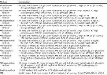
N6, MS and NB media were set as three treatments, and 100 seeds were pre-cultured in each. Rice tissue culturing was carried out following the method of Toki et al. (Reference Toki, Hara and Ono2006). Culture media and ingredients are described in Table 1.
Media used in the study

2,4-D, 2,4-dichlorophenoxyacetic acid; KT, kinetin; NAA, naphthaleneacetic acid.
Induction
Seeds were stripped of glumes, sterilized with 75% alcohol for 3 min, 0.1% HgCl2 for 8 min, and then grown on the induction medium for 6 days at 32°C.
Preparation of Agrobacterium
The Agrobacterium strain AGL-1 harboring the binary vector was inoculated into 10 ml of liquid YEB medium containing 50 mg/l kanamycin and 50 mg/l rifampicin, and cultured in a rotary shaker for 36 h at 180 rpm at 28°C. Then, the bacterium suspension was centrifuged at 5000 rpm for 3 min. The precipitate was suspended with liquid co-culture medium and cultured in a shaker for 2–3 h at 180 rpm at 28°C.
Infection and co-culture
Pre-cultured seeds were immersed in the Agrobacterium suspension by gently inverting the tube for 20 min, and then blotted dry with sterilized filter paper. The seeds were co-cultivated for 3 days at 24°C in the dark.
Selection
When co-cultivation was finished, calli were washed with sterilized water containing 500 mg/l cephamycin three times. After blotting with sterilized filter paper, calli were cultured on the selection medium for 15 days under continuous light at 32°C.
Regeneration
Calli were transferred onto the regeneration medium and cultivated for 25 days under a photoperiod of 14 h of light at 28°C and 10 h of darkness at 24°C. Then, plantlets were transferred onto the rooting medium. Rooted seedlings were transplanted to the greenhouse.
Molecular identification of transgenic plants
PCR detection
DNA was isolated from leaves of rice plants of the T0, T1 and T2 generations. The primer sequences were designed for 1190 bp of pemG1 and 723 bp of ubiquitin promoter. The primer sequences were as follows:
pemG1 forward (primer 1): ACCCTGTTGTTTGGTGTTACTT,
pemG1 reverse (primer 2): GGCGTCTCGCATATCTCAT;
ubi forward (primer 3): AGTCTAACGGACACCAAC,
ubi reverse (primer 4): AAAGATGACCCGACAAAC.
Northern blot analysis
Total RNA was extracted from leaves of polymerase chain reaction (PCR)-positive and untransformed plants with Trizol reagent (from Invitrogen Corp., Carlsbad, California, USA). The digoxygenin (DIG)-labelled pemG1 fragment was used as a probe in the Northern blot, following the method of Coca et al. (Reference Coca, Peñas and Gómez2006) and the user manual from the DIG High Prime DNA Labeling and Detection Starter Kit I.
Western blot analysis
Polyclonal antibodies against the PemG1 protein extracted from Magnaporthe grisea were produced in a rabbit. Rice leaves of the T2 generation were ground with liquid nitrogen and suspended in phosphoric acid buffer. Proteins were separated by 12% sodium dodecyl sulphate polyacrylamide gel electrophoresis (SDS-PAGE), and then transferred to a polyvinylidene fluoride (PVDF) membrane with Trans-blot SD cell (from Bio-Rad Laboratories, Hercules, California, USA) for immune blotting (Coca et al., Reference Coca, Peñas and Gómez2006).
Genetic analysis of transgenic progenies
Transgenic progenies were analysed for transgene heredity (Xi et al., Reference Xi, Hou, Zhang and Lu2002). Three transgenic lines were tested: 50 seeds from each line (T0) were stripped of glumes, sterilized with 75% alcohol for 3 min, 0.1% HgCl2 for 8 min, and then were grown on 1/2 MS medium containing 100 mg/l kanamycin for 15 days under a photoperiod of 14 h of light and 10 h of darkness at 28°C. PCR was also conducted for heredity analysis.
Results and analysis
Preparation of transgenic plants and performance of different media
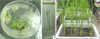
Calli emerged on the sixth day of induction, turned green on the eighth day of regeneration and regenerated kanamycin-resistant plantlets after 25 days (Fig. 2). After rooting, these plantlets were transplanted to the greenhouse for further growth.
Growth of transgenic rice plants: (A) the growth of resistant shoots; (B) the rooting of resistant plantlets; (C) resistant plantlets transplanted to soil. (See online for a colour version of this figure.)

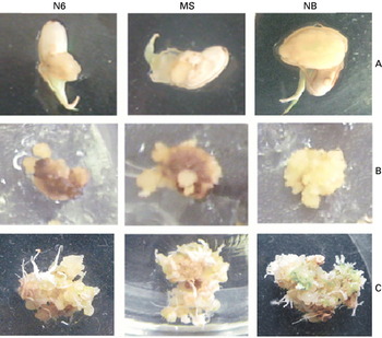
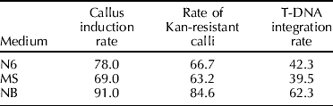
NB worked best among the three kinds of media. Calli induced by the NB medium had bigger bulk and tighter quality when compared to those induced by N6 and MS media (Fig. 3A). The callus induction rates of NB, N6 and MS media were 91%, 78% and 69%, respectively (Table 2).
Comparison of N6, MS and NB media. (A) Growth of calli at the sixth day of induction; (B) growth of calli at the tenth day of selection; (C) growth of calli at the eighth day of regeneration. (See online for a colour version of this figure.)

Comparison of different media
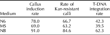
The growth patterns of calli on the three kinds of culture media were also different. Calli on the NB medium kept green constantly, but those on N6 and MS media turned brown gradually with ongoing cultivation. On the eighth day of regeneration, calli turned green on the NB medium, but those on N6 and MS media did not turn green until the tenth day. On the 25th day of regeneration, 77 calli on the NB medium regenerated plantlets, 52 on the N6 medium and 43 on the MS medium, respectively. So, more calli were induced and a higher percentage of calli were kanamycin resistant on the NB medium than on N6 and MS media (Table 2).
Molecular detection
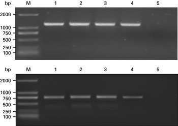
The two pairs of primers showed the correct amplification of the pemG1 gene or the promoter sequence in the T0, T1 and T2 plants, but not in the untransformed plants (Fig. 4), indicating the pemG1 integration into the rice genome and its stable heredity. Among the plantlets regenerated on NB, N6 and MS media, 44, 21 and 17 showed T-DNA integration, respectively. Plantlets regenerated on the NB medium had the highest T-DNA integration rate.
PCR analysis of transgenic rice plants. upper panel, PCR analysis with primer 1 and primer 2; lower panel, PCR analysis with primer 3 and primer 4. Lanes: M, DL2000 mark; 1–4, transgenic plants; 5, non-transgenic plant.

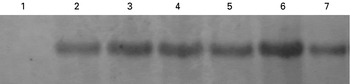
Hybridization with a pemG1-specific probe indicated that the accumulation level of pemG1 mRNA varied in the different transgenic lines. Accumulation of pemG1 mRNA was lacking in the non-transformed plants (Fig. 5).
Northern blot analysis of transgenic plants. Lanes: 1, non-transgenic rice plants; 2–7, transgenic rice plants.

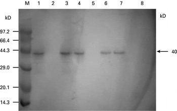
Most of the transgenic lines showed a positive immunological reaction where a 40 kDa band was detected, whereas non-transformed control plants and some transgenic lines showed no immunoblotting band (Fig. 6).
Western blot analysis of transgenic rice plants. Lanes: M, protein molecular weight marker; 1–7, transgenic rice plants; 8, non-transgenic rice plants.

Transgene heredity in progenies
The germination rates of the T1 seeds from the three transgenic lines were 74%, 78% and 72%, respectively, indicating that resistance to kanamycin separated in the T1 generation at a ratio of 3:1. The ratio of PCR-positive and PCR-negative plants was also 3:1, revealing that there was no transgene amplification on the part of the transgenic plants.
Discussion
At present, N6, MS and NB are the most frequently used basic media and scutellum is the most frequently used explant in Agrobacterium-mediated transformation of rice (Raineri et al., Reference Raineri, Bottino, Gordon and Nester1990; Chang, Reference Chang1992; Rashid et al., Reference Rashid, Yokoi, Toriyama and Hinata1996; Hiei et al., Reference Hiei, Komari and Kubo1997). The performance of three media was compared in this study, and the results revealed that the NB medium induced more calli, and regenerated more plantlets with higher T-DNA integration rate than the N6 and MS media. So, NB medium was most suitable for tissue culturing of Nipponbare scutellum.
Relatively speaking, N6 and MS are adopted on more occasions than the medium B5 in rice scutellum tissue culture. In fact, other media were also used besides N6 and MS. Mixed use of major elements and minor elements of different basic media has been reported. NB is a case in point, for it is in fact a mixture of major elements of N6 and minor elements of B5.
N6 medium was usually adopted for calli induction, but MS medium was suitable for regeneration. So, the induction medium, selection medium and regeneration medium of different basic media were often blended for use. Transgenic plants were obtained by using N6 induction medium and MS regeneration medium (Datta et al., Reference Datta, Vasquez and Tu1998); similar results were achieved by mixing MS with N6 basic medium (Pons et al., Reference Pons, Marfà, Melé and Messeguer2000).
A new kind of medium more suitable for rice scutellum culturing is very likely to be obtained by mixed use of components of different basic media. So, to establish a highly efficient transformation system, there are still many things to be done, such as the overcoming of genotypic restriction, improvement of induction, regeneration and transformation efficiency, and shortening of the culture period.
Transgenic technology has been important in working towards the control of rice blast disease, which is an important disease worldwide. Different genetic strategies have been used successfully to increase rice resistance against blast disease, such as the expression of chitinase, maize anthocyanin and antifungal protein (Nishizawa et al., Reference Nishizawa, Nishio and Nakazono1999; Gandikota et al., Reference Gandikota, Kochko, Chen, Ithal, Fauquet and Reddy2001; Coca et al., Reference Coca, Bortolotti and Rufat2004, Reference Coca, Peñas and Gómez2006; Moreno et al., Reference Moreno, Penas and Rufat2005), but there are few reports about the transgenic study of elicitor-encoding genes. Here, the protein elicitor gene pemG1 from Magnaporthe grisea was transferred into Nipponbare via the Agrobacterium-mediated method. The transgene exhibited stable heredity in transgenic progenies.
The protein elicitor has no germicidal action in vitro, but acts through triggering plant defence responses and inducing SAR (systemic acquired resistance). Thus it is likely to develop into a new method in disease and pest control and become an important field of research in the 21st century.
A series of protein elicitors from Magnaporthe grisea, Botrytis cineres, and Alternaria spp. have been isolated in this laboratory in recent years. These elicitors are heat stable, able to induce non-host defence responses and promote crop growth without the occurrence of hypersensitive responses (HR). The expression of elicitor-encoding genes may be another feasible approach for disease control. This strategy may serve as an important component of a multifaceted, ecologically based disease management programme.
Acknowledgments
This work was supported by grants from the Ministry of Science and Technology of China, project numbers 973 (2003CB114204) and 863 (2006AA10A210) and the Beijing Grand Project (D706005040431). We would like to thank Dr Rick Rochester and his organization, the International College of China Agricultural University, for proofreading the English manuscript of this paper.