Introduction
The co-occurrence of papillary and medullary thyroid carcinomas within the thyroid gland has been reported since the 1980s, but the cause remains unknown.Reference Machens and Dralle1 At present, there is only one reported case of simultaneous medullary thyroid carcinoma, papillary thyroid carcinoma and secondary hyperparathyroidism.Reference Behrend, von Wasielewski and Brabant2
We present the first case report of papillary and medullary thyroid carcinomas associated with primary hyperparathyroidism in the context of a negative test for RET (encoding a receptor tyrosine kinase) gene mutation.
Case report
A 59-year-old female was referred to the otolaryngology clinic with general malaise, nocturia and raised serum calcium (2.86 mmol/l; reference range 2.10–2.55) and parathyroid hormone levels (186 ng/l; reference range 10–65). She had a history of recently diagnosed myxoedema that was well controlled with thyroxine. Her clinical history was otherwise unremarkable and a physical examination was normal.
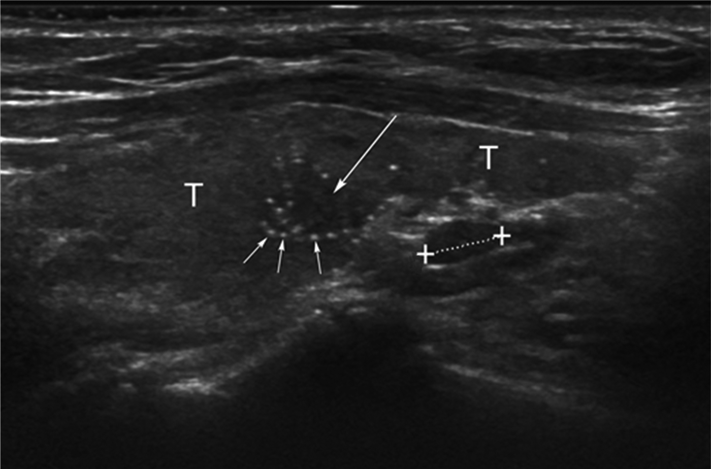
99 mTc-sestamibi scanning showed increased isotope uptake in the region of the right inferior parathyroid gland, suggesting a parathyroid adenoma. Ultrasonography of the neck revealed a single 10 mm nodule containing small calcifications within the right lobe and some scattered microcalcifications within both lobes of the thyroid gland (Figure 1). Fine needle aspiration of the nodule within the right lobe led to a classification of Thy4 (i.e. suspicious for malignancy), indicating a possible medullary carcinoma. Serum calcitonin was found to be elevated to 72 µg/l (reference, less than 4.8 µg/l). This prompted 24-hour urinary catecholamine and metanephrine testing, the results of which were normal. A subsequent normal [131I]metaiodobenzylguanidine radioisotope scan excluded the presence of a phaeochromocytoma.
A longitudinal sonogram through the right lobe of thyroid (T), showing an ill-defined hypoechoic lesion in the midpole (long arrow), which underwent fine needle aspiration. Note also the diffuse associated microcalcifications (small arrows). A further hypoechoic nodule is seen lying posterior and separate to the lower pole of the gland (dotted line), consistent with a parathyroid adenoma at this site

Further ultrasonography and staging computed tomography revealed indeterminate bilateral bulky nodes at level II, with smaller inferior nodes within the neck and superior mediastinal lymph nodes measuring up to 11 mm in the short axis. Mediastinal nodes were thought not to represent tumour spread. Histological analysis of an ultrasound-guided core biopsy of a left level II node showed it to be a reactive lymph node with no evidence of malignancy.
The case was discussed at the regional thyroid cancer multidisciplinary meeting, where surgery was recommended. The patient underwent total thyroidectomy, right inferior parathyroidectomy and bilateral level VI selective neck dissection.
Histological analysis confirmed a right inferior parathyroid adenoma (Figure 2).
Parathyroid adenoma within the inferior parathyroid gland. (H&E; ×40)
Evaluation of the thyroid tissue showed medullary thyroid carcinoma foci within the right lobe specimen, with the largest focus in one block measuring 7 mm in the largest dimension (Figure 3). Tumour cells showed positive staining with the immunohistochemical markers calcitonin, chromogranin A, synaptophysin, cytokeratin 19 (CK19), carcinoembryonic antigen and thyroid transcription factor (TTF1), but were negative for thyroglobulin. There was also papillary thyroid carcinoma within the right lobe, the left lobe and the isthmus measuring 2 mm, 1 mm and 4 mm respectively (Figure 4). Psammoma bodies were located in the smallest tumour focus within the left thyroid lobe. The background thyroid tissue showed benign thyroid follicles with lymphocytic thyroiditis.
Medullary carcinoma of the right thyroid lobe, measuring 7 mm in the largest dimension (a) (H&E; ×200), confirmed by (b) calcitonin immunohistochemistry highlighting the tumour islands of the same tumour. (×200)
A nidus of papillary carcinoma within the left thyroid lobe. (H&E; ×100)
Sixteen lymph nodes were removed in a right level VI neck dissection. Four of these nodes were infiltrated by medullary thyroid carcinoma: they showed positive staining with immunohistochemical markers TTF1 and CK19, but were negative for thyroglobulin. One node was infiltrated by papillary thyroid carcinoma, with tumour cells showing positive staining for thyroglobulin, CK19 and TTF1. A left level VI neck dissection removed eight nodes, all of which were free of malignancy.
The patient had transient post-operative left-sided vocal fold paresis that resolved within two months; she completed a course of radioactive iodine ablative therapy. No abnormalities were found on genetic testing of RET exons 5, 8, 10, 11 and 13–16.
The patient remained well 15 months later. Serum calcitonin and thyroglobulin levels continue to be monitored and have recently both been recorded as well within their normal reference ranges.
Discussion
Incidental thyroid disease has been discovered in 57 per cent of patients with primary hyperparathyroidism, and 9.7 per cent of those cases were malignant.Reference Onkendi, Richards, Thompson, Farley, Peller and Grant3, Reference Arciero, Shiue, Gates, Peoples, Dackiw and Tufano4 The incidental discovery of two thyroid malignancies in our patient reinforces the importance of a thorough radiological investigation for patients presenting with primary hyperparathyroidism.
Ten per cent of cases of parathyroid adenoma are hereditary, usually the result of multiple endocrine neoplasia syndromes.Reference Pallan, Rahman and Khan5 The coexistence of medullary thyroid carcinoma with primary hyperparathyroidism more specifically suggests multiple endocrine neoplasia type two syndrome. In 1998, a case of simultaneous papillary thyroid carcinoma and parathyroid adenoma with a raised calcitonin level was reported. However, the cause of the raised calcitonin was not found, and there was no evidence of medullary thyroid carcinoma. Furthermore, this case tested positive for a mutation in the RET gene, suggesting a variant of multiple endocrine neoplasia syndrome.Reference Rone, Lane and Grinkemeyer6 Our patient had a histologically confirmed diagnosis of medullary thyroid carcinoma, papillary thyroid carcinoma and parathyroid adenoma, and tested negative for all RET genetic mutations currently known to be associated with multiple endocrine neoplasia and familial medullary thyroid cancer syndromes. Phaeochromocytoma was effectively excluded through negative [131I]metaiodobenzylguanidine scanning and 2 rounds of normal 24-hour urinary catecholamine and metanephrine tests. RET mutations are the only recognised genetic associations with multiple endocrine neoplasia and familial medullary thyroid cancer that follow a classic familial inheritance pattern. RET mutations are also found in 7–8 per cent of sporadic medullary thyroid carcinoma cases. It should be noted that new mutation loci within the RET gene continue to be discovered and mapped to cases of both hereditary and sporadic cases of medullary thyroid carcinoma.Reference Elisei, Romei, Cosci, Agate, Bottici and Molinaro7, Reference Romei, Mariotti, Fugazzola, Taccaliti, Pacini and Opocher8 Since our patient's genetic testing involved only those loci known to date, it is possible that a genetic component, perhaps even a ‘RET-negative’ variant of a multiple endocrine neoplasia syndrome, was not identified in this case.
Non-medullary thyroid cancer occurs in approximately 3 per cent of primary hyperparathyroidism cases.Reference Leitha and Staudenherz9 Hypercalcaemia is postulated to have a role in inducing thyroid neoplasia via an initial stage of tissue hyperplasia.Reference Triggs and Williams10, Reference LiVolsi and Feind11 The coexistence of papillary and medullary thyroid carcinomas is also well documented.Reference Machens and Dralle1, Reference Kim, Gong, Kim, Kim, Hong and Kim12–Reference Rossi, Lugazzola, De Pasquale, Braidotti, Cirello and Beck-Peccoz14 Papillary thyroid carcinoma is reported to occur in up to 19 per cent of cases of medullary thyroid carcinoma.Reference Machens and Dralle1, Reference Kim, Gong, Kim, Kim, Hong and Kim12, Reference Biscolla, Ugolini, Sculli, Bottici, Castagna and Romei13 Papillary microcarcinomas are common incidental findings at autopsy, and occult tumours of less than 1 cm in diameter are found in up to 35 per cent of post-mortem examinations.Reference Lang, Borrusch and Bauer15 It is therefore reasonable to suggest that papillary thyroid carcinoma occurs in the presence of medullary thyroid carcinoma through coincidence, as previously suggested.Reference Kim, Gong, Kim, Kim, Hong and Kim12–Reference Rossi, Lugazzola, De Pasquale, Braidotti, Cirello and Beck-Peccoz14
• Thyroid carcinoma can occur synchronously with parathyroid adenoma
• Multiple concurrent thyroid carcinomas may be present
• A thorough radiological investigation is required in all cases of primary hyperparathyroidism to avoid missing concurrent thyroid carcinomas
In the case of simultaneous medullary and papillary thyroid carcinoma reported in 2002, the patient had previously undergone surgery for secondary hyperparathyroidism.Reference Behrend, von Wasielewski and Brabant2 This patient had a history of renal transplant surgery and chronic hepatitis C, both of which were highlighted as possible explanations for the development of thyroid pathology. Our patient had a history of recently diagnosed hypothyroidism that was well controlled; however, a histological examination revealed evidence of background lymphocytic thyroiditis. Background chronic lymphocytic thyroiditis is associated with approximately 30 per cent of all papillary thyroid carcinoma cases, but the cause of this association is unclear.Reference Jeong, Kim, Lee, Park, Park and Kang16, Reference Kebebew, Treseler, Ituarte and Clark17 Isolated hypothyroidism has been shown to induce hyperplasia of follicular and parafollicular cells in experimental animals,Reference Galera-Davison, Fernandez, Salguera, Martin-Lacave and Gonzalez-Campora18 but there is no definitive evidence that this occurs in humans. Increased serum thyroid-stimulating hormone (TSH) levels are associated with a higher incidence of well-differentiated thyroid cancerReference Boelaert19; however, the question of whether raised serum TSH has a causal role in thyroid carcinogenesis is undetermined. Prior to her first referral to the otolaryngology clinic, our patient had recently been diagnosed with hypothyroidism. On review of her biochemical records, the highest serum TSH level measured was 46.95 mU/l, together with a thyroxine (T4) level of 3 pmol/l, 1 year before her first assessment. According to her records, both serum TSH and T4 levels had returned to the normal range within two months of starting treatment with levothyroxine, and had remained well controlled. From the available literature, it is unclear whether this played a role in the tumourigenesis of either of the two types of cancer diagnosed in our patient.Reference Boelaert19 Furthermore, this would not explain their coexistence with parathyroid adenoma.
Conclusion
The simultaneous occurrence of papillary and medullary thyroid carcinomas with concurrent parathyroid adenoma is presented in this case report. This could represent a variant of multiple endocrine neoplasia syndrome type two, which is not associated with a genetic mutation in RET. Overall, there is no single explanation for the co-occurrence of these three distinct pathologies. We reiterate the importance of a thorough radiological investigation for all cases of primary hyperparathyroidism to identify coexisting thyroid disease.