Introduction
In this work, the occurrence, crystal structure and properties of belomarinaite, a new mineral from Tolbachik volcano, are reported.
In the system Na2SO4–K2SO4, eight phases have been described: K3Na(SO4)2 (P  $ {\bar 3} $m1), KNaSO4 (P3m1), α-K2SO4 (P63/mmc), β-K2SO4 (Pmcn), Na2SO4 (Fddd), Na2SO4 (Cmcm), Na2SO4 (P63/mmc) and Na2SO4 (Pbnm). But only five of them have been discovered as minerals: aphthitalite (K3Na(SO4)2), thénardite (Na2SO4), metathénardite (Na2SO4), arcanite (K2SO4), and now belomarinaite (KNaSO4). Moreover, metathénardite and belomarinaite were only discovered in the last two years. The crystal chemistry of aphthitalite and related structures has been reviewed by Moore (Reference Moore1973, Reference Moore1976, Reference Moore1981), Eysel (Reference Eysel1973), Egorov-Tismenko et al. (Reference Egorov-Tismenko, Sokolova, Smirnova and Yamnova1984) and Hawthorne et al. (Reference Hawthorne, Krivovichev, Burns, Alpers, Jambor and Nordstrom2000).
Belomarinaite was discovered on the Toludskoe lava field, formed during the Tolbachik Fissure eruption in 2012–2013 (55°45'1.32”N, 160°19'40.74”E, altitude 1480 m). The specimen was found in the skylight of a lava tube that emanated volcanic gases with temperatures up to 1024°C (Fig. 1). According to our unpublished high-temperature X-ray diffraction data, belomarinaite exists in the temperature range 20–460°C.
(a) Photo of the lava fields of the 2012–2013 Tolbachik eruption showing the source location for belomarinaite (Toludskoye lava field) (Belousov et al. Reference Belousov, Belousova, Edwards, Volynets and Melnikov2015, with modifications by A. B. Belousov). (b) The skylight of a lava tube where the belomarinaite mineral was found (in the greenish-yellow deposit on the roof of the tube). Photo by M. G. Belousova.

Belomarinaite has been approved by the Commission on New Minerals, Nomenclature and Classification of the International Mineralogical Association (IMA2017-69a, Filatov et al., Reference Filatov, Shablinskii, Vergasova, Saprikina, Bubnova, Moskaleva and Belousov2018). The mineral was named in honour of Russian volcanologist Marina Gennadievna Belousova (b. 1960) (Institute of Volcanology and Seismology, Russian Academy of Sciences, Petropavlovsk–Kamchatskiy, Russia) for her significant contributions to the monitoring of the Tolbachik Fissure eruption. The type material is deposited in the Mineralogical Museum of Saint Petersburg State University (catalogue number 1/19678).
Experimental methods and results
General appearance and physical properties
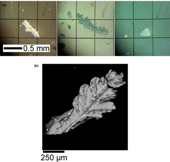
Belomarinaite occurs as arborescent aggregates of tabular crystals up to 1 mm × 0.3 mm × 0.1 mm (Fig. 2). The average linear size of the aggregates is 0.5–0.7 mm, the colour is pale blue to green, and the streak is white. It has a vitreous lustre. No cleavage is observed in the arborescent aggregates. Fracture is irregular to uneven. Parting is not observed. The Mohs’ hardness is 2–3. The calculated density based on empirical formula and single-crystal unit cell parameters is 2.687 g/cm3.
(a) Optical images of belomarinaite. (b) Electron microscope image of an arborescent aggregate of belomarinaite.

The mineral is optically uniaxial (+), ω = 1.485(3) and ε = 1.488(3) (λ = 589 nm). No dispersion is observed. It does not exhibit pleochroism. The Gladstone–Dale compatibility index (Mandarino, Reference Mandarino1981) based on empirical formula and unit-cell parameters from powder X-ray diffraction data is calculated as 1 – (K P/K C) = 0.007 (superior).
Chemical composition
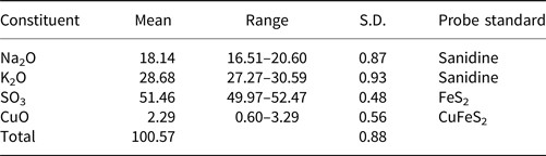
Fifty-eight analyses were carried out using a TESCAN ‘Vega3’ electron microprobe equipped with an Oxford Instruments X-Max 50 silicon drift energy-dispersive spectroscopy system, operated at 20 kV and 0.730 nA, with a beam size of 0.22 μm. Analytical results are given in Table 1. Processing was performed using AZtec software and an X-MAX-80 mm2 detector (Institute of Volcanology and Seismology, Petropavlovsk–Kamchatsky, Russia). The empirical formula based on 4 O atoms per formula unit (apfu) is (K0.95Na0.92Cu0.04)Σ1.91S1.01O4. The ideal formula is KNaSO4, which requires Na2O 19.60, K2O 29.78 and SO3 50.62 wt.%.
Chemical composition of belomarinaite.

S.D. – Standard deviation
Powder X-ray diffraction data
Powder X-ray diffraction data were collected using a Rigaku MiniFlex II diffractometer (CuKα radiation; R = 150 mm). According to this data, belomarinaite is trigonal, with the space group P3m1 and the unit-cell parameters a = 5.607(1), c = 7.178(1) Å, V = 195.25(6) Å3 and Z = 2.
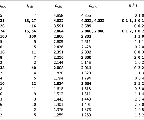
The experimental and calculated powder-diffraction patterns are given in Table 2. The strongest lines are [d, Å (I, %) (hkl)]: 4.022(31)(101); 3.591(26)(002); 2.884(74)(102); 2.800(100)(110); 2.391(16)(003); 2.296(8)201; 2.008(38)(022); and 1.634(10)(212).
Powder X-ray diffraction data for belomarinaite.

The strongest lines are given in bold
Single-crystal X-ray diffraction data
Single-crystal X-ray diffraction data were collected using a Bruker Kappa APEX DUO diffractometer equipped with an image plate using MoKα radiation. A hemisphere of three-dimensional data was collected using a frame width of 0.5° in ω, with 50 s used to acquire each frame. The data were corrected for Lorentz, polarisation and background effects using Bruker programs APEX and XPREP. A semi-empirical absorption-correction based on the intensities of equivalent reflections was applied in the SADABS program.
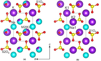
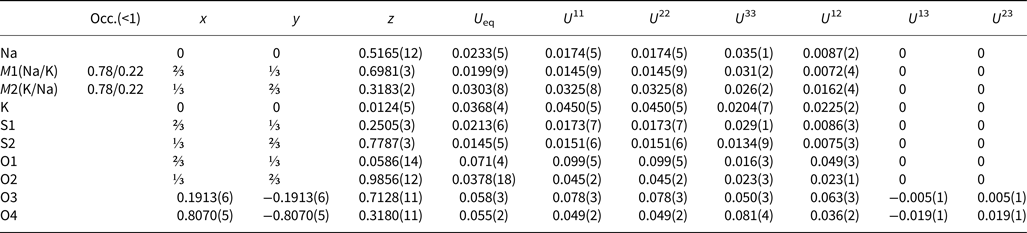
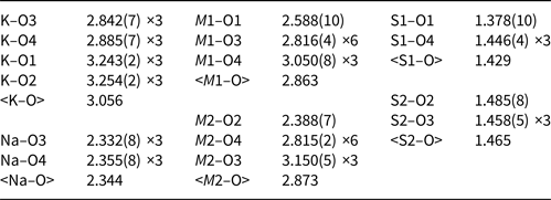
The crystal structure of belomarinaite was solved by charge flipping and refined on the basis of 713 unique observed reflections using the Jana 2006 program suite (Petříček et al., Reference Petříček, Dusek and Palatinus2006) (Table 3). Atomic coordinates, atomic anisotropic displacement parameters and selected bond distances are given in Tables 4, 5. The crystallographic information file has been deposited with the Principal Editor of Mineralogical Magazine and is available as Supplementary material (see below). The crystal structure of belomarinaite and coordination polyhedra of alkali cations are given in Figs 3, 4.
Crystal structure of: (a) belomarinaite; and (b) aphthitalite.

The linkage of the inherited polyhedra: (a) BaO9 and NaO9 polyhedra in the BaNaMe(BO3)2 (Me = Sc or Y) crystal structures (Volkov et al., Reference Volkov, Filatov, Bubnova, Ugolkov, Svetlyakova and Kokh2012); and (b) Na2O10 and K2O10 polyhedra in the belomarinaite crystal structure (this work). Solid lines denote normal Na polyhedra.

Crystal data, data collection information and structure refinement details for belomarinaite.

Structural occupancy factors, atomic coordinates, and equivalent isotropic and anisotropic displacement parameters (Å2) for belomarinaite.

Selected interatomic distances (Å) in the structure of belomarinaite.

Discussion
Crystal structure of belomarinaite
Belomarinaite is isostructural with the synthetic compound KNaSO4 [space group P3m1, a = 5.6066(7), c = 7.177(1) Å, Z = 2 and R 1 = 4.2%] (Okada and Ossaka, Reference Okada and Ossaka1980). In the crystal structure of belomarinaite, there are six cation sites: [4]S1 and [4]S2 sites occupied by S; [6]Na and [12]K sites; and the [10]M1 and [10]M2 sites. The S1–O and S2–O distances are 1.378–1.446 and 1.458–1.485 Å, respectively. The Baur (Reference Baur1974) distortion index in belomarinaite is 0.018 for S1O4 and 0.007 for S2O4, thus distortions of the SO4 tetrahedra are relatively high. The K and Na sites are occupied by one alkali atom, K and Na, respectively, with a standard deviation of ± 0.02–0.03. The Na atom is octahedrally coordinated by six O atoms with Na–O = 2.332–2.355 Å (Table 5). The K–O distances vary from 2.842 to 3.254 Å (average 3.06 Å). The M2–O distances vary from 2.388 to 3.150 Å (average distance is 2.87 Å) and the M1–O distances vary from 2.588 to 3.050 Å (average distance is 2.86 Å). The crystal structure refinement performed in the current work let us conclude that the M1 site is occupied 78% by Na and 22% by K atoms, Na0.78K0.22, i.e. Na0.39K0.11 apfu and the M2 site is occupied 78% by K and 22% by Na atoms, K0.78Na0.22, i.e. K0.39Na0.11 apfu (Table 4). The M2 and M1 polyhedra share common faces of 2.4 × 3.4 Å2. The size of these faces is suitable for atomic migration and for K–Na order–disorder processes as well (Fig. 4). The crystal structure is a framework of SO4 tetrahedra, Na octahedra and K, M1 and M2 polyhedra. The apices of neighbouring SO4 tetrahedra point in opposite directions along the c axis.
Okada and Ossaka (Reference Okada and Ossaka1980) showed that the K and Na atoms are ordered at the M2 and M1 sites. We conclude that: (1) belomarinaite is isostructural with its synthetic analogue KNaSO4; and (2) in belomarinaite, Na and K are disordered over M1 and M2 sites; in its synthetic analogue, Na and K are ordered over M1 and M2 sites, respectively.
Belomarinaite is also related structurally to aphthitalite, möhnite, bubnovaite, hanksite and metathénardite (Table 6).
Crystallographic data for belomarinaite and related minerals (based on Gorelova et al. Reference Gorelova, Vergasova, Krivovichev, Avdontseva, Moskaleva, Karpov and Filatov2016).

The inheritance of the cation polyhedra during the cooling of the high-temperature solid solutions
The bond lengths in the Na-dominant [10]M1 polyhedron (<M1–O> = 2.83 Å) are too large for Na compared to the mean value of 2.34 Å (International Tables for Crystallography, 2006). To understand the nature of this phenomenon, let us consider similar examples in BaNaMe(BO3)2 (Me = Sc or Y) borates described by Volkov et al. (Reference Volkov, Filatov, Bubnova, Ugolkov, Svetlyakova and Kokh2012). According to the data (International Tables for Crystallography, 2006), the mean <Ba–O> and <Na–O> bond lengths are 2.738 and 2.336 Å, respectively. At high temperatures, these cations (Ba2+ and Na+) are disordered over the same cation sites. Further, upon the cooling, the Ba and Na atoms become ordered at 775°C (Sc) and 375°C (Y), respectively. Moreover, cation ordering leads to a slight rearrangement of the BaO9 (<Ba–O> = 2.796 Å (Sc) and 2.822 Å (Y)) and NaO9 (<Na–O> = 2.873 Å (Sc) and 2.867 Å (Y)) coordination polyhedra (Fig. 4). In the NaO6 polyhedra (Fig. 4, solid lines), the bonds are also large, 2.642 and 2.714 Å. The BaO9 and NaO9 polyhedra are linked between each other by shared faces of 3.4 × 3.4 Å2.
These examples can be used to the investigation of the mineral inheritance occurred in the belomarinaite crystal structure upon cooling. The mineral was found in the skylight of a lava tube at ~1000°С. High-temperature polymorphs of K2SO4, K3Na(SO4)2, KNaSO4 and Na2SO4 are isotypic and crystallise in the P63/mmc space group (Fischmeister, Reference Fischmeister1962). At room temperature, K3Na(SO4)2 and KNaSO4 are structurally similar to their high-temperature polymorphs (Gorelova et al., Reference Gorelova, Vergasova, Krivovichev, Avdontseva, Moskaleva, Karpov and Filatov2016). Also in the crystal structure of room- and high-temperature polymorphs the K and Na coordination polyhedra are similar. In the crystal structure of belomarinaite the K atoms form regular coordination polyhedra with mean bond lengths equal to 2.87 and 3.06 Å, while the Na atoms form one regular coordination polyhedron with bond lengths equal to 2.332–2.355 Å and one unusual, M1O10, in which M–O chemical bonds are much longer (2.588–3.050 Å). The K and Na atoms are ordered over two [10]-coordinated M1 and M2 sites upon cooling due to its difference in ionic radii. One can suggest that the longer Na–O bonds of the unusual NaO10 polyhedron are inherited from the high-temperature polymorph KNaSO4 (P63/mmc).
Conclusions
As a result of the investigation of the chemical composition, crystal structure and physical properties of samples from Tolbachik volcano (Kamchatka Peninsula), a new mineral belomarinaite, ideally KNaSO4, was discovered. The consideration of crystal structure lets one conclude that belomarinaite is an example of a phase, which forms in accordance with Ostwald's rule (Ostwald, Reference Ostwald1897; Krivovichev et al., Reference Krivovichev, Hawthorne and Williams2017). As a result of high-temperature exhalation processes, structural fragments of phases crystallised first are structurally similar to the atomic and molecular structure of volcanic gases, which produce these phases.
The K and Na atoms are ordered in the crystal structure of belomarinaite. These atoms are disordered over two sites in the crystal structure of high-temperature KNaSO4 which crystallises in the P63/mmc space group (Fischmeister, Reference Fischmeister1962). The differences between the atoms ionic radii increase with the decrease in temperature. According to this, the M1O10 coordination polyhedron with the very long Na–O bonds in the crystal structure of belomarinaite are inherited from the high-temperature polymorph of KNaSO4. At the early stages of the cooling process, the solid solutions, which contain the small cations in its chemical composition, can be crystallised as the phases that contain inherited polyhedra with relatively long bond lengths.
Acknowledgements
This work was financially supported by the Russian Found of Basic Research, grant no. 18-29-12106. Technical support by the SPbSU X-ray Diffraction Centre is gratefully acknowledged.
Supplementary material
To view supplementary material for this article, please visit https://doi.org/10.1180/mgm.2018.170.