Tetralogy of Fallot is the most common form of congenital cyanotic heart disease. It is characterised by right ventricular outflow obstruction, a ventricular septal defect, right ventricular hypertrophy, and an over-riding aorta. After repair, long-term survival is reported to be excellent but still lower than in the normal population,Reference Murphy, Gersh and Mair 1 with morbidity and mortality increasing with age, patients presenting with symptomatic right ventricular failure or arrhythmias, and the risk of sudden cardiac death. Pulmonary regurgitation is the most common post-repair lesion and has been associated with enlargement of the right ventricle, exercise intolerance, arrhythmia, and sudden cardiac death.Reference Vliegen, van Straten and de Roos 2 – Reference Fox, Devendra, Hart and Krasuski 4 Pulmonary valve replacement may lead to improved right ventricular volumes and function, improved functional class, and a reduction in arrhythmias.Reference Eyskens, Reybrouck and Bogaert 5 – Reference Discigil, Dearani and Puga 9
Former studies suggest that fibrosis could be the cause of right ventricular dysfunction and arrhythmias,Reference Babu-Narayan, Kilner and Li 10 , Reference Deanfield, Ho, Anderson, McKenna, Allwork and Hallidie-Smith 11 and myocardial fibrosis has been demonstrated in patients after repair and in animal models of tetralogy of Fallot.Reference Jones and Ferrans 12 , Reference Garson 13
Delayed enhancement cardiac MRI has been established as a reliable tool to visualise myocardial necrosis and scar in clinical practice with modern phase-sensitive inversion recovery sequences providing consistently high-quality images for both ventricles.Reference Plaisier, Burgmans and Vonken 14
The aim of this study was to reliably detect or exclude myocardial scars of the right and left ventricles after complete repair of tetralogy of Fallot using phase-sensitive inversion recovery delayed enhancement imaging. We hypothesise that myocardial scars are common in these patients and are related to an impaired cardiac function and adverse clinical outcome.
Materials and methods
Patient population and study design
A total of 53 consecutive patients (24 female and 29 male) were retrospectively enrolled after total correction of tetralogy of Fallot from October 2007 to December 2009. All patients were seen for regular outpatient follow-up visits at our tertiary care institution and were routinely referred for clinically indicated cardiac MRI. Informed consent was obtained for the MRI examinations and for scientific use of the anonymised data. This retrospective study was performed according to the guidelines of the ethics committee and complies with the Declaration of Helsinki. Exclusion criteria were standard magnetic resonance contraindications, such as implanted defibrillators, pacemakers, and ferromagnetic intracranial metallic implants.
The NYHA functional class was determined by patient interrogation. A 12-channel electrocardiogram was performed on the same day as the cardiac magnetic resonance examination and the width of the QRS complex was measured. Cardiopulmonary exercise testing was performed within 2 days of the cardiac magnetic resonance examination, and maximum VO2, peak exercise heart rate, and duration of exercise were recorded. Clinically relevant arrhythmias associated with symptoms or syncope were obtained from patient records and Holter electrocardiogram.
Cardiac MRI
All examinations were performed on a 1.5 T scanner (Intera CV, Philips Medical Systems, Best, The Netherlands) with the patients in the supine position, using a dedicated five-channel phased-array surface cardiac coil. Black-blood images in axial orientations were acquired to visualise morphology. Standard breath-hold cine steady-state free precession sequences in axial, short-axis, and four-chamber views were acquired for volumetric and functional imaging. Cine short-axis imaging was determined from the four-chamber view parallel to the atrioventricular valve covering the whole heart, gapless from the apex to the base, with slice thickness of 8 mm. The echo time was 1.8 ms, and the repetition time was 3.6 ms with a flip angle of 50°. Typical in-plane resolution was 1.8×1.5 mm. In all, 30 phases per heart cycle were acquired.
To determine pulmonary regurgitation fraction, two-dimensional phase-contrast flow measurements perpendicular to the main pulmonary artery were recorded using a free breathing sequence with a standard encoding velocity of 150 cm/second. In case of phase wrapping, the velocity encoding was adjusted. The echo time was 6.5 ms, repetition time was 15 ms, flip angle was 30°, slice thickness was 6 mm, field of view was 300 mm, and matrix was 256, for one acquisition with 30 reconstructed phases. Pulmonary insufficiency was graded as follows: grade 1, <20% regurgitation fraction; grade 2, 20–40%; grade 3, 40–60%; and grade 4, >60%.
Delayed enhancement images, also covering both ventricles gapless in short-axis and four-chamber views, were acquired for visualising the myocardial scar 10–15 minutes after application of 0.2 mmol per kg bodyweight gadobutrol (Gadovist, Bayer HealthCare Pharmaceuticals, Berlin, Germany) using a multi-breath-hold two-dimension T1-weighted phase-sensitive inversion recovery turbo gradient echo sequence with a standard inversion time of 270 ms and slice thickness of 10 mm. The repetition time was 4.7 ms, and the echo time was 2.3 ms with a flip angle of 15°. Typical in-plane resolution was 1.88×2.5 mm.
Cardiac magnetic resonance image analysis
Image analysis was independently performed offline by two experienced observers blinded to the patient status (U.P. and M.G.), using a Philips View Forum workstation (Version 4.2;Cardiac Evaluation Package). Our cardiac magnetic resonance laboratory has expertise in delayed enhancement imaging of the right ventricle.Reference Preim, Hoffmann and Lehmkuhl 15 , Reference Grothoff, Elpert and Hoffmann 16 Reproducibility of scar tissue after correction of tetralogy of Fallot has been shown before.Reference Babu-Narayan, Kilner and Li 10
Biventricular endocardial and epicardial borders were manually traced in end systole and end diastole in each slice. Right ventricular trabeculations were excluded from the myocardial mass, as this has shown better reproducibility in systemic right ventricles compared with their inclusion.Reference Winter, Bernink and Groenink 17 End-diastolic and systolic volumes and ejection fraction were calculated for both ventricles. The volumes were normalised to body surface area.
The specific density of 1.05 g/ml was used to calculate myocardial mass.
The pulmonary regurgitation fraction was obtained from two-dimensional phase-contrast flow measurements.
In phase sensitive inversion recovery delayed enhancement imaging, myocardial enhancement was considered present if the signal intensity of the hyperenhanced myocardium was more than five standard deviations above the mean signal intensity of the remote myocardium.Reference Bondarenko, Beek and Hofman 18 To determine the extent of scar tissue, we performed the volumetry manually using a Philips View Forum workstation. To describe the location of delayed enhancement, standardised myocardial segmentation and nomenclature for tomographic imaging of the heart published by the American Heart Association was used.Reference Cerqueira, Weissman and Dilsizian 19
Statistics
Categorical variables were expressed as number and percentage. According to their distribution, continuous variables were presented as mean±standard deviation or as median and range, or both, for comparability with previous studies. Normality of the data was assessed by the Kolmogorov–Smirnov test. Continuous variables were analysed by either a two-sample independent t-test or the Mann–Whitney test. Fisher’s exact test was used to assess group differences for categorical variables. A probability value <0.05 was considered statistically significant. SPSS version 19 was used for data analysis.
Results
Patient characteristics
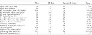
A summary of patient characteristics is shown in Table 1.
Patient characteristics.

LV=left ventricle; RV=right ventricle
Over a period of 26 months (October, 2007 to December, 2009) we studied 53 patients (24 female and 29 male) after total correction of tetralogy of Fallot. The mean patient age was 20 years (median 20, range 2–48). The oldest patient in the study was born in 1961, and the youngest in 2007. The earliest date of repair surgery was 1968 (one patient). Three patients underwent repair surgery in the 1970s. The other patients underwent corrective surgery in later years. The interval between repair and the MR examination ranged from 2 to 42 years (median 14 years).
Most patients (83%) presented with a good self-reported ability (NYHA class grade I). Six of the 53 patients (11%) were classified as NYHA class 2, two (4%) as class 3, and one patient (2%) as class 4. Exercise testing data were available on 21/53 of the patients (Table 1). All but two patients had pulmonary insufficiency: 15 patients (28%) with grade 1, 27 (51%) with grade 2, eight (15%) with grade 3, and one (2%) patient with grade 4.
Surgical techniques
Of the 53 patients, eight (15%) underwent palliative surgery before definitive repair, 15 (28%) underwent repeat surgery, which included early and late pulmonary valve replacement, and 10 (19%) underwent repeat transcatheter intervention for pulmonary valve replacement. A Blalock–Taussig shunt or Waterston–Cooley anastomosis was used in cyanotic symptomatic patients before 6 months of age. Outflow tract repair was carried out as muscular resection and pulmonary valvotomy (n=2), distal to the pulmonary valve patch (n=1) or a transannular patch (n=25). In 25 patients, other surgical techniques were applied or the technique was unknown.
Delayed enhancement
Diagnostic image quality was achieved in all patients. Enhancement at the anterior or inferior insertion point was detected in 35 patients (66%).
Adhesion of contrast agent to prosthetic patches was seen in eight of the cases (15%). Two of eight patients with patch enhancement at the right ventricular outflow tract had clinically relevant arrhythmias (p=0.6).
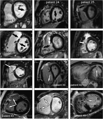
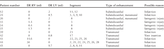
Excluding enhancement of the septal insertion and prosthetic patches, delayed enhancement was seen in 11/53 cases (21%): in six cases (11%) in the right ventricle and in five in the left ventricle (9%). Small scars (⩽4 ml) were seen in seven cases (13%) and extensive scars (⩾9 ml) in 4/53 patients (8%). The distribution of the affected segments and the extent of delayed enhancement is shown in Table 2. The most often affected area was segment 8 (36%, 4/11 cases). Images of patients with delayed enhancement are shown in Figure 1.
Images of patients with a positive delayed enhancement imaging. Possible reasons for scarring are myocardial infarction in patients 12, 24, 39, 43, 44, and 45 and iatrogenic injury in patients 26, 27, 34, and 35. In patient 42 a scar is seen in the left ventricular apex consistent with vent insertion during surgery. In patient 25, adhesion of contrast agent to the patch is seen, which was not interpreted as positive delayed enhancement.

Distribution of delayed enhancement.
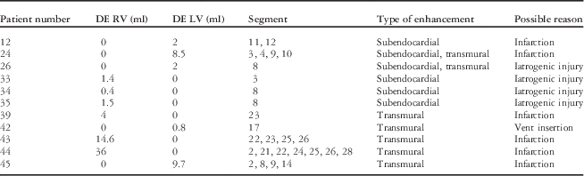
DE=delayed enhancement; LV=left ventricle; RV=right ventricle
Relationship between delayed enhancement and clinical data
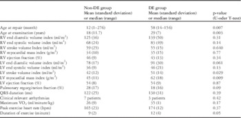
The patient group with delayed enhancement was significantly older at the time of the examination (p=0.003), had a later repair (p=0.007), a higher left ventricular stroke volume index (p=0.029), and a higher left ventricular myocardial mass index (p=0.009).
Right ventricular volumes and function, pulmonary regurgitation fraction, QRS width, clinically relevant arrhythmias, NYHA class, and parameters of cardiopulmonary exercise tolerance showed no significant differences between the groups. Table 3 details the mean, median, standard deviation, and p-values of volumetric and clinical parameters for both groups.
Comparison between patients with and those without delayed enhancement.

DE=delayed enhancement; LV=left ventricle; RV=right ventricle
Discussion
Study aims
The aim of this study was to precisely detect myocardial scarring in patients with repaired tetralogy of Fallot, and to test the hypothesis that myocardial scars are related to an impaired cardiac function and adverse clinical outcome.
We used a state-of-the-art two-dimensional phase-sensitive inversion recovery delayed enhancement sequence that is less dependent on the choice of the correct inversion time to precisely null the intact myocardium compared with delayed enhancement imaging with magnitude detection.Reference Huber, Schoenberg and Hayes 20 With phase-sensitive reconstruction, it is possible to use a nominal value of TI and achieve consistent contrast and good image quality. Phase-sensitive inversion recovery imaging avoids the loss of contrast by restoring signal polarity. This results in consistent image quality without polarity artefacts, a better reproducibility of infarct size, and a reduction in background noise.Reference Plaisier, Burgmans and Vonken 14 , Reference Kellman, Arai, McVeigh and Aletras 21 Using this sequence we have had good experience in detecting necrosis and scar tissue in post-ischaemic, post-inflammatory, and CHD.Reference Preim, Hoffmann and Lehmkuhl 15 , Reference Grothoff, Elpert and Hoffmann 16
Corrected tetralogy of Fallot and scar tissue
Our study indicates that scarring is a frequent sequela in patients with corrected tetralogy of Fallot (21%). Small scars due to iatrogenic injury and small subendocardial infarctions, however, are more frequent (13%) compared with extensive areas of infarction (8%). Patients with myocardial scars were mostly older or had undergone a later repair. The reason might be a more refined surgical technique in recent years. In addition, with early age at surgery, preoperative ischaemia and damage from pressure overload can be avoided. Other authors also found that the trend towards early primary repair may reduce the risk for endomyocardial fibrosis and that an older age at surgery is a powerful predictor of poorer late survival.Reference Murphy, Gersh and Mair 1 , Reference Gatzoulis, Clark, Cullen, Newman and Redington 22
Delayed enhancement has previously been shown to be a predictor of adverse clinical outcome. We found considerable differences between a previous study by Babu-Narayan and our patient cohort. Babu-Narayan et alReference Babu-Narayan, Kilner and Li 10 used a two-dimensional segmented fast low-angle shot inversion recovery sequence with imaging performed 5 minutes after an intravenous injection of gadolinium-DTPA (0.1 mmol/kg). The inversion time was adjusted to maintain nulling of healthy myocardium, typically increasing from 310 to 420 ms. Despite our modern imaging method, we found fewer scars compared with the study by Babu-Narayan,Reference Babu-Narayan, Kilner and Li 10 which evaluated scars in 92 patients with tetralogy of Fallot. We found scars in the right ventricle in 11% compared with 24% and scars in the left ventricle in 9% compared with 62% in the cited study. In our study, eight patients (15%) showed adhesion of contrast agent to the right ventricular outflow patch compared with 91 of 92 patients (99%) in the study by Babu-Narayan. Adhesion of contrast agent to patches was not interpreted as scar tissue in our study. A possible explanation for more scarring in the former study is the fact that the patient cohort of Babu-Narayan represents an earlier era of surgery with more aggressive reconstruction techniques. Their patient cohort was also older at the time of examination (mean age 20 versus 32 years) and had undergone later repairs (2 versus 5 years). The incidence of prior palliative surgery (15 versus 39%) and of a left ventricular apical vent (9 versus 53%) was higher in the study by Babu-Narayan. We therefore conclude that our patient cohort is more contemporary.
In our study, scars in the right ventricle were probably caused by iatrogenic injury in four and myocardial infarction in three patients. Scars in the left ventricle were probably caused by vent insertion (one patient) and by infarction (three patients). Left ventricular scars can result from perioperative embolic infarction, volume overload from arterial shunts in palliated patients, or preoperative relative hypoxia.Reference Davlouros, Kilner and Hornung 3
Delayed enhancement frequently occurred at the inferior or anterior insertion point (66%). In this location, delayed enhancement is a usual finding in right ventricular hypertrophy and volume overload and does not correlate with clinical findings.Reference Babu-Narayan, Kilner and Li 10 , Reference Moon, McKenna, McCrohon, Elliott, Smith and Pennell 23 We therefore excluded this pattern of enhancement from analysis.
Impact of scar on arrhythmias
Earlier studies have suggested that fibrosis of the right ventricle can be the cause of arrhythmiasReference Babu-Narayan, Kilner and Li 10 , Reference Deanfield, Ho, Anderson, McKenna, Allwork and Hallidie-Smith 11 and right ventricular delayed enhancement was identified as a predictor of arrhythmias.Reference Babu-Narayan, Kilner and Li 10 In addition, the risk of symptomatic arrhythmias is high when marked right ventricular enlargement and QRS prolongation develops.Reference Gatzoulis, Till, Somerville and Redington 24 In contrast, an intraoperative electrophysiological mapping study in four patients after tetralogy of Fallot repair showed that ventricular tachycardia starts in the right ventricular outflow tract, and at these sites no visible scar could be identified.Reference Downar, Harris and Kimber 25 Cullen et al detected arrhythmias in up to 50% of patients after tetralogy repair, which were mostly asymptomatic, and non-sustained ventricular arrhythmias did not identify those patients with sudden cardiac death. This implies that the occurrence of non-sustained ventricular arrhythmias may be a marker of patients at risk, rather than a causal agent.Reference Murphy, Gersh and Mair 1 , Reference Cullen, Celermajer, Franklin, Hallidie-Smith and Deanfield 26
In our study, there was no significant difference in the occurrence of clinically relevant arrhythmias between patients with and those without myocardial scars. There was also no significant difference in the occurrence of clinically relevant arrhythmias between patients with and those without patch enhancement at the right ventricular outflow tract. This implies that visible myocardial scars as detected by delayed enhancement imaging may not be a strong prognostic parameter in more contemporary patients who have undergone correction of tetralogy of Fallot. Further studies should address the role of diffuse fibrosis in the development of arrhythmias.
Impact of scar on right ventricular function
Our patients had dilated right ventricles and impaired right ventricular function. There was, however, no significant difference between the groups with and those without myocardial scars. We cannot exclude other factors such as diffuse fibrosis, not seen in delayed enhancement imaging, as an additional underlying cause.
In the study by Babu-Narayan, patients with supramedian right ventricular delayed enhancement had higher NYHA-class grades, more right ventricular dysfunction, and exercise intolerance. In our study, there were no differences between patients with and those without scars with respect to right ventricular dysfunction, NYHA class, or exercise tolerance. To some degree, perhaps, scars protect against right ventricular dilatation because of restriction. Norgård et al found that, late after repair, restrictive right ventricular physiology was associated with superior exercise performance, less ventricular dilation, and fewer arrhythmias. In addition, restrictive right ventricular physiology was associated with less pulmonary regurgitation.Reference Norgård, Gatzoulis and Moraes 27 In our study, patients without fibrosis had a slightly higher pulmonary regurgitation fraction (28 versus 18%) without reaching statistical significance (p=0.09). Patients with delayed enhancement showed a slightly longer exercise duration, which almost reached significance. We believe, however, that this is not the result of an underlying different physiology but rather the result of the relatively small sample size. This assumption is supported by the observation that no other exercise parameter (max VO2, VE/VCO2 slope, peak exercise heart rate) reached significance and that biventricular ejection fractions are similar in both patient groups. As we did not examine restrictive physiology by measuring end-diastolic forward flow in the pulmonary artery, we cannot decide whether right ventricular function and pulmonary regurgitation depends on restriction.
Study limitations
Our study is limited by the small patient cohort. In addition, the prevalence of right ventricular outflow tract fibrosis and arrhythmias was low, and we did not perform a power analysis. The patient cohort represents all surgical eras and is inhomogeneous with respect to age and surgical technique.
The retrospective design is a further drawback, in that case histories and some clinical data were not available. Another limitation of our study is that analysis of intraobserver variability was not performed.
We did not perform modern T1 mapping sequences that might be able to quantify diffuse fibrosis. Future studies will have to address the role of diffuse myocardial fibrosis in the development of right ventricular failure and the genesis of arrhythmia in patients after correction of tetralogy of Fallot.
There is no consensus on the optimal delayed enhancement imaging of the hypertrophied right ventricle and the optimal dose of gadolinium. We administered our standard dose of gadobutrol (0.2 mmol/kg), which was appropriate for visualising myocardial scars in right ventricular infarction.Reference Grothoff, Hoffmann and Lehmkuhl 28
We compared our study results with a similar study by Babu-Narayan et al.Reference Babu-Narayan, Kilner and Li 10 Yet, the methodology of the two studies was different as the cited study used a semi-quantitative score.
Conclusions
Our study shows that scarring is a frequent sequelae in patients after surgically repaired tetralogy of Fallot. Small scars due to iatrogenic injury and small subendocardial infarction were more common than extensive areas of infarction. In our study, older patients had more scar tissue compared with younger patients, presumably because of less refined surgical techniques in earlier years. However, there was no difference in right ventricular function, NYHA class, or occurrence of clinically relevant arrhythmias between patients with and those without myocardial scars.
Acknowledgements
None.
Financial Support
This research received no specific grant from any funding agency, commercial, or not-for-profit sectors.
Conflicts of Interest
None.
Ethical Standards
The study was approved by the local ethics committee and complies with the Declaration of Helsinki.