I. INTRODUCTION
Encapsulation of enzymes has become a routine technique to increase their environmental stability and reuse. Numerous techniques have been used for entrapment, using a variety of host materials.Reference May, Li and Chang1–Reference Walde and Ichikawa7 An inorganic oxide matrix has been the material of choice for encapsulation, especially silicon dioxide (silica) because of its ease of formation under biofriendly conditions and its physical and chemical stability.Reference Livage, Coradin and Roux4, Reference Li and Yip8–Reference Buisson, Hernandez, Pierre and Pierre19 The advantages of this method include providing a convenient platform for recognition elements and increased protection from high heat and caustic pH levels.Reference Livage, Coradin and Roux4, Reference Luckarift, Spain, Naik and Stone10, Reference Naik, Tomczak, Luckarift, Spain and Stone11, Reference Chen, Yi, Brennan and Brook20–Reference Campas and Marty23
The development of biomimetic approaches to silica formation is based on research pertaining to the mechanism used by organisms to direct the precipitation of this oxide as an exoskeleton. Using either the enzyme silicatein or a combination of the peptide silaffin and natural polyamines, organisms form silica under aqueous conditions, at neutral pH and atmospheric pressure and temperatures.Reference Shimizu, Cha, Stucky and Morse24–Reference Sumper and Kröger27 The naturally occurring silica-forming peptides and polyamines have functional groups that can act as Lewis bases to create complexes with silicic acid, which rapidly hydrolyze and condense as silica. Biomimetic agents that closely mimic the structure of native silica-forming peptides have been identified and have shown the capability to precipitate silica in vitro.Reference Luckarift, Spain, Naik and Stone10, Reference Naik, Tomczak, Luckarift, Spain and Stone11, Reference Zhang, Wu, Li, Li, Jiang, Jiang and Jiang18, Reference Cha, Stucky, Morse and Deming28–Reference Belton, Patwardhan, Annenkov, Danilovtseva and Perry34 These studies have facilitated the use of silica as the favored material for enzyme encapsulation.
For certain applications, a reactive oxide, in addition to an entrapped reactive enzyme, could maximize the effectiveness of the overall material. Silica does not have sufficient reactivity for applications such as the decontamination of chemical compounds. Further processing after formation would be required to supply the oxide matrix with a robust reactive capability.Reference Singh, Saxena, Nigama, Ganesan and Pandeya35–Reference Gude, Gun’ko and Blitz37 Other oxides have demonstrated greater inherent reactivity; of particular interest is titanium dioxide (titania), which has the ability to carry out oxidative breakdown of toxic chemicals.Reference Davit, Martra and Coluccia38–Reference Malato, Blanco, Alarcón, Maldonado, Fernández-Ibáñez and Gernjak40 The industrial procedure for precipitating titania uses organic solvents and/or harsh reaction conditions, which would likely result in the deactivation of most enzymes. Unlike silica, there does not exist any naturally occurring biological mechanisms for the formation of titania from which a model biomimetic method could be derived. However, a few biotemplating and biomineralization methods have been identified, using organisms and biological compounds, that can precipitate titania in laboratory settings from water-soluble precursors and are not associated with titania formation in nature.Reference Luckarift, Dickerson, Sandhage and Spain41–Reference Jiang, Yang, Zhang, Li, Sun, Zhang, Li and Jiang47
One such example uses the protein protamine, which contains a high percentage of basic residues, in the production of titania. The presence of enzymes in the reaction allowed encapsulation to occur within the forming oxide matrix. This biocompatible approach led to the formation of a stable titania powder containing enzymes that retain their activity postencapsulation.Reference Jiang, Yang, Zhang, Li, Sun, Zhang, Li and Jiang47, Reference Jiang, Sun, Jiang, Zhang, Li, Li and Sun48 Though encouraging, the cost of using organisms and naturally occurring biomolecules for titania precipitation can be prohibitive to large-scale commercialization. In addition, though powders have a higher surface area than coatings, application of powders to surfaces requires multiple reactive steps and/or adherents, which can mitigate this advantage. A biomimetic template that can precipitate the oxide as a coating, using reaction conditions amenable to environmentally sensitive biomaterials in a one-step process, would eliminate the need for additional adhesive agents for surface application.
Using the active components of silica-precipitating agents as a starting point, efforts have been made to identify biomimetic templates for precipitating titanium-containing coatings, including: surface-modified poly(styrene) beads, amine-containing polymers, and patterned surfaces.Reference Imhof49–Reference Kisailus, Truong, Amemiya, Weaver and Morse55 One of the more promising materials for titania templating is Sephadex beads because of their relative stability and the high concentration of reactive groups present throughout the material.
One recent study has been reported, in which the hydroxyl groups of Sephadex G-100 beads proved to be adequate initiators for titania growth from an aqueous titanium tetrachloride (TiCl4) solution, and the templated titania showed photocatalytic activity against a Rhodamine dye, after calcinations of 500–700 °C.Reference Zhang, Yang, Zhang, Lu and Qi56 The precursor solution, however, is not buffered in this effort, and the highly acidic TiCl4 solution would deactivate most proteins and enzymes as soon as it was added to the solution for in situ encapsulation. Any enzyme that survived the acidic conditions of precipitation would be rendered inactive by the subsequent calcination procedure. A precipitation on a reactive template, at neutral pH, would provide control over the deposition in an environment suitable to sensitive biological materials for encapsulation. A survey of the functional materials used for biomimetic precipitation of titania listed earlier suggests that an amine-containing template would be well suited to this task.
In this report, a biomimetic method is described for the deposition of a titania coating, from a water-soluble precursor, at neutral pH, onto an amine-containing polymer bead surface (DEAE Sephadex). Other Sephadex beads will be tested, under the same environmental conditions, for reactivity against the same precursor to determine the effect of the Lewis basicity of the reactive group on precipitation. This biofriendly approach is amenable to the addition of environmentally sensitive decontaminating enzymes, which can compliment or enhance the ability of the overall material to break down toxic chemicals. Specifically, diisopropylfluorophosphatase (DFPase), an enzyme that catalyzes the defluorination of toxic chemical compounds,Reference Mounter, Baxter and Chanutin57, Reference Hoskin58 is encapsulated in situ within the titania coating. The activity of the encapsulated enzyme is compared with unencapsulated DFPase in solution. In addition, the titania precipitate itself was tested for oxidative activity under ultraviolet light against an oxidatively sensitive dye, ethidium bromide.
II. EXPERIMENTAL PROCEDURES
A. General materials and instrumentation
Trizma hydrochloride (Tris–HCl), titanium (IV) bis-(ammonium lactate)-dihydroxide (TBALD), CM Sephadex C-25, DEAE Sephadex A-50, Sephadex LH20, Sephadex G-100, SP Sephadex C-25, Piperazine-N,N′-bis(2-ethanesulfonic acid) (PIPES), diisopropylfluorophosphate (DFP), ethidium bromide (EtBr), and ammonium persulfate (APS) were purchased from Sigma-Aldrich (St. Louis, MO) and used as received. Crude recombinant DFPase at 40% purity was purchased from Codexis (Redwood City, CA) and used without further purification. The bichinchoninic acid (BCA) protein assay kit was purchased from ThermoFisher Scientific (Waltham, MA), and used as directed. Nanoactive TiO2 powder was obtained from NanoScale Corp. (Manhattan, KS) and used as received. All scanning electron microscopy (SEM) and energy dispersive x-ray spectroscopy (EDS) data were obtained on an EVO 60 SEM (Zeiss, Peabody, MA) equipped with an EDAX Genesis EDS x-ray detector. Fluoride concentration measurements were obtained with an Orion 9609 ionplus fluoride electrode purchased from Thermo Fisher Scientific. Freeze drying of the samples was performed on a Freezone 6 system (LABCONCO, Kansas City, MO). The BCA assay was analyzed on a Thermomax microplate reader (Molecular Devices, Sunnyvale, CA). Ultraviolet light was provided using a Blak-Ray B-100 AP long wave ultraviolet lamp (UVP, LLC, Upland, CA) (100 W, 365 nm). Absorbance measurements at 482 nm were made using an 8453 UV–visible spectrophotometer (Agilent Technologies, Santa Clara, CA). X-ray diffraction studies were performed using a BTX X-Ray Diffractometer (inXitu, Campbell, CA).
B. Precipitation of titania with Sephadex beads
Aliquots of 50 mg of polymer beads were freeze dried overnight to remove residual absorbed water, and then accurately weighed before being swelled in 1600 μL of a 25 mM Tris–HCl buffer (pH 7.2) for 24 h. After swelling, 400 μL of a 50 wt./vol.% solution of buffered TBALD was added to the beads, and the mixture was allowed to react at room temperature for 24 h with mild agitation along with a control containing no TBALD. After completion, the reaction samples were pelleted via centrifugation at 10,000 rpm for 2 min at room temperature, washed, and re-pelleted five times in distilled, deionized (MQ) water to remove the buffer and unreacted TBALD and then freeze-dried before analysis by SEM and EDS. In addition, the dried samples were weighed to determine the total amount of titania produced. Subsequent studies described below were performed with 25 mg of polymer beads to minimize swelling in smaller reaction volumes.
C. Entrapment of DFPase
The enzyme DFPase was dissolved in 25 mM Tris–HCl, pH 7.2, to form a stock solution at a concentration of 100 mg/mL protein for entrapment studies. In a typical entrapment reaction, 100 μL of the DFPase solution was added to 700 μL of Tris–HCl buffer, pH 7.2, and a 100 μL aliquot of DEAE Sephadex A-50 beads. To this solution, a 100 μL of a 50 wt./vol.% solution of TBALD was added and allowed to react at room temperature for 24 h with mild agitation. After completion, the reaction beads were collected via centrifugation and prepared for analysis as described earlier. The amount of DFPase entrapped was estimated by measuring total protein content in the supernatant using the standard microplate BCA assay protocol and subtracting that amount from the initial addition.
D. DFPase activity assay
The ability of entrapped DFPase to defluorinate DFP was determined using a previously described method with a fluoride probe,Reference Hoskin, Walker and Stote59 and was compared with free and adsorbed enzyme. Briefly, after a full set of washings, samples were resuspended in 7 mL of 0.010 M PIPES, pH 7.2, and the probe was allowed to equilibrate. Adsorbed enzyme samples were prepared by immersing 25 mg of DEAE Sephadex A-50 beads or Nanoactive TiO2 in 900 μL of a 25 mM Tris–HCl, pH 7.2, buffer overnight and then by adding 100 μL of a 100 mg/mL DFPase solution. DFP solutions were prepared in MQ water at a concentration of 0.010 M. Then, in triplicate, a 3.00 mL aliquot of a DFP solution in MQ water was added to an equilibrated sample and fluoride ion measurements were recorded every 10 s for 3 min. Controls for this assay were established by adding a 3.00 mL aliquot of DFP to 6.90 mL of 0.010 M PIPES, pH 7.2, and allowing it to equilibrate, taking fluoride measurements every 10 s to determine the extent of unassisted defluorination (negative control). A 100 μL aliquot of DFPase (100 mg/mL) was then added (positive control) and measurements were taken every 10 s. The first 30 s of data was used to determine initial activity rates for the enzyme when free in solution, adsorbed to Sephadex beads and Nanoactive TiO2, and entrapped within a titania matrix.
E. Photocatalyzed dye degradation using titania precipitates
The degradation of EtBr was studied in the presence of UV light, using titania as the active material, based on a method previously reported.Reference Faisal, Abu Tariq and Muneer60 Briefly, stock aqueous solutions of EtBr and APS (radical scavenger), at concentrations of 0.040 mM, were prepared and then mixed in equal portions to produce a final concentration of 0.020 mM in each. In triplicate, aliquots of this mixture were dispensed into quartz cuvettes containing a stir bar and the equivalent of 25 mg of titania was added to each cuvette. The solution was allowed to equilibrate for 30 min, with stirring, in order for the dye to reach an adsorption equilibrium with the titania. Before taking any reading, the mixture was centrifuged at 10,000 rpm for 5 min to remove any solids from the solution. After an initial absorbance reading at 482 nm, the samples were exposed to a UV light source over 24 h, stirring constantly. At discrete time intervals, the exposure was halted and residual EtBr absorbance was measured at 482 nm in solution to determine degree of degradation. These results were compared with the commercially available product, Nanoactive TiO2, a powder of titania nanoparticles.
F. Stability of DFPase in the presence of UV light
The stability of DFPase in the presence of UV light was tested, dissolved in water, adsorbed to Nanoactive TiO2 nanoparticles, and encapsulated in Titania-coated beads. DFPase activity against DFP was measured at 0, 6, and 24 h after exposure to a UV light source by stirring constantly. Fluoride measurements were obtained for 3 min as described earlier.
III. RESULTS
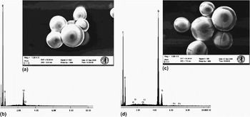
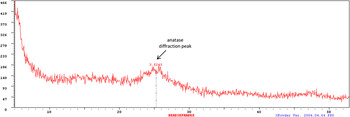
Polymer beads derivatized with amine (Sephadex DEAE), alcohol (Sephadex G-100), carboxymethyl (CM Sephadex), sulfopropyl (SP Sephadex), and hydroxypropyl (Sephadex LH20) groups were swelled in buffer and exposed to a solution of TBALD, a water soluble titanium complex. After reacting for 24 h, each bead sample was washed, dried, and examined for evidence of titania precipitation. DEAE Sephadex and Sephadex G-100 beads gained on average 53 and 10 mg, respectively. However, no appreciable weight gain was recorded for CM Sephadex, SP Sephadex, and Sephadex LH20 beads. SEM coupled with EDS confirmed that a titania coating was present only on the DEAE Sephadex and Sephadex G-100 beads; there was no evidence of a coating on CM Sephadex, SP Sephadex, and Sephadex LH20 beads. Figure 1 shows the SEM and EDS results for DEAE Sephadex. Figure 1a displays the SEM image of the DEAE beads before the reaction with TBALD. The EDS of the unreacted Sephadex beads (Fig. 1b) showed no evidence of titanium on the polymer particles. Upon exposure to TBALD for 24 h, the SEM image revealed a shell of material around the dried DEAE Sephadex beads (Fig. 1c), and the EDS confirmed the presence of titanium on the particles (Fig. 1d). Similar SEM/EDS results were observed with the Sephadex G-100 beads and DEAE beads with encapsulated DFPase (data not shown). XRD analysis of the material showed that the precipitate is primarily amorphous with regions of nano-crystalline anatase (Fig. 2).
SEM images of DEAE Sephadex with and without exposure to TBALD at pH 7.2. (a) DEAE resin with no TBALD exposure; (b) EDS results for (a); (c) DEAE Sephadex with TBALD exposure; and (d) EDS results for (c).

X-ray diffraction pattern for DEAE Sephadex coated with titania. A characteristic diffraction peak for anatase TiO2 is seen around 25.25° corresponding to a d-spacing [(101) plane] of 3.524 Å. There is no diffraction peak seen at 27.5°, indicating no rutile TiO2 was produced by the reaction.

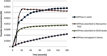
The conditions in which the titania coatings are formed should be amenable to biological macromolecules. The enzyme DFPase, which catalyzes the defluorination of DFP, was utilized to illustrate the biofriendly nature of this encapsulation procedure. The DFPase was added to the DEAE bead solution before exposure with TBALD. After the precipitations were completed and washed, a BCA assay was performed, which showed 60% of the initial protein addition was encapsulated by the titania. Of that amount, no more than 40% (2.4 mg) is DFPase, based on the purity of the crude cell isolate provided by the manufacturer. The activity of the DFPase was measured against DFP under four different exposure conditions: alone in water, adsorbed to DEAE beads, adsorbed to Nanoactive TiO2, and encapsulated in titania (Fig. 3). The activity rate was determined using these curves. Initially, DFP alone was monitored for 3 min to determine the level of fluoride, as well as the rate of unassisted defluorination, already present in solution. In the absence of DFP, fluoride concentrations were negligible in the presence of DFPase and encapsulated DFPase alone.
Graph of DFPase activity against DFP in different local environments: alone in water (-■-), entrapped in DEAE titania (-●-), adsorbed to DEAE Sephadex beads (-▲-), and adsorbed to Nanoactive TiO2 (-♦-). The reaction was run at ambient temperature over 3 min.

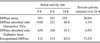
Addition of the DFPase enzyme to DFP alone in water after equilibration caused an immediate and dramatic increase in fluoride concentration, as expected. The kinetic analysis of the free enzyme produced an initial activity rate of 971 nmol (F−)/min/mg protein. DFPase encapsulated in titania coating also exhibited immediate defluorination of DFP at all concentration levels, but at a lower rate (112 nmol (F−)/min/mg protein). The initial rates for adsorbed DFPase onto Sephadex beads and Nanoactive TiO2 also showed higher activity rates than the encapsulated enzyme (Table I). A titania coating containing no DFPase (negative control) was not able to defluorinate DFP in the absence of light, illustrating the need for a material such as an enzyme, which can be activated through alternate means for specific targeting of toxic chemical compounds.
Initial activity rate of DFPase in nmol (F−)/min/mg protein adsorbed on Sephadex beads and Nanoactive TiO2 compared with encapsulate DFPase upon exposure to UV light.

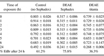
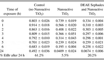
One of the primary reasons for using titania as the encapsulation matrix is its ability to degrade many toxic compounds through photocatalytic activation.Reference Davit, Martra and Coluccia38–Reference Malato, Blanco, Alarcón, Maldonado, Fernández-Ibáñez and Gernjak40 To determine whether the biomimetically precipitated titania would be active against toxic compounds, a dilute solution of EtBr in MQ was exposed to UV light in the presence of a radical scavenger (APS) and titania over a 24-h period (Table II). There was little evidence of EtBr degradation, as measured by a decrease in absorbance at 482 nm, in the samples containing the DEAE titania over the first 6 h of the experiment. The solution did quickly become slurry-like, as did the negative control containing a DEAE titania sample with no EtBr present (data not shown). In addition, this slurry became thicker as the experiment proceeded. After 24 h, there was little evidence of intact Sephadex beads in either of the samples that created a slurry, and the DEAE titania sample containing EtBr did measure a loss of absorption at 482 nm of greater than 60% (Table II). Negative controls containing titania showed no evidence of absorption at 482 nm because of bead degradation, even after 24 h. By contrast, DEAE no titania, in the presence of EtBr and a radical scavenger, exhibited about a 25% decrease in absorbance at 482 nm over a 24 h period, and the EtBr–APS solution alone showed almost a 40% loss of absorption. This is significantly less than that with the addition of the DEAE titania into the sample. Nanoactive TiO2 was also tested in the presence and absence of DEAE Sephadex beads and a similar pattern emerged. The sample containing Nanoactive TiO2 by itself began degrading the EtBr immediately, as evidenced in Table III, by a decrease of more than 95% in the absorbance at 482 nm over 24 h. In the presence of DEAE Sephadex beads, however, the progress of EtBr degradation by NanoTiO2 was drastically slowed over the short term (<25% over 6 h), and the solution quickly formed a slurry, similar to the results with DEAE titania. Over 24 h, the structural integrity of the DEAE Sephadex beads was compromised and the EtBr absorption at 482 nm had dropped by 80%.
Average ethidium bromide absorbance at 482 nm upon exposure to UV light in the presence of unmodified and titania-modified DEAE Sephadex beads.

Ethidium bromide absorbance remaining at 482 nm upon exposure to UV light in the presence of commercially available titania powder (Nanoactive TiO2) by itself and with DEAE Sephadex beads, compared with a control.

The photocatalytic activity of titania could be harmful to the enzyme it is meant to protect. DFPase was tested alone, encapsulated in a titania coating, and adsorbed to Nanoactive TiO2 under UV light for 24 h. Rates of degradation were taken at 0, 6, and 24 h of exposure (Table I). In water by itself, DFPase retained 36.6% of its activity after 24 h of exposure at room temperature. However, in the presence of Nanoactive TiO2, 3.3% activity was retained after 24 h when adsorbed to the nanoparticles in water. When adsorbed onto Sephadex beads, 6.9% of the activity was retained. When the enzyme is fully encapsulated, the loss of activity is significantly less than that through adsorption. After 24 h, 73.2% of the initial activity of the encapsulated enzyme was present.
IV. DISCUSSION
Biological production of inorganic oxides such as silica under aqueous, pH neutral conditions involved the evolution of peptides and polymers that could both locally concentrate the reactive precursors (e.g., silicic acid) and catalyze the condensation of these precursors to silica. Mechanistic studies, such as those mentioned in the introduction, have shown that one of the key components of the natural biomolecules driving this reaction is the functional groups (e.g., amines) involved in the reaction. These groups can behave as Lewis bases under ambient environmental conditions to form transient coordination complexes with the precursors, which at a critical concentration can condense to form the inorganic oxide.Reference Kroger, Deutzmann and Sumper61–Reference Lopez, Gautier, Livage and Coradin63 Based on these studies, the reactivity of a series of functionalized polymer beads with a range of Lewis acid/base strengths was tested on a water-soluble titanium complex at pH 7.2. Specifically, five different Sephadex resins were utilized: lipophilic Sephadex LH20, a resin functionalized with hydroxypropyl groups (soft Lewis base); Sephadex SP, a resin functionalized with sulfopropyl groups (hard Lewis acid); Sephadex CM, a resin functionalized with carboxymethyl groups (soft Lewis acid); DEAE Sephadex, a resin functionalized with tertiary amine groups (hard Lewis base); and Sephadex G-100, a resin functionalized with hydroxyl groups (soft Lewis base). From the studies mentioned in the introduction, in which precipitation occurred using reactive materials containing either an amine or hydroxyl group, it was expected that those resins exhibiting Lewis base character (Sephadex LH20, DEAE Sephadex, and Sephadex G-100) would be able to precipitate titania. However, after exposure to TBALD for 24 h, SEM and EDS confirmed that only DEAE Sephadex and Sephadex G-100 beads showed evidence of a titania coating. XRD analysis confirmed that the precipitate is primarily amorphous titania containing regions of nanocrystalline anatase, with a domain size of about 5 nm, calculated using Scherrer’s formula.Reference Patterson64 These results are consistent with published XRD results for nanocrystalline domains of anatase titania formed at room temperature.Reference Jagtap, Bhagwat, Awati and Ramaswamy65 The cracks observed in the coating are likely a result of freeze drying the sample or the high vacuum of the SEM. Because the Sephadex beads are swelled in buffer before reaction, the size of the coating that forms will be dependent on the swelled size of the beads. As they dry, the beads shrink from the coating walls, causing stress at the coating surface, which is relieved through cracking.
Unlike the results for DEAE and G-100 Sephadex, there was no visible change after 24 h to the Sephadex LH20, SP, or CM beads. SEM and EDS data confirmed that there was no titanium deposited on these beads. Though LH20 beads have structurally similar side chains to G-100, the introduction of a more lipophilic character to the beads negatively impacts the interaction with dissolved species in the solution. In addition, the hydroxypropyl structure is more sterically hindered, with hydroxyl groups farther apart from one another than on the unmodified Sephadex beads (G-100); this would slow down a condensation reaction that is highly dependent on locally high concentrations of the transient coordination intermediate. The SP and CM beads results were expected, as these beads have reactive groups that do not act as Lewis bases.
The oxidative reactivity of titania has generated a significant amount of research interest in this material, though it does not specifically target toxic chemical species. In addition, this activity is dependent on photoactivation, which may not be available in all scenarios. The technique for titania coating formation described here is performed in aqueous buffer (pH 7.2) and at room temperature, which are favorable conditions for retaining enzymatic activity postencapsulation. The enzyme DFPase was selected for testing because it is environmentally sensitive and can complement the general oxidizing capability of titania through specific targeting of fluorophosphates. DFPase was originally isolated from squid and has subsequently been incorporated recombinantly into bacterial DNA.Reference Hartleib and Ruterjans66 However, activity studies have dramatic variability between recombinantly synthesized and naturally occurring enzymes. To the authors’ knowledge, there have been no studies to determine the difference in enzyme kinetics between the recombinant enzyme and enzyme isolated from squid.Reference Mounter, Baxter and Chanutin57, Reference Hoskin58, Reference Hoskin and Roush67–Reference Drevon, Danielmeier, Federspiel, Stolz, Wicks, Yu and Russell70 Therefore, the kinetics of encapsulated and unencapsulated recombinant enzyme was compared together to determine the effects of the encapsulation on activity.
Previous encapsulation studies with this enzyme in polyurethane coatings involved chemical modification of the enzyme and/or irreversible attachment to the matrix.Reference Drevon, Danielmeier, Federspiel, Stolz, Wicks, Yu and Russell70, Reference Drevon, Hartleib, Scharff, Rüterjans and Russell71 Though the enzyme retained activity and was more thermostable in the polymer coating, it is unclear to what extent immobilization adversely affects the activity of the enzyme. In contrast, this encapsulation procedure does not require enzyme modification, and the oxide should surround the enzyme in porous pockets that allow the free flow of substrates through the matrix. The DEAE Sephadex beads were chosen for further study because of the rapid rate at which titania formation occurs at the surface, maximizing the encapsulation of the enzyme. The procedure was split into two steps to ensure the enzyme was well distributed in the bead suspension before addition of TBALD. After washing to remove any residual TBALD and unencapsulated DFPase, the beads were examined under the SEM with no noticeable difference in the structure of the titania coating. Published research has shown that oxide entrapped enzymes can change the morphology of the resulting precipitate.Reference Naik, Tomczak, Luckarift, Spain and Stone11, Reference Luckarift, Dickerson, Sandhage and Spain72 However, as with the finding of Cole et al., addition of the DFPase enzyme to the reaction did not affect the morphology of the resulting precipitate.Reference Cole, Ortiz, Schoonen and Valentine9 The size of the beads when swelled (>50 μm), compared to the smaller enzyme (∼10 nm), accounts for the lack of morphological difference observed.
In the presence of DFP, the titania coating with no DFPase (negative control) was not able to defluorinate the compound, illustrating that some toxic chemical compounds may need to be specifically targeted. When DFPase is encapsulated in the coating, there is immediate defluorination of DFP at all concentration levels (Fig. 3). The encapsulated DFPase was determined to have an initial activity rate of 112 nmol (F−)/min/mg protein, which is significantly slower than the free enzyme [971 nmol (F−)/min/mg protein]. This can be attributed to the decreased exposure of DFP to DFPase within the coating (dependent on the rate of DFP diffusion through the oxide matrix). Comparison of these apparent values with previously published results is difficult, as the origin, handling, and purity of the enzyme can vary widely.Reference Hoskin58, Reference Hoskin and Roush67–Reference Drevon, Danielmeier, Federspiel, Stolz, Wicks, Yu and Russell70 The DFPase obtained for this study is purified to 40%, with the remaining material being an unknown composition of cell lysate debris.
Enzyme encapsulation provides a protective barrier against destabilizing environmental conditions, which allows for targeted breakdown of a particular toxic compound. However, if the biomimetically precipitated titania is not generally active against toxic compounds, there would be no advantage to this system over silica. The biomimetically precipitated titania was tested for oxidative activity using EtBr as the model toxic compound.Reference Faisal, Abu Tariq and Muneer60 The results illustrate that the DEAE titania will break down the EtBr, but at a slower photodegradation rate (Table I) than that reported by Faisal et al.Reference Faisal, Abu Tariq and Muneer60 This can be attributed to a few key differences in our technique: (i) suspensions were stirred rather than continuously purged with air throughout the experiment, replenishing the oxygen in the solution, which would aid in the photodegradation; (ii) the wattage of the UV lamp utilized for this report was 20% lower; (iii) the surface area of a titania coating, and therefore, the amount of reaction sites available, is significantly less than that of the powders used by Faisal et al. These differences did not explain, however, the delayed start of the observed photodegradation of the EtBr in the DEAE titania samples (>6 h).
We attribute the delay to the close proximity and relative amount of the DEAE Sephadex. It is known that Sephadex beads are susceptible to degradation from oxidative environments. The addition of UV light would enhance the oxidative capability of the titania and hence speed up this degradation process. Indeed, the Sephadex beads lost their structural integrity over the course of the 24-h experiment, leaving a thick slurry, limiting the amount of solution that could be recovered from centrifugation. Samples that included DEAE Sephadex beads were measured in cuvettes with smaller volumes to account for this. Apparently, only when much of the cross-linked dextran had been degraded did the biomimetically precipitated titania begin oxidizing the EtBr in solution; after 24 h, significantly more EtBr was degraded in the presence of the biomimetically precipitated titania than in the EtBr/APS solution alone.
To confirm this hypothesis, a commercially available titania powder, Nanoactive TiO2, was tested in the presence and absence of DEAE Sephadex beads to determine whether the susceptibility of the crosslinked dextran beads to photodegradation affects the photodegradation rate of the EtBr (Table II). As expected, the Nanoactive TiO2 by itself began degrading the EtBr immediately. In the presence of DEAE Sephadex beads, however, the progress of EtBr degradation by Nanoactive TiO2 was drastically slowed over the short term and the solution quickly formed a slurry, similar to the results with DEAE titania beads. The data indicate that once the DEAE Sephadex beads had been sufficiently degraded, the Nanoactive TiO2 began degrading the EtBr, resulting in a significant loss in absorption (80%) after 24 h. The addition of a small amount of Sephadex beads was sufficient to retard the short-term photodegradation rate of a powdered form of titania, preferentially degrading the Sephadex beads over the EtBr dye. The DEAE titania coating, which has been templated by the beads and, therefore, is more intricately linked to them, would take longer to degrade the beads. Once largely released from the template, the titania coating can then begin oxidizing the dye, resulting in the observed delay.
This oxidative environment could have a detrimental effect on the activity of the enzyme itself, either through direct action on the enzyme or through by-products of the UV exposure, negating the protective benefits of the encapsulation. Titania-encapsulated and unencapsulated enzymes were tested for activity after exposure to UV at discrete time intervals, looking specifically at initial rates of reaction over the first 30 s. The titania-encapsulated enzyme actually increased in initial rate over the first 6 h, before decreasing by 26.7% from the initial measurement after 24 h of constant UV exposure. All the samples were freeze dried before the initiation of this test, which accounts for this initial increase as the encapsulated enzyme is slowly hydrated. In contrast, the unencapsulated enzyme dissolved in water, upon exposure to UV light, retained 36.6% of its degradation capability after 24 h. Much of this activity loss occurred within 6 h of UV exposure with a gradual loss thereafter. The unknown components of the DFPase material may have a detrimental effect on the enzyme in the initial stages of UV exposure. When adsorbed to DEAE Sephadex beads or Nanoactive TiO2, significant loss of activity was observed (almost 94% and 97%, respectively, after 24 h). Although it is possible that the oxidative environment created by the titania was having a minor negative impact, a test of pH showed there was a significant drop-off after 24 h in the starting pH of the solution, before degradation of DFP had begun. Coupled with the fact that the Sephadex beads were breaking down after 24 h in the presence of titania, a drop in solution pH is not surprising. A previously published kinetic study performed on DFPase showed significant pH dependence below 5.5 on the activity of the enzyme.Reference Kroger, Deutzmann and Sumper61 The reaction rate of the adsorbed enzyme was much more negatively impacted by this drop in pH than the titania encapsulated enzyme, illustrating the protective effects of the coating against environmental changes. Environmental changes are more immediately felt by an adsorbed enzyme and, with no protective shell, can be more quickly deactivated by those changes.
V. CONCLUSIONS
This investigation has illustrated a biomimetic approach to the formation of titania coatings on functionalized beads. The formation of titania under aqueous conditions at room temperature and neutral pH provides a technique to incorporate biomacromolecules that can enhance or complement the decontaminating capabilities inherent to this oxide. Using this biofriendly method, an environmentally sensitive enzyme, DFPase, has been encapsulated within the titania matrix while retaining its activity. In addition, the inherent degradation capability of titania is preserved using this biomimetic precipitation method of formation. This material has the advantage of both nonspecific (oxidative degradation) and specific (enzymatic degradation) targeting of toxic substances, illustrating that the procedure offers numerous possibilities for preparing materials with unique multifunctionality in a single synthetic step. Considering the mild reaction conditions that have been described, this method for enzyme encapsulation is amenable to other reactive surfaces, including oxidatively stable resins and cellulose.
ACKNOWLEDGMENTS
This research was funded by the Defense Threat Reduction Agency. The authors acknowledge Dr. John Walker for his assistance with the DFPase activity assay and Dr. Lian Li and Professor Jayant Kumar from the University of Massachusetts-Lowell for their assistance with x-ray diffraction of the titania-coated Sephadex beads.