Neurodegeneration is a common theme of many nervous system diseases, such as Alzheimer’s disease, Parkinson’s disease, amyotrophic lateral sclerosis, head trauma, epilepsy, and stroke. These disorders are devastating and expensive to treat, with annual costs currently exceeding several hundred billion dollars in the United States alone, and current treatments are inadequate. Adding to the urgency of the problem is the fact that the incidence of these age-related disorders is increasing rapidly as population demographics change. Among nervous system disorders, there are specific neurodegenerative diseases affecting the neuro-retina in the eye. The visual system is the sensory modality which enables living organisms to explore the surrounding visual environment. Thus, quality of vision is tightly linked to quality of life. This link is probably the reason for extensive basic and clinical research throughout centuries, using the visual system as one of the best instruments to understand general principle of neurodevelopment, neurodegeneration, and repair. As a result, there is already a wealth of information on how the visual system is built and how connections from the eye to the cortex enable visual perception. Despite this knowledge, many diseases that impair vision, ranging from retinitis pigmentosa (RP) to age-related macular degeneration (AMD), lack effective treatments.
AMD is a retinal neurodegenerative disease characterized in its early stage by large soft drusen and hypo-hyperpigmentation of the retinal pigment epithelium (RPE). Late AMD, the potentially blinding stage of disease, includes geographic atrophy of the RPE (“dry” AMD), or subretinal neovascular membranes (“wet” AMD) (Bird et al., Reference Bird, Bressler, Bressler, Bressler, Chisholm, Coscas, Davis, de Jong, Klaver, Klein and Klein1995).
AMD is generally considered a multifactorial disease, whose development and progression are the results of a complex interaction between genetic and environmental risk factors. Both oxidative stress (Beatty et al., Reference Beatty, Koh, Phil, Henson and Boulton2000) and chronic inflammation (Buschini et al., Reference Buschini, Piras, Nuzzi and Vercelli2011) seem to play a significant role in the pathogenesis of AMD together with many genetic risk factors (Hollyfield, Reference Hollyfield2010; Jarrett & Boulton, Reference Jarrett and Boulton2012; Swaroop et al., Reference Swaroop, Chew, Rickman and Abecasis2009; see also Marangoni et al., Reference Marangoni, Falsini, Piccardi, Ambrosio, Minnella, Savastano, Bisti, Maccarone, Fadda, Mello, Concolino and Capoluongo2013; Ratnapriya and Chew, Reference Ratnapriya and Chew2013). The final outcome of all neurodegenerative retinal diseases is the death of photoreceptors, consequently visual functions progressively deteriorate. Recently, new strategies that are able to mitigate photoreceptor death using natural products have been explored (Age-Related Eye Disease Study 2 Research Group, 2013). Among the others, Maccarone et al. (Reference Maccarone, Di Marco and Bisti2008) provided data showing that l’Aquila saffron is protective against light damage to photoreceptors in a rat model. A proof-of-principle clinical trial in AMD patients confirmed the potentiality of saffron treatment in neurodegenerative diseases and its consistency in time (Falsini et al., Reference Falsini, Piccardi, Minnella, Savastano, Capoluongo, Fadda, Balestrazzi, Maccarone and Bisti2010; Piccardi et al., Reference Piccardi, Marangoni, Minnella, Savastano, Valentini, Ambrosio, Capoluongo, Maccarone, Bisti and Falsini2012) and in patients carrying genetic mutations (Marangoni et al., Reference Marangoni, Falsini, Piccardi, Ambrosio, Minnella, Savastano, Bisti, Maccarone, Fadda, Mello, Concolino and Capoluongo2013).
Saffron is a well-known spice, which is used widely in different cuisines. It has also long been used in traditional medical practice (reviewed in Giaccio, Reference Giaccio2004; see also Ochiai et al., Reference Ochiai, Shimeno, Mishima, Iwasaki, Fujiwara, Tanaka, Shoyama, Toda, Eyanagi and Soeda2007). Egyptian healers used it to treat gastrointestinal ailments; in Roman times, it was used to promote wound healing and relieve upper respiratory complaints. In more recent times, it has been used as an anti-inflammatory, anticonvulsive, and anti-tumor agent; it has also been investigated in the treatment of cognitive defects. Because of this long history of medical use associated to a variety of different clinical applications, it is essential to make an accurate analysis of relevant data specifically in the field of retinal diseases. Saffron is the commercial name of the dried red stigmas of Crocus sativus L flowers. It is produced in many areas all over the world, but the bulbs may present different genetical characteristics and the cultivar and the drying strategies can be quite different, resulting in each single production to be unique, which could explain the variety of effects and discrepancies found in the literature. Chemically, saffron is known to contain more than 150 volatile and aroma-yielding compounds and many non-volatile biologically active components, including carotenoids (zeaxanthin and crocetin) and various alpha- and beta-carotenes. Its golden yellow-orange color comes from alpha-crocin, a water soluble beta-gentobiose (sugar) ester of crocetin. Its flavor arises from the glycoside picrocrocin, a molecule containing safranal and a carbohydrate. Its most potent antioxidant ingredients appear to be crocin, and crocetin, a carotenoid dicarboxylic acid which forms the core of crocin. Several actions of crocin on mammalian tissues have been reported including anti-apoptotic activity and increased oxygen diffusivity (see Maccarone et al., Reference Maccarone, Di Marco and Bisti2008 and Di Marco et al., Reference Di Marco, Romeo, Nandasena, Purushothuman, Adams, Bisti and Stone2013). Kanakis et al. (Reference Kanakis, Tarantilis, Tajimir-Riahi and Polissiou2007) showed that metabolites of saffron bind directly to DNA and induce its partial conformation to beta-DNA, thereby protecting the cell from damage. Saffron has been shown to have anti-inflammatory actions, including for example the inhibition of tissue necrosis factor (Nam et al., Reference Nam, Park, Jung, Lee, Min, Park, Jung, Cho, Park, Kang, Hong and Lee2010). Based on these observations, it is clear that the saffron extract does not act as a simple antioxidant. The peculiar characteristics of saffron components support the hypothesis that saffron has complex mechanisms of action ranging from antioxidant activity to direct control of gene expression, as also suggested by microarray experiments (Natoli et al., Reference Natoli, Zhu, Valter, Bisti, Eells and Stone2010).
Animal model
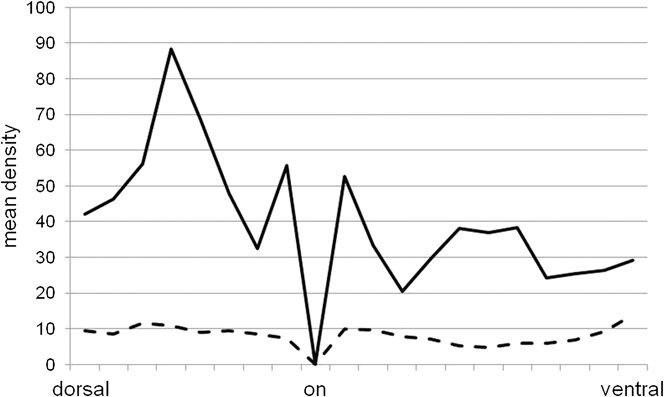
Experiments on saffron treatment in neurodegenerative retinal disease started in animal models with environmentally induced photoreceptors death. The experimental design was 6 weeks (or 2 days) dietary saffron pretreatment (1 mg/kg/day) in albino rats followed by high intensity light exposure (1000 lux/24 h). Subsequently animals were: (1) sacrificed immediately for TUNEL labeling (apoptotic figures); (2) kept alive for one week to monitor changes in outer nuclear layer (ONL) thickness (photoreceptor survival), activation of self-protective mechanism (fibroblast growth factor 2 (FGF2) expression) and to test responsiveness to light with electroretinogram (ERG) recordings. Animals without treatment and exposed to 1000 lux/24 h were used as controls. Published data (Maccarone et al., Reference Maccarone, Di Marco and Bisti2008) showed a good maintenance of both morphology and function. Animals exposed to high intensity light presented a destabilizing area, or “hot spot,” located a few millimeters dorsal to the papilla (Bowers et al., Reference Bowers, Valter, Chan, Walsh, Maslim and Stone2001; Organisciak & Vaughan, Reference Organisciak and Vaughan2010). Light-induced degeneration starts in that “hot spot” and progressively expands to adjacent areas (Rutar et al., Reference Rutar, Provis and Valter2010). Saffron not only reduces photoreceptor death (Maccarone et al., Reference Maccarone, Di Marco and Bisti2008; Di Marco et al., Reference Di Marco, Romeo, Nandasena, Purushothuman, Adams, Bisti and Stone2013) but also stabilizes the morphology of this area by reducing “rosettes” formation and inflammatory reactions (Figs. 1 and 2). Fig. 1A and 1B shows the dorsal to ventral reconstruction of saffron treated and control retinas one week after light-induced damage (LD). The insets (higher magnification view) clearly illustrate that in the saffron treated retina, the hot spot area is not only thicker but also shorter (limits defined by white arrows) than in control. The upregulation of FGF2 in the ONL after light exposure is reduced by saffron pretreatment (see Maccarone et al., Reference Maccarone, Di Marco and Bisti2008). Fig. 2 shows densitometry measurements across the entire ONL in treated and untreated retinas immunolabeled for FGF2. In saffron treated retina FGF2, it is present only in the periphery and close to the papilla as it has been reported in normal retina (Stone et al., Reference Stone, Mervin, Walsh, Valter, Provis, Penfold, Penfold and Provis2005). Interestingly in microarray experiments (Natoli et al., Reference Natoli, Zhu, Valter, Bisti, Eells and Stone2010) FGF gene, upregulated by light, is only mildly modulated by saffron, but the protein is absent, as demonstrated by Western blot analysis and immune labeling. We can therefore formulate the hypothesis that saffron components might regulate gene transcription product. Retinal function was tested by recording ERG responses to light flashes of increasing intensities before and a week after LD. Light responses (both a-and b-waves) were partially preserved (see also Maccarone et al., Reference Maccarone, Di Marco and Bisti2008; Figs. 3 and 4).
Representative reconstruction of saffron treated LD retina (A) and control LD retina (B): bisbenzimide labeling of cryosections from dorsal to ventral retina through the optic nerve. A1 and B1 high magnification field of hot spot. Arrows indicate the extension of the hot spot localized in the dorsal retina. (onl: outer nuclear layer; inl: inner nuclear layer; gcl: ganglion cells layer; LD: light damage). Scale bar: A1) and B1) 50 μm.

Densitometric analysis for FGF2 immunolabeling in the onl from dorsal to ventral retina through the optic nerve. Comparison between two experimental group: control LD continuous line, saffron treated LD dotted line.
Representative ERG recordings before and 1 week after light exposure in two experimental conditions: control LD and saffron treated LD. (a) and (b) show b-wave, (c) and (d) a-wave.
b-wave amplitude of ERG before (pre damage) and 1 week after (post damage) light exposure at 3 different flash luminances from 103 to 105 photometric units (105= 1488 cd/m2). (a) controls LD; (b) saffron treated LD.
Clinical trials
In a masked, placebo controlled, cross-over phase I/II study (Falsini et al., Reference Falsini, Piccardi, Minnella, Savastano, Capoluongo, Fadda, Balestrazzi, Maccarone and Bisti2010), early AMD patients were randomly assigned to oral saffron 20 mg/day or placebo supplementation over a three-month period, and then reverted to placebo or saffron for further three months. Focal electroretinograms (fERGs) and clinical findings were recorded at baseline and after three months of either saffron or placebo supplementation. fERGs were recorded in response to a sinusoidally-modulated (41 Hz) uniform field presented to the macular region (18°) at different modulations between 16.5 and 93.5%. Main outcome measures were fERG amplitude (in µV), phase (in degrees), and modulation thresholds. After saffron, patients’ fERGs were increased in amplitude, compared either to baseline values or to values found after placebo supplementation (mean change after saffron: 0.25 log µV, mean change after placebo: −0.003 log µV, P < 0.01). fERG thresholds were decreased after saffron, but not placebo supplementation compared to baseline (mean change after saffron: −0.26 log units; mean change after placebo: 0.0003 log units). Fundus appearance did not change significantly in either saffron or placebo-treated patients. The results of this study indicate that short-term dietary saffron supplementation improves retinal flicker sensitivity in early AMD. The fERG represents an objective outcome measure of central retinal function, and it has been shown to detect early functional losses associated with early AMD (Falsini et al., Reference Falsini, Fadda, Iarossi, Piccardi, Canu, Minnella, Serrao and Scullica2000). This ERG methodology allows accurate quantification of the effects of disease on retinal function, and how retinal function changes after treatment (Falsini et al., Reference Falsini, Fadda, Iarossi, Piccardi, Canu, Minnella, Serrao and Scullica2000, Reference Falsini, Piccardi, Iarossi, Fadda, Merendino and Valentini2003). Previous studies have also demonstrated a tight relationship between fERG measurements and psychophysical sensitivity in the central retina, in both AMD (Piccardi et al., Reference Piccardi, Ziccardi, Stifano, Montrone, Iarossi, Minnella, Fadda, Balestrazzi and Falsini2009) and inherited retinal degenerations (Iarossi et al., Reference Iarossi, Falsini and Piccardi2003). Although the results of this clinical trial need to be further replicated and the clinical significance is yet to be evaluated, they provide important clues that nutritional saffron carotenoids may impact AMD in novel and unexpected ways, possibly beyond their antioxidant properties (clinicaltrials.gov: NCT00951288).
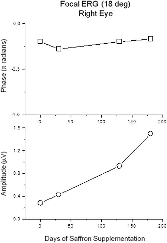
To evaluate whether the observed functional benefits from saffron supplementation may extend over a longer follow-up period, a longitudinal, interventional open-label study was conducted in an outpatient ophthalmology setting. Twenty-nine early AMD patients (age range: 55–85 years) with a baseline visual acuity >0.3 were enrolled. They had dietary saffron supplementation (20 mg/day) over an average period of treatment of 14 (±2) months. Clinical examination and fERG-derived macular (18°) flicker sensitivity estimate (Falsini et al., Reference Falsini, Fadda, Iarossi, Piccardi, Canu, Minnella, Serrao and Scullica2000, Reference Falsini, Piccardi, Iarossi, Fadda, Merendino and Valentini2003, Reference Falsini, Piccardi, Minnella, Savastano, Capoluongo, Fadda, Balestrazzi, Maccarone and Bisti2010) were collected every three months over a follow-up of 14 (±2) months. Retinal sensitivity, the reciprocal value of the estimated fERG amplitude threshold, was the main outcome measure. After three months of supplementation, mean fERG sensitivity improved by 0.3 log units compared to baseline values (P < 0.01) and mean visual acuity improved compared to baseline values (6/9 +2 to 6/6 –2, P < 0.01). These changes remained stable over the follow-up period. An example is reported in Fig. 5. In the same patients, fundus appearance and optical coherence tomography evaluation of the central retina did not show any significant change during follow-up. These results indicate that in early AMD saffron supplementation induces macular function improvements from baseline that are extended over a long-term follow-up.
Amplitude and phase of the fERG recorded from the central retina (18°) in an early AMD patient at baseline and at several time points during saffron supplementation. Note that response amplitude steadily improved throughout the follow-up period.
To determine whether the functional effects of dietary supplementation with saffron are influenced by the known high risk polymorphisms in the complement factor H (CFH) and age-related maculopathy susceptibility 2 (ARMS2) gene loci, a long-term assessment was conducted in another group of early AMD patients who were preliminarily assessed for these gene polymorphisms (Marangoni et al., Reference Marangoni, Falsini, Piccardi, Ambrosio, Minnella, Savastano, Bisti, Maccarone, Fadda, Mello, Concolino and Capoluongo2013). Thirty-three early AMD patients, screened for CFH (rs1061170) and ARMS2 (rs10490924) polymorphisms and receiving saffron oral supplementation (20 mg/day) over an average period of treatment of 11 months (range, 6–12), were longitudinally evaluated by clinical examination and fERG-derived macular (18°) flicker sensitivity estimate. fERG amplitude and macular sensitivity, the reciprocal value of the estimated fERG amplitude threshold, were the main outcome measures. After three months of supplementation, mean fERG amplitude and fERG sensitivity improved significantly when compared to baseline values (P < 0.01). These changes were stable throughout the follow-up period. No significant differences in clinical and fERG improvements were observed across different CFH or ARMS2 genotypes (Marangoni et al., Reference Marangoni, Falsini, Piccardi, Ambrosio, Minnella, Savastano, Bisti, Maccarone, Fadda, Mello, Concolino and Capoluongo2013). These results indicate that the functional effect of saffron supplementation in individual AMD patients is not related to the major risk genotypes of disease.
Pharmacokinetics
It remains an open question which metabolites reach the tissue and are active after oral intake. Numerous studies have demonstrated a variety of pharmacological actions for crocetin and crocins, the supposedly active saffron components, e.g., enhancement of oxygen diffusivity (Giaccio, Reference Giaccio2004), increment of ocular blood flow (Xuan et al., Reference Xuan, Zhou, Li, Min and Chiou1999), inhibition of tumor cell proliferation (Abdullaev et al., Reference Abdullaev, Riveròn-Negrete, Caballero-Ortega, Manuel Hernàndez, Pérez-Lòpez, Pereda-Miranda and Espinosa-Aguirre2003; Amin et al., Reference Amin, Hamza, Bajbouj, Ashraf and Daoud2011; Garcýà-Olmo et al., Reference Garcýà -Olmo, Riese, Escribano, Ontanòn, Fernandez, Atiènzar and Garcýà -Olmo1999; see also Di Marco et al., Reference Di Marco, Romeo, Nandasena, Purushothuman, Adams, Bisti and Stone2013). However, in contrast to these investigations aimed to evaluate biological activities, there are few studies on the absorption and metabolism of crocetin and crocins. Asai et al. (Reference Asai, Nakano, Takahashi and Nagao2005) investigated the absorption of orally administered crocetin and crocins into the blood circulation in mice. Their results suggested that crocins are hydrolyzed to crocetin before or during absorption and then undergo the metabolic pathway of crocetin. Hence, according to these data, orally ingested crocins could not act as bioactive molecules by themselves “in vivo” except in the gastrointestinal tract. Asai et al. (Reference Asai, Nakano, Takahashi and Nagao2005) proposed a model of the metabolic pathway of orally administered crocetin and crocins in mice (Fig. 6 in ref. Asai). Orally administered crocins would be hydrolyzed to crocetin before being incorporated into blood circulation. Thereafter, crocetin would be partly metabolized to mono- and diglucuronide conjugates. Crocetin is likely to be metabolized to the glucuronide conjugates both in the intestinal mucosa and in the liver of mice. The rapid absorption and glucuronidation of crocetin indicate that the metabolic fate of crocetin is quite different from those of common C40 carotenoids. These analyses have been extended to rats by Xi et al. (Reference Xi, Qian, Du and Fu2007) and their results confirm that orally administered crocin remained undetectable in plasma, further indicating that the intact form of crocin was not absorbed from the intestinal tract. In addition, plasma crocetin concentrations do not tend to accumulate with repeated oral doses of crocin, and the intestinal tract serves as an important site for crocin hydrolysis. At present, these are the only available data on the metabolism of ingested crocins and crocetins. In humans, Umigai et al. (Reference Umigai, Murakami, Ulit, Antonio, Shirotori, Morikawa and Nakano2011) clearly demonstrated that crocetin was more rapidly absorbed than the other carotenoids and was subsequently eliminated from human plasma in maximum 8 h time. Considering the neuroprotective effects of oral saffron supplementation both in animal models and AMD patients, we examined whether it was possible to find metabolites leading to saffron components into different tissues. Specifically, we used an animal model with induced photoreceptors degeneration (see Maccarone et al., Reference Maccarone, Di Marco and Bisti2008) pretreated with dietary saffron with a dose five times higher (5 mg/kg/7 day) to verify whether saffron related metabolites were found in the stressed retina, blood, urine, kidney, liver, and cerebral cortex. As a control, we used treated animals with no degeneration. In addition, we tested blood and urine from healthy voluntaries and AMD patients who had undergone long-term (>1 year) saffron treatment.
Analytical methods
We followed the procedure reported in Yamauchi et al. (Reference Yamauchi, Tsuruma, Imai, Nakanishi, Umigai, Shimazawa and Hara2011). Briefly, the tissue samples were mixed with 2.0 ml of methanol and centrifuged (3000 rpm, 10 min) and, following several steps, analytes were extracted The crocins and crocetin were identified by the reversed-phase HPLC method using Synergi 4 µm Fusion-RP column (250 × 150 mm, Phenomenex). The eluate was monitored with a photodiode array detector (210–500 nm). The chromatogram was monitored at 440 nm.
We analyzed altogether 15 animals with saffron treatment and LD and 5 control (treatment without damage). All animals were sacrificed in the morning in comparable conditions, and saffron was supplemented in the drinking water, the daily dose was diluted in a volume that was always consumed in one day. In agreement with previous reports, we found crocetin in all blood samples but no traces of metabolites related to saffron were ever found in any other tissue examined with the exception of degenerating retinas, wherein 7 out of 15 animals we found modest amounts of crocins. We therefore suggest that crocins may be resynthesized from circulating crocetin, which reaches the retina following damage to the blood–brain barrier. In support, we did not find any metabolites of crocin or crocetin in the healthy retina or other parts of the central nervous system. We also examined blood and urine from two AMD patients under saffron treatment for over a year and three healthy volunteers after two weeks of treatment using the same daily dose (20 mg/day). Samples were taken two hours after the intake of the morning saffron pill. Interestingly, while in healthy volunteers it was always possible to quantify the presence of crocetin in both blood and urine the same procedure often yielded negative results in patients, suggesting that the metabolites were immediately absorbed and used. Kanakis et al. (Reference Kanakis, Tarantilis, Tajimir-Riahi and Polissiou2007) in their study on the interaction of crocetin with human serum albumin demonstrated that the binding of crocetin shows a weak ligand–protein interaction. Since weaker binding to plasma protein tends to facilitate distribution to tissues of the body, crocetin is expected to be well distributed in the system. These preliminary results may explain why the positive effects on visual function revert if the intake of saffron is interrupted.
Saffron and its components mainly crocins, crocetin, and safranal have been tested in animal models with neurodegenerative diseases (Ochiai et al., Reference Ochiai, Shimeno, Mishima, Iwasaki, Fujiwara, Tanaka, Shoyama, Toda, Eyanagi and Soeda2007; Hosseinzadeh et al., Reference Hosseinzadeh, Sadeghnia, Ghaeni, Motamedshariaty and Mohajeri2012; Purushothuman et al., Reference Purushothuman, Nandasena, Peoples, El Massri, Johnstone, Mitrofanis and Stone2013), retinal degeneration (Yamauchi et al., Reference Yamauchi, Tsuruma, Imai, Nakanishi, Umigai, Shimazawa and Hara2011; Ohno et al., Reference Ohno, Nakanishi, Umigai, Tsuruma, Shimazawa and Hara2012; Fernández-Sánchez et al., Reference Fernández-Sánchez, Lax, Esquiva, Martín-Nieto, Pinilla and Cuenca2012; Di Marco et al., Reference Di Marco, Romeo, Nandasena, Purushothuman, Adams, Bisti and Stone2013), and in human subjects with Alzheimer and depression (Akhondzadeh et al., Reference Akhondzadeh, Tahmacebi-Pour, Noorbala, Amini, Fallah-Pour, Jamshidi and Khani2005, Reference Akhondzadeh, Shafiee Sabet, Harirchian, Togha, Cheraghmakani, Razeghi, Hejazi, Yousefi, Alimardani, Jamshidi, Rezazadeh, Yousefi, Zare, Moradi and Vossoughi2009, Reference Akhondzadeh, Sabet, Harirchian, Togha, Cheraghmakani, Razeghi, Hejazi, Yousefi, Alimardani, Jamshidi, Zare and Moradi2010). Looking at the chemistry of various molecules, a direct antioxidant activity can certainly be hypothesized, thus reducing the oxidative stress associated to the initiation and progression of the disease. In addition, based on results present in the literature and our own observations, it seems reasonable to assume that neuronal death can be controlled at least in two ways: acting directly on the apoptotic downstream events like caspase activation (see Yamauchi et al., Reference Yamauchi, Tsuruma, Imai, Nakanishi, Umigai, Shimazawa and Hara2011) and by reducing neuroinflammation and, in turn, the activation of programmed cells death. This possibility is suggested by microarrays experiments (Natoli et al., Reference Natoli, Zhu, Valter, Bisti, Eells and Stone2010) and experiments in progress in our laboratory.
It is interesting to note that in some animal models where crocetin was used (Yamauchi et al., Reference Yamauchi, Tsuruma, Imai, Nakanishi, Umigai, Shimazawa and Hara2011; Ohno et al., Reference Ohno, Nakanishi, Umigai, Tsuruma, Shimazawa and Hara2012), the effects were obtained with relatively very high doses (e.g., 100 mg/kg/day) and with safranal treatment the dose was even higher, 400 mg/kg/day (Fernández-Sánchez et al., Reference Fernández-Sánchez, Lax, Esquiva, Martín-Nieto, Pinilla and Cuenca2012). Similarly, when saffron extract was tested the amount was over 50 mg/kg/day (up to 250 mg/kg/day in Hosseinzadeh et al., Reference Hosseinzadeh, Sadeghnia, Ghaeni, Motamedshariaty and Mohajeri2012), while in our experiments (Maccarone et al., Reference Maccarone, Di Marco and Bisti2008; Natoli et al., Reference Natoli, Zhu, Valter, Bisti, Eells and Stone2010; Di Marco et al., Reference Di Marco, Romeo, Nandasena, Purushothuman, Adams, Bisti and Stone2013) we used 1 mg/kg/day of saffron extract where crocin represents about 40–60%. Regardless of the approach used to administer saffron (oral to intraperitoneal and intravenous injection), the outcome was always positive. In patients, the dose used never exceeded 30 mg/day. Data on safety of saffron treatment have been recently reported by Ayatollahi et al. (Reference Ayatollahi, Javan, Khajedaluee, Shahroodian and Hosseinzadeh2013). The study was a double-blind, placebo controlled study consisting of 1-week treatment with 200 mg and 400 mg saffron tablets. Sixty healthy volunteers (age range 20–50 years) were selected for the study. The administration of saffron tablets (200 or 400 mg/day, for 7 days) did not induce any significant modification on coagulation or anticoagulation system. The dose we used in AMD patients is twenty times lower and metabolites seem to be used immediately. Altogether, our results and other studies in the field provide evidence of a treatment with high degree of safety and no side effects at least at the dose used in humans.
In conclusion, saffron treatment appears able to cope with the degenerative progression of AMD. Although the ways of action are still under investigation it seems reasonable to conclude that saffron components are able to reduce photoreceptor death and preserve visual function.
Competing interests
Prof. Bisti and Dr. Maccarone hold a nonremunerative relationship with “Hortus Novus,” the company that provided the saffron pills used in this study.
Acknowledgment
We wish to thank Dr. Maria Maggi for performing the chemical analysis. Supported by a MIUR-PRIN (2010-2011) research grant to SB and Hortus Novus s.r.l.