

Introduction
Qin mausoleums constructed after about 325 BC, often referred to as the Warring States Qin Mausoleums (WSQMs), signify the maturation of the Qin mausoleum system (Liang & Wang Reference Liang and Wang2022). They share a common layout featuring spatially independent mausoleums, which embodies hierarchical organisation (Jiao et al. Reference Jiao2014). The mausoleum complexes are divided into inner and outer areas by tamped-earth walls, with concentrations of small satellite tombs uniformly aligned in the peripheral area. However, the function of the peripheral area remains poorly understood due to the absence of pertinent historical records and the lack of systematic archaeological excavations.
To resolve this lacuna, Chinese archaeologists have focused on interpreting the social identity of individuals buried in the small peripheral tombs, examining grave goods for insights into individual social roles and wider societal organisation. While placement in these satellite tombs is generally seen as indicative of a lower social status, occupants may still have had a direct association with the Qin royal family, acting as imperial concubines, members of Qin nobility or high-ranking officials, palace servants or tomb guardians (Yuan Reference Yuan2002; Tian et al. Reference Tian2010; Liang & Wang Reference Liang and Wang2022), though their definitive identities remain contested.
The concept of identity is complex and expansive, encompassing the intersecting dimensions of gender, age and social status (Knudson & Stojanowski Reference Knudson, Stojanowski, Knudson and Stojanowski2009), all of which are subject to fluidity or superposition in different situations (Torres-Rouff & Knudson Reference Torres-Rouff and Knudson2017; Ellis Reference Ellis2024). Various bioarchaeological analyses can provide information that aids in the reconstruction of some aspects of social identity (Stojanowski et al. Reference Stojanowski, Knudson and Stojanowski2020). Fundamental information of individuals, such as the biological sex, age-at-death, ancestry and kinship, in tandem with historical accounts, can give an indication of the social positioning of individuals within different historic contexts (Harrod & Stone Reference Harrod, Stone and Stone2018; Johnson & Larsen Reference Johnson, Larsen, Knudson and Stojanowski2020; Buikstra et al. Reference Buikstra2022). Consideration of burial contexts and health status of individuals can provide insight into the attitudes of the living toward the deceased, socially mediated access to resources and their perceived social status (Buikstra & Beck Reference Buikstra and Beck2006; Minozzi et al. Reference Minozzi2020). Moreover, social regulations may compel individuals, in line with or regardless of their status, to conform to specific activity patterns. Thus, social identity can be further inferred by rationally examining activity markers and trauma and employing multidisciplinary approaches (Chen et al. Reference Chen2025; Ma et al. Reference Ma2025).
This study focuses on the individuals buried in small peripheral tombs at the Shangzhaoyao Qin mausoleum, a newly discovered WSQM in Shaanxi, China. Through the analysis of biological sex, age-at-death, biomarkers and activity markers, we aim to reconstruct the health status, activity patterns and, ultimately, the social identity of this group of individuals from a bioarchaeological perspective. Our results provide critical evidence for understanding the function of the outer area in WSQMs and the funerary materialisation of Qin hierarchical structure.
Archaeological context
The Qin state won the interstate competition during the Warring States period (475–221 BC) and established the first unified empire of China. Social hierarchy under the Qin state was highly stratified and strict laws regulated all aspects of civilian life, including manufacturing, corvée labour and commerce. With subsistence predominantly agrarian, the residents of the newly unified empire were largely dependent on millet and panicum, supplemented by some livestock (Ling Reference Ling2010). Serving as the final settlement in the nine-capital migration sequence, the city of Xianyang on the northern bank of the Wei River witnessed the transformation from a regional kingdom to a part of a centralised empire. Correspondingly, the Xianyang Plateau Necropolis was established along the western outskirts of the capital (Sima Reference Sima1999).
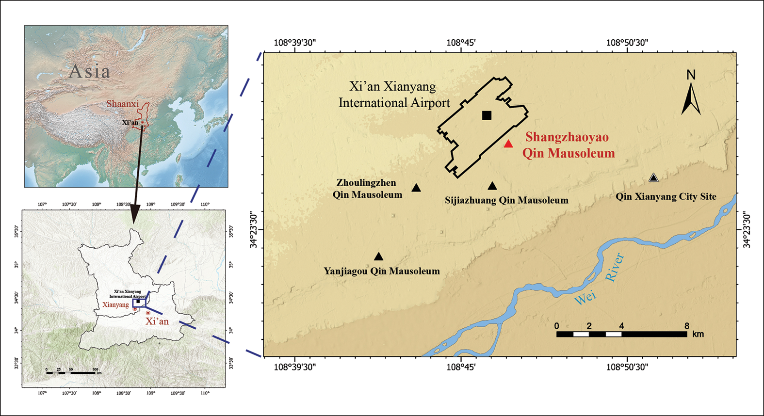
Within the present landscape, the Shangzhaoyao Qin mausoleum lies south-east of Xi’an Xianyang International Airport in Shaanxi Province, about 8km east of the Qin Xianyang city site (Figure 1). At least three other WSQMs were constructed nearby, the Yanjiagou, Zhoulingzhen and Sijiazhuang Qin mausoleums (Li et al. Reference Li2018). The Shangzhaoyao mausoleum has a nested rectangular layout composed of enclosure ditches, tamped-earth walls, satellite tombs, subsidiary pits and architectural remains (foundations), consistent with the plan observed in other WSQMs (see online supplementary material (OSM) Figure S1). The central tomb is a ‘Ya’ (亚)-shaped grand tomb with four ritual passageways oriented along the cardinal axes, a tomb typology used exclusively by rulers (Liang & Wang Reference Liang and Wang2022). The Shangzhaoyao mausoleum can, therefore, confidently be identified as the royal tomb of a Qin king, queen or dowager.
Location of the Shangzhaoyao Qin mausoleum and other currently identified Warring States Qin mausoleums (figure by W.H. Cheng).

Figure 1 Long description
The map displays the geographic distribution of various Qin mausoleums in the Shaanxi province of China. Key locations marked include the Shangzhaoyao Qin mausoleum, Zhoulingzhen Qin mausoleum, Sijiazhuang Qin mausoleum, Yanjiagou Qin mausoleum, and the Qin Xianyang City Site. The Xi'an Xianyang International Airport is also highlighted. The map uses symbols such as triangles and squares to denote the specific mausoleum sites. The Wei River is shown flowing through the region, providing a natural boundary. The map is part of a larger context that includes Asia and a detailed view of Shaanxi province, illustrating the broader setting of these historical sites.
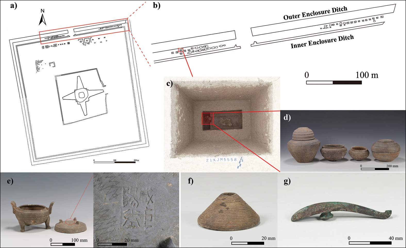
The peripheral area lies between the walls and the outer enclosure ditch. During rescue excavations, 45 Warring States Qin tombs were unearthed between the inner and outer enclosure ditches in the northern section of the peripheral area (hereafter referred to as ‘inter-ditch’ tombs) (Figure 2). All tombs were constructed in vertical pits or earth caves, with single wooden coffins or without burial furniture, and individuals were primarily interred along an east–west orientation with legs tightly flexed (Figure 2c). Most tombs lack luxury burial goods and are primarily accompanied by utilitarian pottery, tools and decorations. Some of the pottery is marked with the inscriptions of Xianyang city government pottery workshop (Figure 2e), which reflects the Qin artisan accountability system (物勒工名), confirming the late Warring States date (Yuan Reference Yuan2008). A manual phalanx from tomb XJM5558 was submitted for radiocarbon dating, returning a calibrated age of 389–208 cal BC (95.4% confidence), which falls within the mid–late Warring States period (Figure S2).
Excavation of the Shangzhaoyao Qin mausoleum: a) plan view of the Shangzhaoyao Qin mausoleum; b) planimetric layout of small satellite tombs between the inner and outer enclosure ditches; c) tomb XJM5558 during excavation; d) pottery from tomb XJM5558; e) inscriptions on the lid of a pottery tripod from tomb XJM5554; f) spindle whorl from tomb XJM5540; g) belt hook from tomb XJM5557 (photographs by H.Q. Zhao; figure by W.H. Cheng & H.Q. Zhao).

Figure 2 Long description
The image contains one diagram, one photo, and five illustrations. The diagram shows the plan view of the Shangzhaoyao Qin mausoleum and the planimetric layout of small satellite tombs between the inner and outer enclosure ditches. The diagram includes labels for the outer enclosure ditch and inner enclosure ditch, with a scale of zero to one hundred meters. The photo shows tomb XJM5558 during excavation, with a red box highlighting a specific area. The illustrations depict various artifacts: pottery from tomb XJM5558, including a large jar and smaller bowls with a scale of zero to two hundred millimeters; inscriptions on the lid of a pottery tripod from tomb XJM5554 with a scale of zero to one hundred millimeters; a spindle whorl from tomb XJM5540 with a scale of zero to twenty millimeters; and a belt hook from tomb XJM5557 with a scale of zero to forty millimeters.
Materials and methods
The skeletal remains of 43 individuals from the 45 inter-ditch tombs were included in analysis. Overall, the skeletal preservation is generally good, with 33 individuals well preserved and 10 individuals represented by fragments. Determination of biological sex and age-at-death estimation followed standard procedures (Lovejoy et al. Reference Lovejoy1985; Buikstra & Ubelaker Reference Buikstra and Ubelaker1994). Based on age estimations, individuals were categorised into six age intervals for ease of analysis: 0–10, 10–20, 20–30, 30–40, 40–50 and 50+ years.
‘Frailty’ is defined as physiological stress accrued throughout an individual’s lifespan, which predisposes them to increased risk of morbidity and mortality (Zedda et al. Reference Zedda2021; Yaussy et al. Reference Yaussy2024). As a proxy for the general health status of the inter-ditch group, we evaluated 11 biomarkers—short stature, low body mass, linear enamel hypoplasia, periodontal disease, rickets, cribra orbitalia, porotic hyperostosis, periostitis, osteoarthritis and other joint disease, vertebral diseases and trauma (see Table S1)—following the criteria for the biological index of frailty (BIF) (Zedda et al. Reference Zedda2021). For application of the BIF, at least three biomarkers should be observable. Each biomarker receives a weighted score, dependent on presence/absence and appearance, and summed scores fall into three frailty categories: low (0 ≤ BIF < 21), medium (21 ≤ BIF < 53) and high (53 ≤ BIF ≤ 100).
To reconstruct activity patterns, we observed two primary skeletal activity markers: degenerative joint disease (including osteoarthritis (OA) and vertebral degeneration) and entheseal changes (Waldron Reference Waldron and Buikstra2019). The large synovial joints of the shoulder, elbow, wrist, hip, knee and ankle were examined for OA, based on the criteria proposed by Waldron (Reference Waldron2008) (Table S2). Vertebral degeneration was assessed by recording the grades of vertebral osteophytes (bony spurs projecting from the vertebral body) and ossification of ligamentum flavum (connecting the laminae of adjacent vertebrae), following the standards of Hou and colleagues (Reference Hou2017). Observations were made in the cervical, upper thoracic (T1–T9), lower thoracic (T10–T12) and lumbar vertebrae. This enabled the calculation of a mean degenerative joint disease severity score for each anatomical region (see Table S2). Entheseal changes refer to macroscopic alterations to the bone surface at the sites of tendonous or ligamentous attachments. Using the method of Villotte et al. (Reference Villotte2010), these changes were categorised as either ‘healthy’ or ‘enthesopathy’ (Table S2). Morphological changes were recorded at 21 fibrocartilaginous entheses (Villotte & Knüsel Reference Villotte and Knüsel2013; Weiss Reference Weiss2015), which were grouped by the biomechanical functions of associated joints (Table S3). Individuals aged greater than 50 years or otherwise displaying osteogenic pathologies were excluded from analysis to minimise age-related influence among activity markers.
Statistical analyses were conducted using SPSS Statistics v.27. As sample data were non-normal, we used the Kruskal–Wallis test to compare mean BIF values across different age groups. The Wilcoxon signed-rank test was applied to assess symmetry in OA and entheseal changes for bilateral joints and between the upper and lower limbs.
Results
Biological sex and age-at-death
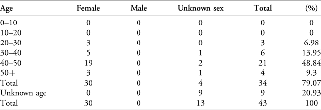
Biological sex was not osteologically determinable for 13 individuals, the remaining 30 individuals are determined to be female. Age-at-death was estimated for 34 individuals: most died between the ages of 40 and 50 years, no individuals under 20 years are recorded (Table 1).
Gender and age-at-death distribution of the inter-ditch group.

Table 1 Long description
The table presents the gender and age-at-death distribution of the inter-ditch group, consisting of 43 individuals. The table has 10 rows and 5 columns. The columns are labeled Age, Female, Male, Unknown sex, and Total (percentage). The rows are labeled with age ranges: 0-10, 10-20, 20-30, 30-40, 40-50, 50+, Total, Unknown age, and Total. The data shows that no individuals under 20 years are recorded. Most individuals died between the ages of 40 and 50 years, with 19 females and 2 unknown sex individuals in this age range, totaling 21 individuals or 48.84 percentage. Biological sex was not determinable for 13 individuals. The remaining 30 individuals are determined to be female.
Frailty assessment
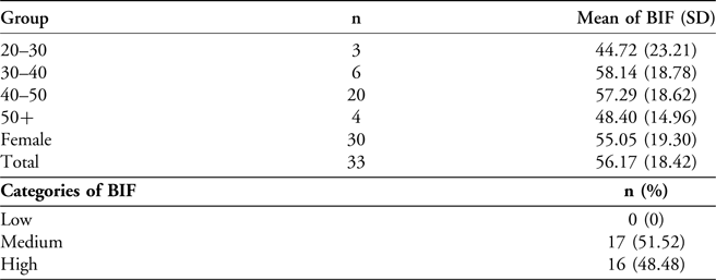
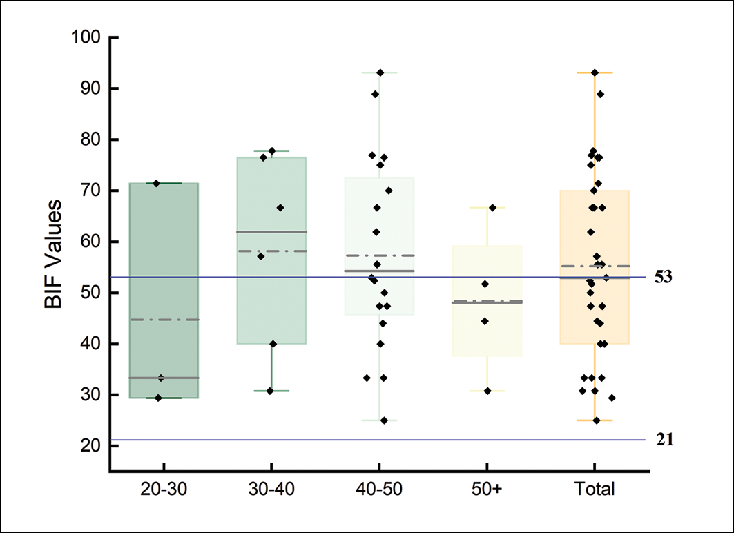
Thirty-three individuals met the criteria for BIF evaluation. A mean BIF indicative of high frailty is observed in the 30–40 and 40–50 age groups, while the mean BIF of the 20–30 and over 50 age groups indicates medium frailty (Tables 2 & S4). No significant differences in BIF were found across age cohorts (p = 0.538). Overall, the inter-ditch group exhibited a high mean BIF, with almost half of individuals classified as high frailty and none as low frailty.
Comparisons between mean values and categories of BIF.

Table 2 Long description
The table presents data on 33 individuals evaluated for BIF, divided into age groups: 20-30, 30-40, 40-50, and 50+. It includes the number of individuals in each group and the mean BIF with standard deviation. The age groups are 20-30, 30-40, 40-50, and 50+, with respective mean BIF values of 44.72, 58.14, 57.29, and 48.40. The table also categorizes BIF into low, medium, and high, with no individuals in the low category, 17 in the medium category, and 16 in the high category. The overall mean BIF is 56.17. The table highlights that the 30-40 and 40-50 age groups show high frailty, while the 20-30 and over 50 age groups indicate medium frailty. No significant differences in BIF were found across age cohorts.
Abbreviation: SD, standard deviation.
A high (exceeding 85%) prevalence of vertebral degenerative diseases, periostitis, cribra orbitalia and periodontal disease is observed in the inter-ditch group. Over 70 per cent of individuals exhibit porotic hyperostosis, OA and other joint diseases (Table S5, Figures S3 & S5), and 10 observations of healed antemortem trauma in eight individuals can be made (Table S5, Figure S4). No cases of rickets are identified.
Activity markers
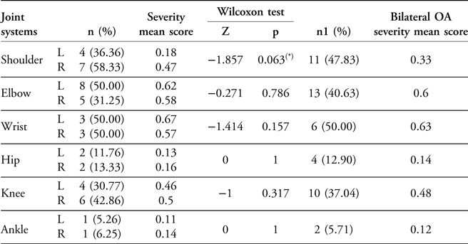
Most individuals (n = 25, 72%) exhibited OA during their lifetime. No statistically significant asymmetry in OA severity was detected (Tables 3 & S6, Figure S5), although slight bilateral differences are observed at the shoulder joints. Therefore, subsequent analyses combined both sides. OA severity is higher in the elbows, wrists, knees and right shoulder than in other joints, and is greater in the upper limbs than the lower limbs, though this latter difference is not statistically significant (Z = −1.79, p = 0.073, n = 19).
The prevalence and severity of OA at joint systems in the inter-ditch group.

Table 3 Long description
The table presents data on the prevalence and severity of osteoarthritis (OA) in different joint systems within the inter-ditch group. It includes columns for joint systems, the number and percentage of cases, severity mean score, Wilcoxon test results, and bilateral OA severity mean score. The joint systems analyzed are shoulder, elbow, wrist, hip, knee, and ankle. The table shows that the shoulder has 4 left and 7 right cases with a severity mean score of 0.18 and 0.47 respectively. The elbow has 8 left and 5 right cases with a severity mean score of 0.62 and 0.58 respectively. The wrist has 3 left and 3 right cases with a severity mean score of 0.67 and 0.57 respectively. The hip has 2 left and 2 right cases with a severity mean score of 0.13 and 0.16 respectively. The knee has 4 left and 6 right cases with a severity mean score of 0.46 and 0.5 respectively. The ankle has 1 left and 1 right case with a severity mean score of 0.11 and 0.14 respectively. The Wilcoxon test results indicate no statistically significant asymmetry in OA severity, although slight bilateral differences are observed at the shoulder joints.
Note: (*) almost significant p value
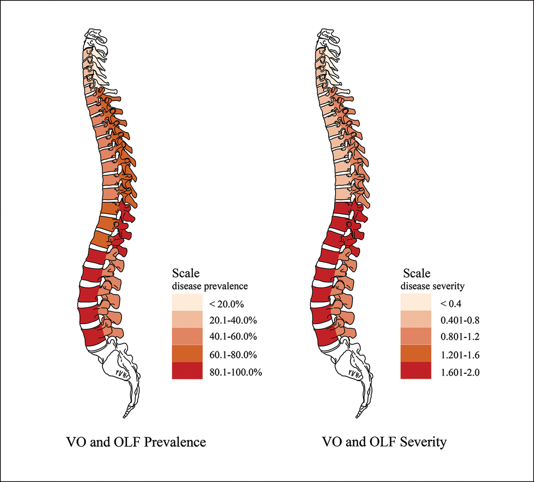
Vertebral osteophytes affect 94.74% of examined adults (n = 19), and ossification of ligamentum flavum affects all assessable individuals (n = 14). More than half of assessable vertebral body margins and pedicles display vertebral osteophytes (57.41%) and ossification of ligamentum flavum (57.78%), respectively. Vertebral degeneration is most frequent and severe in the lower thoracic vertebrae, followed by the lumbar and cervical regions (Figure 3). Specifically, vertebral osteophytes are more prevalent and severe in the lumbar and lower thoracic regions, while ossification of ligamentum flavum is common throughout the thoracic vertebrae, with greater severity in the lower thoracic region (Tables S7 & S8, Figure S5).
The prevalence and severity of vertebral osteophytes (VO; n = 18) and ossification of ligamentum flavum (OLF; n = 14) in the inter-ditch group (figure by W.H. Cheng).

Figure 3 Long description
The heat map illustrates the prevalence and severity of vertebral osteophytes (VO) and ossification of ligamentum flavum (OLF) in the inter-ditch group. The left side of the heat map shows the prevalence of VO and OLF, with a color scale ranging from light peach to dark red. Light peach indicates a prevalence of less than 20 percent, while dark red indicates a prevalence of 80.1 to 100 percent. The right side of the heat map shows the severity of VO and OLF, with a color scale ranging from light peach to dark red. Light peach indicates a severity of less than 0.4, while dark red indicates a severity of 1.601 to 2.0. The heat map uses a grid layout to represent the spine, with each segment of the spine color-coded according to the prevalence and severity of VO and OLF. The heat map provides a visual representation of the distribution and intensity of these conditions across different segments of the spine.
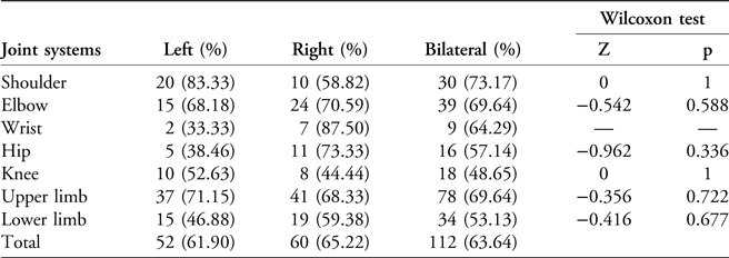
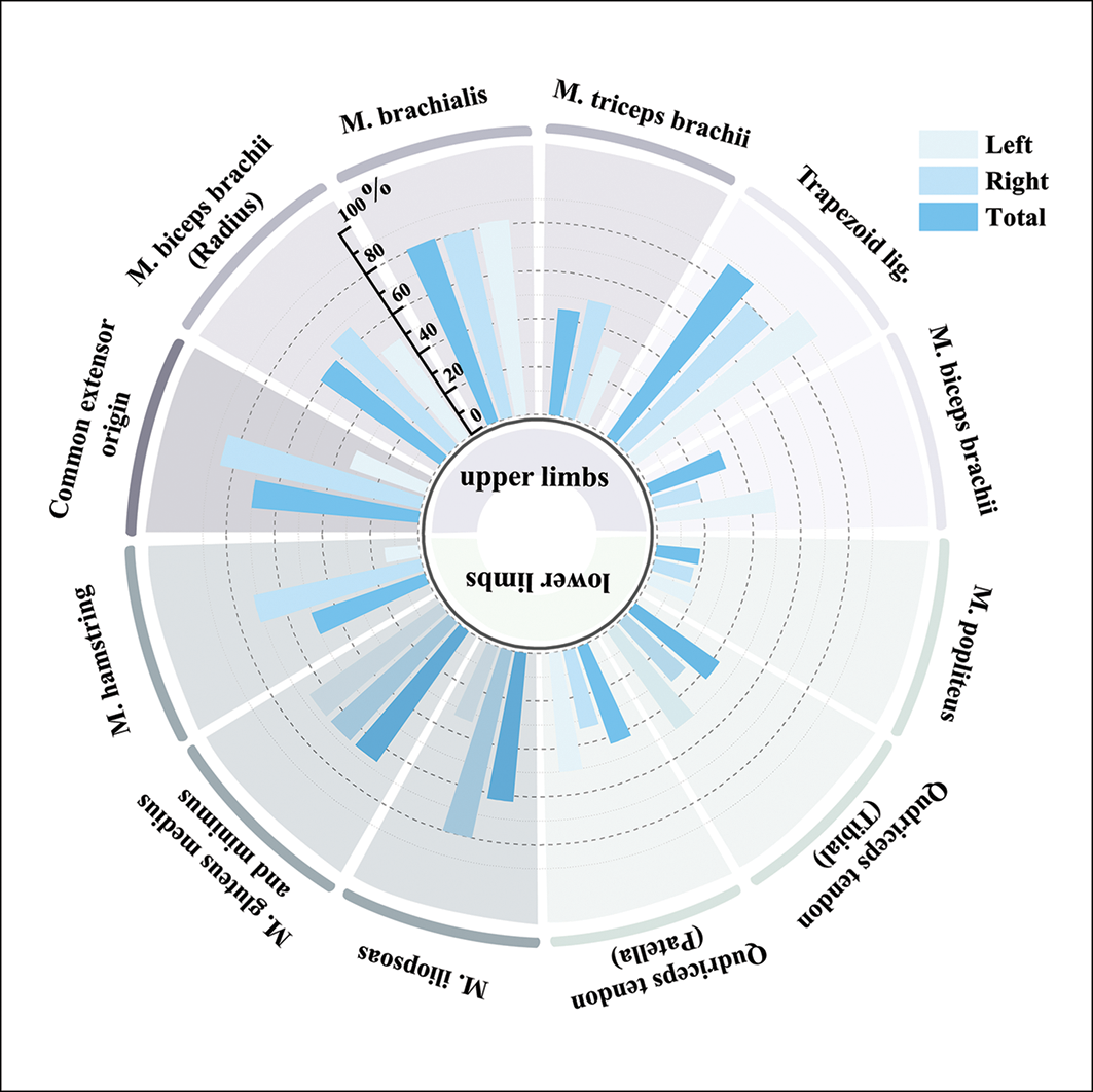
Most individuals (n = 25, 96%) exhibit enthesopathies, with 63.64% of examined entheses showing pathological alterations. No statistically significant asymmetry was detected at any bilateral joint (Table 4), allowing the combination of data from both sides for subsequent analysis. Entheses associated with upper-limb joint activity exhibit more severe pathological changes than those in the lower limbs (Z = −2.63, p = 0.009, n = 17; Table S9). To improve the reliability results, we also conducted frequency analyses on adequately preserved entheses (Figure 4). The results revealed a higher prevalence of pathological changes at the attachments for the trapezoid ligament, the brachialis muscle and the common extensor tendon.
The prevalence of enthesopathy at joint systems in the inter-ditch group.

Table 4 Long description
The table presents data on the prevalence of enthesopathy at different joint systems in the inter-ditch group. It includes percentages for left, right, and bilateral joints, as well as results from the Wilcoxon test. The joint systems analyzed are shoulder, elbow, wrist, hip, knee, upper limb, and lower limb. The table shows that the shoulder has a higher prevalence on the left side (83.33 percentage) compared to the right side (58.82 percentage), with a bilateral prevalence of 73.17 percentage. The elbow shows a similar prevalence on both sides, with 68.18 percentage on the left and 70.59 percentage on the right, resulting in a bilateral prevalence of 69.64 percentage. The wrist has a lower prevalence on the left side (33.33 percentage) compared to the right side (87.50 percentage), with a bilateral prevalence of 64.29 percentage. The hip shows a prevalence of 38.46 percentage on the left and 73.33 percentage on the right, with a bilateral prevalence of 57.14 percentage. The knee has a prevalence of 52.63 percentage on the left and 44.44 percentage on the right, with a bilateral prevalence of 48.65 percentage. The upper limb shows a higher prevalence on the left side (71.15 percentage) compared to the right side (68.33 percentage), with a bilateral prevalence of 69.64 percentage. The lower limb shows a prevalence of 46.88 percentage on the left and 59.38 percentage on the right, with a bilateral prevalence of 53.13 percentage. Overall, the total prevalence is 61.90 percentage on the left, 65.22 percentage on the right, and 63.64 percentage bilaterally. The Wilcoxon test results indicate no statistically significant asymmetry at any bilateral joint.
The prevalence of enthesopathy at muscle origin/insertion sites in the inter-ditch group (only including sites for which n≥3) (figure by W.H. Cheng).

Figure 4 Long description
A radar chart displays the prevalence of enthesopathy at muscle origin and insertion sites in the inter-ditch group. The chart is divided into two main sections: upper limbs and lower limbs. Each section is further divided into specific muscle groups, such as M. biceps brachii, M. triceps brachii, M. brachialis, and others for the upper limbs, and M. iliopsoas, M. gluteus maximus, and others for the lower limbs. The chart uses three different shades of blue to represent data for the left side, right side, and total prevalence. Each muscle group has corresponding bars indicating the percentage prevalence of enthesopathy, with the scale ranging from 0 to 100 percent. The chart highlights variations in the prevalence of enthesopathy across different muscle groups and sides of the body.
Discussion
Sex, age-at-death and health status
The female bias in the inter-ditch tombs is unusual, contrasting with sex composition at contemporaneous and geographically proximate cemeteries (e.g. Pujiazhai; Han Reference Han2023). This bias could indicate a shared activity-based identity among the interred individuals, as observed in wartime mass graves (Viva et al. Reference Viva2020), consort burial areas within royal cemeteries (Ouyang & Song Reference Ouyang and Song1975) and certain areas in monastic cemeteries (Judd Reference Judd2020). The organised spatial planning within the burial array supports the designation of different areas for specific funerary purposes.
Superposition of some tombs suggests that individuals were not buried simultaneously but probably died of natural causes and were subsequently interred in predetermined locations. The age-at-death profile therefore reflects health status before death. Notably, the inter-ditch group displays a left-skewed normal distribution (skewness = −0.457, kurtosis = 0.017) that could result from multiple factors. Limitations of the burial environment may prevent the preservation of younger individuals. And socioenvironmental conditions may have provided favourable living standards or healthcare resources that reduced early-life mortality.
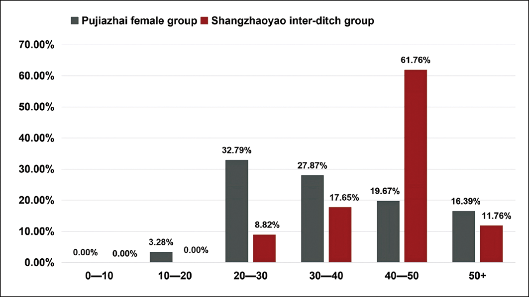
Comparison with females from the predominantly agricultural Pujiazhai cemetery (Han Reference Han2023) reveals distinct differences in the age-at-death distribution (Figure 5). The inter-ditch group shows lower mortality rates before age 40, with a relative postponement in the mortality peak of approximately two decades. This longevity difference is likely related to microenvironmental advantages of the Qin palace. Relatively stable medical conditions and food supplies (Shuihudi Qin Tomb Bamboo Slips Editorial Group 1990) would reduce the adverse risks of disease, accidents and childbirth for palace inhabitants. Moreover, the royal court may have implemented age- or health-based selection criteria for those entering, leading to the observed left-skewed age distribution.
Comparison of age-at-death profiles of the Shangzhaoyao inter-ditch group and Pujiazhai females (figure by W.H. Cheng).

Figure 5 Long description
The bar graph compares age-at-death profiles of two groups: Pujiazhai females and Shangzhaoyao inter-ditch group. The x-axis represents age ranges: 0-10, 10-20, 20-30, 30-40, 40-50, and 50+. The y-axis represents the percentage of individuals in each age range, from 0% to 70%. The Pujiazhai female group is represented by gray bars, and the Shangzhaoyao inter-ditch group is represented by red bars. For the 0-10 age range, both groups have 0% representation. In the 10-20 age range, the Pujiazhai female group has 3.28%, while the Shangzhaoyao inter-ditch group has 0%. In the 20-30 age range, the Pujiazhai female group has 32.79%, and the Shangzhaoyao inter-ditch group has 8.82%. For the 30-40 age range, the Pujiazhai female group has 27.87%, and the Shangzhaoyao inter-ditch group has 17.65%. In the 40-50 age range, the Pujiazhai female group has 19.67%, while the Shangzhaoyao inter-ditch group has 61.76%. For the 50+ age range, the Pujiazhai female group has 16.39%, and the Shangzhaoyao inter-ditch group has 11.76%. All values are approximated.
The overall high mean BIF of the inter-ditch group does, however, reflect generally poor lifetime health. These individuals experienced multiple stress events and episodes of frailty throughout their lifetimes, resulting in an accumulation of skeletal biomarkers (Marklein et al. Reference Marklein2016). Through detailed analysis of specific biomarkers (Table S5) focusing on early-life health status, we find that nearly half of inter-ditch individuals exhibit linear enamel hypoplasia, suggesting they experienced nutritional deficiency or disease during infancy or early childhood. Similarly, the widespread presence of cribra orbitalia and porotic hyperostosis suggests that individuals endured prolonged or recurrent nutritional stress in their early life. Moreover, the high prevalence of OA and vertebral diseases indicates that the group experienced repeated mechanical stress during adulthood.
Prolonged exposure to universal environmental stressors and similar dietary, labour and living conditions could result in comparable rates of frailty accumulation across different age groups. The consistent presence of individuals with medium to high levels of frailty across all age groups within the inter-ditch tombs, regardless of sample size (Figure 6), suggests that the health status of this group has both homogeneity (common environmental stress) and heterogeneity (individual differences in resilience and stress-buffering capacity). In the same stressful environment, frail individuals are more sensitive to external hazards and more prone to premature death than those individuals with higher adaptive capacity, who are more likely to survive these stresses into middle–late adulthood (Wood et al. Reference Wood1992). This filtration process creates a convergence of health status across different age cohorts, as observed here.
Boxplot with jittered points for BIF values across age groups in the inter-ditch group (figure by W.H. Cheng).

Figure 6 Long description
A box-and-whisker plot with jittered points displays BIF values across different age groups in the inter-ditch group. The x-axis represents age groups: 20-30, 30-40, 40-50, 50+, and Total. The y-axis represents BIF values ranging from 20 to 100. Each box plot shows the distribution of BIF values for the respective age group. The boxes indicate the interquartile range (IQR), with the lower and upper quartiles marked by the edges of the boxes. The median values are represented by the horizontal lines within the boxes. The whiskers extend to the minimum and maximum values within 1.5 times the IQR from the quartiles. Outliers are shown as individual points outside the whiskers. The 20-30 age group has a median around 50, with values ranging from approximately 30 to 70. The 30-40 age group has a median around 60, with values ranging from approximately 40 to 80. The 40-50 age group has a median around 50, with values ranging from approximately 30 to 80. The 50+ age group has a median around 40, with values ranging from approximately 20 to 60. The Total group has a median around 53, with values ranging from approximately 21 to 90. The plot indicates variability in BIF values across different age groups, with some overlap in the ranges. All values are approximated.
Signs of healing are also observable in approximately half of individuals exhibiting cribra orbitalia and porotic hyperostosis and in almost all cases of periostitis. These findings could suggest that most individuals had physiological resilience that enabled them to survive and recover from episodic stress (McFadden & Oxenham Reference McFadden and Oxenham2020; DeWitte Reference DeWitte2024). Alternatively, individuals may have later lived in a relatively stable environment with access to communal care (Tilley Reference Tilley2015), which is also evidenced by the healed ante-mortem trauma.
Activity patterns
Everyday biomechanical loading associated with an upright posture and bipedal locomotion contributes to the degeneration of the joints of the lower limbs and lower vertebrae (Zhang et al. Reference Zhang2017). Prolonged exposure to additional biomechanical stress, for example through repetitive actions during labour or the maintenance of abnormal postures, can result in different patterns of degeneration and entheseal change (Klaus et al. Reference Klaus2009). The inter-ditch group exhibits degeneration in the thoracolumbar region, upper limbs and knee joints, and changes to the entheses of the upper limbs are more severe than those of the lower limbs, reflecting habitual actions.
Individuals may have engaged in daily activities that involved the repetitive flexion of the thoracolumbar region or the maintenance of a flexed posture. The presence of vertebral osteophytes and ossification of ligamentum flavum indicate chronic loading of the intervertebral discs, possibly linked to repetitive spinal flexion (Waldron Reference Waldron2008; Hirabayashi Reference Hirabayashi2017). A wedge-shaped compression fracture, observed in an upper thoracic vertebra from individual XJM5519 (Figure S4c), also suggests prolonged flexion of the spine (Curate et al. Reference Curate2016).
Pathological changes to the bones of the upper limb indicate that the joints and muscles of the arms and shoulders were frequently engaged or remained tense during daily tasks. OA in these non-weight-bearing joints may indicate repetitive mechanical loading (Palmer et al. Reference Palmer2016), as may entheseal changes (Villotte et al. Reference Villotte2010). Though severity was lower than the elbow and wrist joints, the shoulder joints showed a relatively high prevalence. And the prevalence of enthesopathy at the trapezius ligament, which is primarily involved in scapular elevation and upward rotation (Mansfield & Neumann, Reference Mansfield and Neumann2018), exceeded 80% bilaterally. These together suggest inter-ditch group’s long-term engagement in activities involving frequent shoulder movements. Entheseal changes associated with the brachialis muscle and common extensor tendon further suggest that the elbow and wrist joints were subject to frequent and prolonged use.
The general lack of significant asymmetry in the expression of OA and entheseal changes suggests engagement in activities that resulted in relatively balanced bilateral loading. However, slight right-sided bias is observed in both the severity of shoulder OA and the frequency of enthesopathy at the wrist extensor tendon. Together, these might suggest frequent use of the right forearm, possibly in conjunction with the shoulder for repetitive wrist dorsiflexion or radial abduction (Schünke et al. Reference Schünke2011).
The knee joints also indicate frequent movement and high-stress loading in the inter-ditch group. In addition to the effects of daily ambulation, prevalent kneeling-sitting behaviour influenced by ritual practices can impact degeneration (Zhao et al. Reference Zhao2020). Kneeling-sitting with the hips resting against the heels, a culturally ritual resting position in ancient China (Lin Reference Lin1993), would hold the knees in a hyper-flexed attitude for extended periods, exacerbating knee OA and enthesopathies.
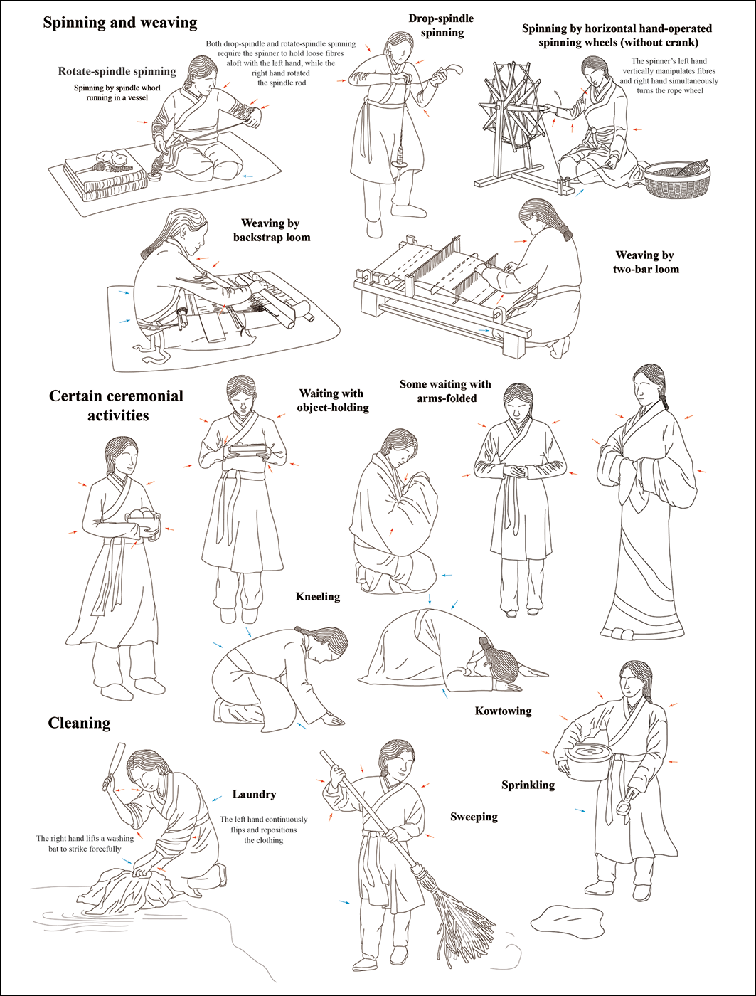
Considering the bioarchaeological evidence alongside archaeological findings and historical records, we hypothesise that the females from the inter-ditch group engaged in daily activities such as spinning and weaving (Figure 7). Spindle whorls were included as burial goods in two tombs (XJM5540 and XJM5556), strongly suggesting that spinning formed an essential part of daily life for at least some individuals. The dominant spinning techniques in the Warring States period included drop-spindle and rotate-spindle spinning, while horizontal hand-operated spinning wheels (without a crank) were just emerging (Xia Reference Xia1972; Li et al. Reference Li2020). These spinning processes require bilateral shoulder mobility and elbow flexion, with right forearm-driven wrist abduction or internal rotation. Weaving activities during the Warring States period were primarily carried out on backstrap looms (Li et al. Reference Li2020), with some evidence suggesting the emergence of two-bar looms (see Figure 7) (Saitaer & Li Reference Saitaer and Li2019). Upper-limb activity patterns for operating either type of loom are similar: beating the weft requires continuous bilateral elbow flexion and wrist extension. In addition, operation of the backstrap loom requires the use of a backstrap and tension created by holding the end bar with the feet, which would amplify loading on the thoracolumbar regions.
Reconstruction of possible habitual activities of the inter-ditch group (joint systems involved in upper-limb activity are indicated by red arrows, while those involved in spine and lower-limb activities are marked with blue arrows) (figure by W.H. Cheng).

Figure 7 Long description
The illustration depicts individuals performing different tasks. In the spinning and weaving section, people are shown using various tools like the rotate-spindle, drop-spindle, and horizontal hand-operated spinning wheels. They are also weaving using backstrap looms and two-bar looms. The certain ceremonial activities section shows individuals in different poses such as waiting with object-holding, kneeling, and some with arms folded. The cleaning section illustrates people doing laundry, sprinkling, and sweeping. Red and blue arrows indicate joint systems involved in upper-limb, spine, and lower-limb activities.
Other potential activities include cleaning and ritual practices. The Rites of Zhou (周礼) record that court servants were primarily responsible for palace cleaning duties (Xu & Chang Reference Xu and Chang2014), such as sprinkling, sweeping or laundry. Furthermore, the inter-ditch group might frequently participate in certain ceremonial mausoleum rituals involving waiting with arms-folded or object-holding and kneeling kowtow (Shuihudi Qin Tomb Bamboo Slips Editorial Group 1990). These activities all require co-ordinated use of both upper limbs to regulate joint movements and distribute load, while prolonged kneeling postures exacerbate mechanical loading of the spine and knees.
Social identity
According to historical records, women of the Qin court were primarily either royal consorts or palace servants, the latter further subdivided into female administrators, handmaidens and menials (Sima Reference Sima1999; Liang & Wang Reference Liang and Wang2022). Royal consorts typically came from elite families or political marriage unions (Liu Reference Liu2018) and served political and reproductive roles. Before entering the palace, they generally enjoyed high social status and maintained robust health. After entering the court hierarchy, consorts received salaries commensurate with their rank and were exempt from heavy labour (Liang & Wang Reference Liang and Wang2022). However, only queens and dowagers received high-status burials; other consorts were interred in small tombs (Ban Reference Ban1962). Palace servants typically endured hardships in early life, entering service through sale, demotion, confiscation or provision by subordinate states (Liu Reference Liu1984; Shuihudi Qin Tomb Bamboo Slips Editorial Group 1990). Female administrators and handmaidens engaged in light domestic duties for the ruling class, while menials undertook more miscellaneous tasks.
The relatively poor health status of the individuals from the inter-ditch tombs indicates unstable nutritional support during early life. Archaeologically, their peripheral burial location, tomb typology and limited grave goods collectively indicate a low social status (Teng Reference Teng2003). These individuals are therefore more likely to have been palace servants. Activity patterns, reflecting repetitive and high-intensity daily activities involving the upper limbs, also support this inference, suggesting an identity as lower-ranking female menials.
Bamboo slips from the Shuihudi Qin Tomb document the role of ‘Dianren’ (甸人), “those who guard or attended the mausoleums of Duke Xiao and Duke Xian of Qin” (“守孝公、(献)公冢者殹”; Shuihudi Qin Tomb Bamboo Slips Editorial Group 1990). This indicates that the Qin state had already recognised the need to look after the mausoleum during the late Warring States period. Partial building foundations uncovered in the northern part of the Shangzhaoyao mausoleum possibly served ritual purposes or as residences for mausoleum-related personnel (Shang Reference Shang1994). Given the diachronic burial pattern of the individuals studied here, we infer that they were likely involved in the maintenance of the mausoleum before their death.
Reconstructions of social identity must, however, consider the complex interplay between identity and imperial authority. Before these individuals were associated with mausoleum affairs, they may have held various roles as palace females, with certain court interventions potentially enforcing identity transitions. Archaeological interventions primarily reveal aspects of social identity at the time of death, but shifts in identity throughout an individual’s lifetime and the minutia of status are more difficult to discern. Therefore, it is important to avoid oversimplified or excessive interpretation regarding the social identity of an individual or a group.
It is likely that the individuals of the inter-ditch group served the Qin royal household during their lifetimes and were buried alongside the mausoleum in death, eternally attending to their ruler. This arrangement not only reflects their function as societal tools but also reveals the rigid hierarchical and ritual systems of the Qin state, where some individuals existed to satisfy the needs of the ruling class, their social identities eternally bound to power service frameworks.
Conclusion
The reconstruction of health status, activity patterns and social identity in a group of 43 individuals buried between the inner and outer enclosure ditches in the northern sector of the Shangzhaoyao Qin mausoleum provides important bioarchaeological evidence for understanding the layout of WSQMs.
The relatively poor health with prolonged engagement in high-intensity daily labour, placing considerable stress on the upper limbs and thoracolumbar region, suggest that the inter-ditch individuals were lower-ranking palace menials. Probably deriving from low-status origins, they nevertheless achieved a structural survival advantage over contemporaneous agricultural cohorts through the palace’s stable basic support. However, this social status afforded them a perpetual servile association with their ruler.
Meanwhile, the absolute female-biased composition of the inter-ditch group also reflects deliberate choices in mausoleum layout based on hierarchical ranking and functional zoning. This ‘order in death’ is not only an extension of the Qin power structure, it also embodies the Qin funerary ideology that the dead are treated as the living.
Acknowledgements
We thank the Shaanxi Academy of Archaeology for providing the skeletal material.
Funding statement
Work was funded by the National Social Science Fund of China (25AKG003) and supported by the Archaeological Talent Promotion Program of China (2024-270; 2025-187), the Shandong Social Science Planning Fund Program (21CKGJ03) and the Institute of Cultural Heritage of Shandong University Academic Research Fund (12360050080200).
Online supplementary material (OSM)
To view supplementary material for this article, please visit https://doi.org/10.15184/aqy.2026.10307 and select the supplementary materials tab.