




No CrossRef data available.
Article contents
Percutaneous transluminal coronary angioplasty for anastomotic stenosis after coronary arterial bypass grafting in Kawasaki disease
Published online by Cambridge University Press: 24 May 2005
Abstract
Objectives: We evaluated the efficacy of percutaneous transluminal coronary angioplasty for anastomotic stenosis after coronary arterial bypass grafting using the internal thoracic artery in patients with coronary arterial lesions due to Kawasaki disease. Subjects and Methods: From July 1997 to April 2000, four boys and one girl underwent percutaneous transluminal coronary angioplasty for 6 anastomotic lesions following coronary arterial bypass grafting using the left or right internal thoracic artery. Progressive severe stenosis of the grafts in the follow-up angiograms after grafting, and evidence of ischemia, were regarded as indications for percutaneous transluminal coronary angioplasty. Age at coronary angioplasty ranged from 4.2 to 16.7 years, with a median of 6.9 years, while the interval from operation ranged from 0.3 to 3.0 years, with a median of 1.1 years. The diameter of the balloon catheter employed varied from 1.5 to 2.5 mm, and the pressure of inflation ranged from 8 to 16 atmospheres. Results: The degree of stenosis decreased from 63 to 99%, with a median of 88%, to 0 to 40%, with a median of 17% immediately after angioplasty. A follow-up angiogram either 3 months or 1 year later revealed no restenosis in any patient. Conclusion: Percutaneous transluminal coronary angioplasty is a feasible and useful procedure for treating anastomotic stenosis following coronary arterial bypass grafting using the internal thoracic artery in patients with coronary arterial lesions due to Kawasaki disease.
Information
- Type
- Original Article
- Information
- Copyright
- © 2003 Cambridge University Press
Percutaneous transluminal coronary angioplasty is a standard treatment for adults with an anastomotic stenosis after coronary arterial bypass grafting.1–4 In children with a history of Kawasaki disease, however, to the best of our knowledge there is but one case report5 describing this procedure. Percutaneous transluminal coronary angioplasty, and percutaneous transluminal coronary rotational ablation, have recently been performed for severe localized stenosis.6–10 Experience with interventional catheterisation in Kawasaki disease, nonetheless, is very limited. It is coronary arterial bypass grafting that is the essential procedure for revascularization of stenotic lesions due to Kawasaki disease. Though bypass grafting using the internal thoracic artery is the most feasible procedure, the patency rate in our institution had declined with time, being 90.9%, 74.1%, and 74.1% at 1, 5, and 10 years, respectively.11 In small children in particular, patency rates are even lower.12–13 Occlusion of the grafts often occurs early after operation. To relieve the degree of anastomotic stenosis, and to prevent the occlusion of grafts in patients with a history of Kawasaki disease, we performed percutaneous transluminal coronary angioplasty for anastomotic stenosis seen after coronary arterial bypass grafting.
Methods
Patients
From July 1997 to April 2000, four boys and one girl with Kawasaki disease underwent percutaneous transluminal coronary angioplasty for 6 anastomotic stenoses following coronary arterial bypass grafting using the left or right internal thoracic artery. Three branches of either the left anterior descending or the right coronary arteries had already occluded before coronary arterial bypass grafting. Three other branches of the left anterior descending artery showed severe localized stenoses. The clinical characteristics of the patients are summarized in Table 1.
Table 1. Characteristics of patients.

Age at percutaneous transluminal coronary angioplasty ranged from 4.2 to 16.7 years, with a median of 6.9 years, while the interval from operation ranged from 0.3 to 3.0 years, with a median of 1.1 years. In all patients, progressive severe stenosis of the grafts was revealed by follow-up angiograms. Dipyridamole stress 99mTc myocardial imaging, and treadmill stress testing, were performed before and after coronary angioplasty. Ischemic signs were detected in 5 patients before coronary angioplasty (Table 2). Aspirin, at 1–1.5 mg/kg, or cilostazol had been administered.
Table 2. Signs of ischemia.

Percutaneous transluminal coronary angioplasty
Nifedipine, at 0.3 to 0.6 mg/kg per day, was administered for 3 days before the procedure. In three patients, the procedure was performed under general anesthesia. A 12-lead electrocardiogram was used for monitoring throughout the procedure. A 6 or 5 French size sheath was placed in either the right or left femoral artery, and 100 units of heparin per kilogram were administered intravenously. The graft was cannulated with a 5 or 6 French size Judkins right guiding catheter, and nitroglycerin at 3 to 5 μg/kg was administered prior to the procedure. The balloon was selected according to the diameter of the coronary artery adjacent to the stenotic lesion in the angiogram. The balloon catheter was passed across the lesion under biplane fluoroscopy. The diameter ranged from 1.5 to 2.5 mm, and the pressure used for inflation ranged from 8 to 16 atmospheres (Table 3).
Table 3. Results of angioplasty.

Follow-up coronary angiography was performed 3 months after coronary angioplasty in 4 patients, and at 1 year in 2 patients. One patient was studied at both 3 months and 1 year. Flow in the internal thoracic arterial graft was studied in 4 patients at 3 years after coronary angioplasty using thransthoracic color Doppler echocardiography to check the patency of the graft.
Results
The angioplasty was effective, and the stenosis was successfully relieved, in all the patients. The degree of stenosis decreased from 63 to 99%, with a median, of 88%, to 0 to 40%, with a median of 17% (Table 3). During the procedure, transient ischemic changes appeared in the electrocardiogram in two patients, but none of the patients had decreased blood pressure. There were no other complications.
At follow-up angiography, the degree of stenosis ranged from 0 to 20%, with a median of 12%. Significant restenosis was not present in any patient (Fig. 1).

Figure 1. Degree of stenosis (%).
In our first patient, although the graft was almost occluded prior to coronary angioplasty, the stenosis remained completely relieved after one year (Fig. 2). In the second patient, the flow across the graft was good, but the native coronary artery was occluded after coronary angioplasty (Fig. 3). In the third patient, a new stenosis, of 54%, appeared at the distal anastomosis to the coronary artery, although the stenosis in the graft had been improved (Fig. 4). In this patient, it was hard to pass the guide wire across the target lesion during the procedure.

Figure 2. Serial angiograms of the left internal thoracic artery in our first patient. (a) Before percutaneous transluminal coronary angioplasty when the degree of stenosis was 99%, the graft being almost totally occluded. (b) After percutaneous transluminal coronary angioplasty, the degree of stenosis had reduced to 40%. (c) After 1 year, the angiogram revealed no stenosis at the site of anastomosis with the coronary arterial bypass graft.

Figure 3. Angiograms of the left internal thoracic artery (upper) and the left coronary artery (lower) in our second patient. (a) Before percutaneous transluminal coronary angioplasty. Native flow is seen in the left anterior descending artery. (b) Angiogram after 3 months. Though the left internal thoracic arterial graft was patent, the left anterior descending artery was occluded at the proximal end of the anastomosis.

Figure 4. Serial angiograms of the left internal thoracic artery in our third patient. (a) Before percutaneous transluminal coronary angioplasty. It was difficult to pass the guide wire across the lesion in this patient. (b) After percutaneous transluminal coronary angioplasty. (c) A follow-up angiogram at 3 months. There is a new stenosis at the distal portion of the anastomosis.
Signs of ischemia had diminished in three of the five patients (Table 2)
Flow through the left internal thoracic artery, as evidenced by transthoracic color Doppler echocardiography, was detected in our first 4 patients 3 years after the angioplasty. The pattern of flow in the internal thoracic artery revealed good patency in the midterm after the angioplasty.
Discussion
According to a recent national survey in Japan, about 6000 patients are affected by Kawasaki disease each year, while about 30 patients require coronary arterial grafting because of the coronary arterial lesions that are part and parcel of Kawasaki disease. Stenotic lesions due to the Kawasaki disease may progress to ischemic coronary arterial heart disease, and coronary arterial bypass grafting is the standard therapy for these patients.14–16 Although long-term patency of the internal thoracic artery graft is the goal, early occlusion occurs in about one-tenth of the patients.11
It is suspected that anastomotic stenosis after bypass grafting is the reason for occlusion of the graft, resulting from formation of scars at the suture line, and from competition with native coronary arterial flow. Because the diameters of the left internal thoracic artery and coronary artery are small in children, early occlusion is a risk. These are very important factors when we consider the indications for bypass grafting in patients with a history of Kawasaki disease.
Our results with percutaneous transluminal coronary angioplasty for these problems are good, in particular for the two lesions where there was competition with native coronary arterial flow. The degree of stenosis was relieved in the remaining four lesions in which the native coronary artery was occluded.
Follow-up angiograms revealed no cases with significant restenosis of the target vessel, although a new stenosis had appeared in the native artery distal to the anastomosis in one patient. Because it was difficult to advance the guiding wire through this lesion, the guiding wire might have produced some intimal damage to the distal segment of the artery.
Though the reasons for graft stenosis may differ from those in adults,2 we confirm that percutaneous transluminal coronary angioplasty is feasible for dilating anastomotic stenoses after coronary artery bypass grafting in children. The process is effective, and can be done without severe complications.
Occlusion of the graft often occurs within 1 year of the initial operation. Progressive and severe stenosis as seen in the follow-up angiograms, along with signs of ischemia, are regarded as the indications for angioplasty. It has been reported that the length and the diameter of the internal thoracic arterial graft grows in proportion to somatic growth.12, 17 We consider that early relief of anastomotic stenosis is more effective, and believe that timely angioplasty can prevent occlusion of the graft and improve the rate of patency. The midterm results after angioplasty were good, but prolonged follow-up will evaluated with great interest.
Acknowledgments
We thank the cardiologists of the coronary care unit at the National Cardiovascular Center for their support. We also thank Professor Peter and Dr Setsuko Olley for their kind help with the English language.
References
Dimas A, Arora R, Whitlow P, et al. Percutaneous transluminal angioplasty involving internal mammary artery grafts. Am Heart J 1991; 122: 423–429.Google Scholar
Shimshak T, Giorgi L, Johnson W, et al. Application of percutaneous transluminal coronary angioplasty to the internal mammary artery graft. J Am Coll Cardiol 1988; 12: 1205–1214.Google Scholar
Smith S, Dove J, Jacobs A, et al. ACC/AHA guidelines for percutaneous coronary intervention (revision of the 1993 PTCA guidelines) – executive summary. Circulation 2001; 103: 3019–3041.Google Scholar
Suwaidi J, Velianou J, Berger P, et al. Primary percutaneous coronary interventions in patients with acute myocardial infarction and prior coronary artery bypass grafting. Am Heart J 2001; 142: 452–459.Google Scholar
Kawata T, Hasegawa J, Yoshida Y, Yoshikawa Y, Kawachi K, Kitamura S. Percutaneous transluminal coronary angioplasty of the left internal thoracic artery graft: a case report in a child. Cathet Cardiovasc Diagn 1994; 32: 340–342.Google Scholar
Akagi T, Ogawa S, Ino T, et al. Catheter interventional treatment in Kawasaki disease: A report from the Japanese pediatric interventional cardiology investigation group. J Pediatr 2000; 137: 181–186.Google Scholar
Ino T, Akimoto K, Ohkubo M, et al. Application of percutaneous transluminal coronary angioplasty to coronary arterial stenosis in Kawasaki disease. Circulation 1996; 93: 1709–1715.Google Scholar
Kuramochi Y, Ohkubo T, Takechi N, Ogawa S. Feasibility of percutaneous transluminal coronary angioplasty to patients with Kawasaki disease as early management strategy. Pediatr Cardiol 2001; 22: 183–188.Google Scholar
Ogawa S, Fukazawa R, Ohkubo T, et al. Silent myocardial ischemia in Kawasaki disease: evaluation of percutaneous transluminal coronary angioplasty by dobutamine stress testing. Circulation 1997; 96: 3384–3389.Google Scholar
Sugimura T, Yokoi H, Sato N, et al. Interventional treatment for children with severe coronary artery stenosis with calcification after long-term Kawasaki disease. Circulation 1997; 96: 3928–3933.Google Scholar
Tsuda E, Ono Y, Tsukano S, Arakaki Y, Kamiya T, Yagihara T. Long-term results of coronary artery bypass grafting for coronary arterial lesions due to Kawasaki disease: Cases with new appearance of localized stenosis after the surgery. In: Imai Y, Momma K (eds). Proceedings of the second world congress of pediatric cardiology and cardiac surgery. Futura Publishing Co. Inc., Armonk, New York, 1997, pp 1117–1119.
Kitamura S, Kameda Y, Seki T, et al. Long-term outcome of myocardial revascularization in patients with Kawasaki coronary artery disease. A multicenter cooperative study. J Thorac Cardiovasc Surg 1994; 107: 663–673.Google Scholar
Yoshikawa Y, Yagihara T, Kameda Y, et al. Result of surgical treatments in patients with coronary-artery obstructive disease after Kawasaki disease. Eur J Cardio-thorac Surg 2000; 17: 515–519.Google Scholar
Kato H, Ichinose E, Yoshioka F, et al. Fate of coronary aneurysms in Kawasaki disease: serial coronary angiography and long term follow up study. Am J Cardiol 1982; 49: 1758–1766.Google Scholar
Kato H, Koike S, Yamamoto M, Ito Y, Yano E. Coronary artery aneurysms in infants and young children with acute febrile mucocutaneous lymph node syndrome. J Pediatr 1975; 86: 892–898.Google Scholar
Kato H, Sugimura T, Akagi T, et al. Long-term consequences of Kawasaki disease: a 10- to 21-year follow up study of 594 patients. Circulation 1996; 94: 1379–1385.Google Scholar
Kameda Y, Kitamura S, Taniguchi S, et al. Differences in adaptation to growth of children between internal thoracic artery and saphenous vein coronary bypass grafts. J Cardiovasc Surg 2001; 42: 9–16.Google Scholar
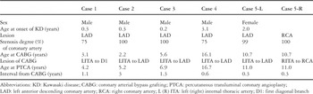
Table 1.
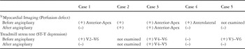
Table 2.
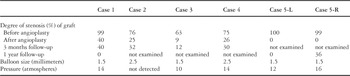
Table 3.

Degree of stenosis (%).

Serial angiograms of the left internal thoracic artery in our first patient. (a) Before percutaneous transluminal coronary angioplasty when the degree of stenosis was 99%, the graft being almost totally occluded. (b) After percutaneous transluminal coronary angioplasty, the degree of stenosis had reduced to 40%. (c) After 1 year, the angiogram revealed no stenosis at the site of anastomosis with the coronary arterial bypass graft.

Angiograms of the left internal thoracic artery (upper) and the left coronary artery (lower) in our second patient. (a) Before percutaneous transluminal coronary angioplasty. Native flow is seen in the left anterior descending artery. (b) Angiogram after 3 months. Though the left internal thoracic arterial graft was patent, the left anterior descending artery was occluded at the proximal end of the anastomosis.

Serial angiograms of the left internal thoracic artery in our third patient. (a) Before percutaneous transluminal coronary angioplasty. It was difficult to pass the guide wire across the lesion in this patient. (b) After percutaneous transluminal coronary angioplasty. (c) A follow-up angiogram at 3 months. There is a new stenosis at the distal portion of the anastomosis.
 You have
Access
- 12
- Cited by