The prevalence of rheumatic fever has declined significantly in most of the developed world. It continues, nonetheless, to be the commonest cause of acquired heart disease in the children and adolescent population of most developing countries.1 Awareness regarding the early onset of rheumatic mitral stenosis in India was provided by Roy et al.,2 who coined the term “juvenile” mitral stenosis for patients presenting with mitral stenosis below the age of 20 years. Such juvenile stenosis constitutes a quarter of all patients with rheumatic mitral stenosis.2 A few studies conducted in developing countries report rates of incidence ranging from 1.0 per 100,000 children of school-age in Costa-Rica, 72.2 per 100,000 in French Polynesia, 100 per 100,000 in Sudan, to 150 per 100,000 in China.3 The disease in these patients results in severe pulmonary arterial hypertension, with the appearance of symptoms at a very young age.4
Evolution of valvoplasty
Until the early 1980s, the only option for patients with mitral stenosis uncontrollable by medical therapy was surgery. Surgical options included open or closed mitral commissurotomy, and replacement of the mitral valve. A major breakthrough in treatment of stenotic valves came in 1982, when Kan et al.5 performed the first non-surgical dilation of the pulmonary valve, using a balloon introduced percutaneously.5
The technique soon developed to such an extent that dilation of the mitral valve was attempted, initially using a single balloon.6 The original procedure, known as the Block Technique, then underwent several modifications, including trans-septal puncture and simultaneous inflation of two balloons positioned side by side within the valve, the so-called double balloon technique.7 This procedure, however, required several catheter changes, and was technically demanding and time-consuming. At the same time, Inoue et al.8 used a single rubber nylon balloon for open mitral commissurotomy under direct vision, and following that, in 1984, he and his colleagues first reported successful percutaneous transvenous mitral commissurotomy.8 Subsequently, the use of the Inoue balloon has gained widespread acceptance, is widely acknowledged as the preferred technique for treating symptomatic patients of rheumatic mitral stenosis, and is applicable to patients of all ages.9 Comparative studies have shown similar haemodynamic results following the use of either the Inoue balloon or double balloons.10–12 As the Inoue balloon was approved by Food and Drug Administration of the United States of America in June, 1994, there is no longer the need to use double balloons in most of the patients.13
Selection of patients
All symptomatic patients of mitral stenosis with significant stenosis should be considered for percutaneous transvenous mitral commissurotomy. Prior to the procedure, a detailed clinical and echocardiographic examination should be undertaken. On clinical examination, it is important to document the staging with the functional classification of the New York Heart Association, any history of systemic embolism or haemoptysis, and any continuing features of rheumatic activity. Another important aspect to be established is the presence of any severe cardiothoracic deformity, which may make difficult fluoroscopic guided trans-septal puncture. In such cases, septal puncture may be performed under transthoracic14 or transoesophageal echocardiographic guidance.15
Indeed, cross-sectional and Doppler echocardiographic techniques have emerged as very useful non-invasive tools for assessing the structure of the mitral valvar apparatus, along with the haemodynamic significance of the stenosis. The assessment of valvar and subvalvar anatomy is frequently expressed as a score,16 with higher scores indicating more severe pathological deformities.
Though the echocardiographic score gives an indication of the severity of the deformity, the predictive accuracy of the score in defining the likely success of the procedure,17 or the chance of development of complications like severe mitral regurgitation, is limited.18 As the principle mechanism by which valvoplasty helps in increasing the mitral valvar area is by splitting the fused ends of the solitary zone of apposition between the valvar leaflets, bilateral calcification of the so-called “commissures”, in reality the ends of the zone of apposition, is a better predictor of a suboptimal result, and if present should be taken as a relative contraindication of valvoplasty. On the other hand, in presence of extensive sub valvar fusion, it is possible to proceed by graded dilation, starting with a balloon measuring 2 to 4 millimetres less than the predicted diameter based on height. Echocardiographic evaluation should then be performed after each dilation to assess the extent of the splitting of the ends of the zone of apposition. There may be persistence of significant gradient across the mitral valve in spite of split “commissures”, as fibrosis of the subvalvar apparatus may be as much responsible for stenosis of inflow into the left ventricle as the valvar stenosis itself.19 Apart from assessing the structure of the valvar apparatus, colour Doppler interrogation is used to assess the presence and severity of associated mitral regurgitation (Fig. 1). While patients with mild or moderate mitral regurgitation can be suitable candidates for percutaneous dilation, the presence of severe mitral regurgitation is conventionally considered a contraindication. But, in the younger population, when it is not always possible to insert a prosthetic mitral valve of adequate size, percutaneous dilation may need to be attempted in spite of the presence of severe central mitral regurgitation.20 In patients with mild or moderate mitral regurgitation, it is justified to follow the principle of graded dilation, with any increase in severity of mitral regurgitation following dilation being taken as the end point, even if it is not possible to obtain a desirable haemodynamic effect, such as achieving a mean gradient across the valve of less than 5 millimetres of mercury. Another important aspect assessed by echocardiography is the presence of thrombus in the left atrium or left atrial appendage. While presence of a clot in the left atrium is an absolute contraindication for trans-septal puncture, there are reports of the procedure being performed with caution in severely symptomatic patients with clots localized in the left atrial appendage.21

Figure 1. Cross-sectional echocardiographic, Doppler, and Colour flow imaging showing (a) the evaluation of the mitral valvar area and the degree of mitral regurgitation, and (b) a large left atrial clot (arrow).
Technical aspects of the procedure
Mitral valvoplasty can be achieved via:
- The transvenous antegrade approach
- The transfemoral approach
- The transjugular approach
- A retrograde arterial approach
The various balloons that have been widely used for mitral valvoplasty through either the transvenous or transarterial approaches are the Inoue balloon (Toray, Japan), and the Mansfield balloon (Mansfield Scientific Inc. Mass.), using either a single or a double balloon technique.
Antegrade valvoplasty using the Inoue balloon inserted through a trans-femoral venous route
The technique does not differ in children from that used in adults, except that the haemostatic sheath used for femoral arterial access is smaller, and usually 4 French, so as to minimize the local complication of arterial occlusion. Dilation of the valvar orifice is performed on fasting patients under local anaesthesia, using additional intravenous sedation, such as ketamine or morphine, as required. With the advent of Doppler echocardiography, which provides excellent haemodynamic parameters non-invasively, such as the peak and mean gradients across the mitral valve, and pulmonary arterial systolic pressure, complete haemodynamic studies are not performed systematically prior to attempting dilation except in the following cases:
- To provide evidence of the severity of mitral stenosis in cases of discrepancies between different echo measurements.
- To assess the severity of mitral regurgitation in patients being considered for dilation when clinical and echocardiographic data are discordant.
- To assess pulmonary arterial pressure, and left atrial and left ventricular diastolic pressures, when symptoms and/or estimated pulmonary arterial pressures are discordant with the severity of mitral stenosis as predicted from cross-sectional and Doppler echocardiographic evaluation.
- To assess the haemodynamic response of pulmonary arterial and left atrial pressures so as to elucidate any discrepancies between clinical symptoms and resting haemodynamics.
Trans-septal puncture is the first and most crucial step of the procedure. This is done via the right femoral vein, monitoring arterial pressure by placing a pigtail catheter at the aortic root through the right femoral artery, usually through a sheath of 4 French dimensions. After accessing the femoral vein, a paediatric or 7 French Mullins dilator is passed over a exchange length wire of 0.025′′ or 0.032′′ diameter into the superior caval vein. The Brockenbrough needle is then passed to its tip into the Mullins dilator. With the index of the Brockenbrough needle pointing posteromedially, at 4 o'clock or 5 o'clock at the groin, the entire assembly is pulled down from the superior caval vein in anteroposterior view until it engages the atrial septum in the region of the oval fossa. Following this, the assembly of the Mullins dilator, with the needle just proximal to its tip, is manipulated in the lateral view, probing for the fossa. Once the assembly is fixed in the fossa, the floor is punctured by firm pressure on the dilator. As soon as the whole assembly enters the left atrium, atrial entry is confirmed by recording the waveform of the left atrial pressure, by oximetry, and by injection of contrast into the left atrium. After confirming entry to the left atrium, the gradient across the mitral valve is recorded by simultaneous recording of left atrial and left ventricular pressures (Fig. 2). A long stainless steel guide wire with a coiled floppy tip, 180 centimetres long, and of 0.025 inches in diameter, is then inserted into the left atrium through the dilator. At this point, 100 units of heparin per kilogram of body weight are given in order to minimize the risk of formation of thrombus on the guidewires and catheters while they remain in the body. Following this, the site of septal puncture is dilated by passing a 14 French polyethylene dilator over the guidewire. At this stage, the Inoue balloon catheter is prepared for insertion. This is achieved by purging the catheter of air using contrast medium diluted by 1 in 3 with normal saline. A syringe containing diluted contrast is then connected to the injection port in order to pre-test the size of the balloon. Because the extent of inflation is controlled by adjusting the volume in the syringe, the volume and the corresponding inflated size of the balloon should be marked clearly on the syringe. It is vital that the operator confirms these measurements, and calculates the size of the balloon to be used in the first dilation.

Figure 2. Simultaneous pressure tracings taken in the left atrium and left ventricle, showing a gradient across the mitral valve.
The formula used for calculation of the size of the balloon in adults22 cannot be used for children. This is because, when using this formula, a high incidence of mitral regurgitation and restenosis was reported in a small series of children aged 12 years or less.23 Kothari et al.,17 therefore, started dilation in children less than 12 years of age using an initial balloon sized at 2 to 4 millimetres less than what would be recommended based on adult nomograms. Similarly, unlike in adults, where the success of the procedure is defined by creating a mitral valvar area greater than 1.5 centimetres squared, the objective in children should be to create a valvar area greater than 1 centimetre squared per metre squared of body surface area, either that or achieving an increase in area more than or equal to half the baseline measurement.
Once a balloon of appropriate size is chosen, it is deflated, elongated, and “slenderized” using an 18 gauge metal cannula of 80 centimetres in length. The balloon catheter is then inserted over the guidewire, through the septum, and into the left atrium. The metal cannula is then released and withdrawn slightly, leaving the tip of the balloon flexible. The balloon catheter is then advanced further until it is well within the left atrium, with its tip near the mitral valve. The guidewire and metal cannula are then removed.
Following septal puncture, entering the left ventricle is the second crucial step. This can also be achieved in several ways:
- Directly, by inflating partially the distal balloon to a diameter of 10 to 15 millimetres with 1 to 2 millilitres of diluted contrast. Inflation of the balloon in the left atrium helps first in facilitating entry to the left ventricle by directing the balloon towards the mitral valvar orifice, analogous to the manoeuvre used for a Swan-Ganz catheter, and secondly prevents inadvertent prolapse of the balloon from left atrium to right atrium during its manipulation for left ventricular entry. A J shaped spring wire stylet is inserted to the tip of the balloon catheter, which results in the distal segment of the catheter forming a J shape, with its tip directed downwards. Then, with a counter-clockwise twisting of the stylet of around 180°, the balloon catheter is directed anteriorly towards the mitral valvar orifice. Once the balloon catheter aligns with the mitral orifice, the balloon is seen to move back and forth along the long axis of the left ventricle, and is deflated. At this point, the stylet is pulled back 4 to 5 centimetres, and the deflated balloon is simultaneously advanced forward for 4 to 5 centimetres. This manoeuvre causes the balloon to cross the mitral valve into the left ventricle (Fig. 3).
- By using the reversed loop technique. Puncture of the septum through the oval fossa usually helps in aligning the balloon by the direct method. But, in patients having mitral stenosis with increased left atrial pressures, the septal anatomy is distorted. This occasionally leads to puncture of the atrial walls either cephalically or more anterior to the oval fossa. The reverse loop technique is useful in such cases. In this technique, the balloon catheter is inserted into the left atrium to make a large catheter loop. Then the J shaped stylet is inserted to a point 2 to 3 centimetres proximal to the base of the balloon. The stylet is then rotated clockwise for about 360 degrees, which results in formation of a loop in the left atrium, with the tip of the catheter aligned with mitral valvar orifice. With the stylet held firm, only the balloon catheter is advanced, allowing the balloon to move across the mitral valvar orifice (Fig. 4).

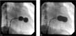
Figure 3. Entry of the Inoue balloon from the left atrium to the left ventricle by the direct method. (a) shows the waist at the mitral valve, while (b) shows the fully inflated balloon.

Figure 4. Entry of the Inoue balloon from the left atrium to the left ventricle by the reverse loop method.
Dilation of the mitral valvar orifice
Once the balloon enters the left ventricle, the distal part of the balloon is partially inflated with diluted contrast and moved back and forth two or three times inside the left ventricle to be certain that it is free in the left ventricular cavity and not straddled among the tendinous cords. The inflated balloon is then pulled back until a resistance is felt, when the balloon gets fixed at the mitral valve. At this point, the balloon is fully inflated by the predetermined amount of diluted contrast in the syringe. During inflation, the distal balloon is inflated, followed by the proximal balloon, and finally the middle portion across the constricted valvar orifice, producing an hourglass shape. At full inflation, the middle constriction disappears, suggesting the ends of the zone of apposition have split, and producing a barrel shape to the balloon.
So as to assess the efficiency of dilation, we follow a principle of staged balloon dilation, starting with a balloon of 2 to 4 millimetres less than the recommended diameter, and using progressive dilation with increments of 1 to 2 millimetres until the end point is reached.
The end point during staged dilation should be any of the following:
- mitral valvar area greater than 1 centimetre squared per metre squared body surface area with split ends of the zone of apposition between the valvar leaflets. It is important to estimate the extent of splitting of the zone of apposition by using cross-sectional echocardiography in the short axis view. If both ends of the zone of apposition are sufficiently split to a point near the annulus, further dilation will cause significant mitral regurgitation. Similarly, if only one end is adequately split, further dilation requires extra care because of risk of excessive avulsion of that area to the mitral annulus, resulting in severe mitral regurgitation.
- mean gradient across mitral valve of less than 5 millimetres of mercury.
- development of severe mitral regurgitation or an increase in severity of mitral regurgitation by one grade in patients with pre-existing mild to moderate mitral regurgitation.
Antegrade “over the wire” technique
In this technique, one or two balloons (Mansfield Scientific Inc. Mass.) are placed side-by-side across the stenosed mitral valve, using the appropriate number of wires. Various authors have proposed means of assessing the diameter of the balloon to be used for achieving an optimal mitral valvar area without increasing the risk of development of severe mitral regurgitation by disrupting the valvar apparatus. In adults, while Carlos et al.24 had used balloons with a diameter of 10 to 37% larger than the annular diameter, with a mean of 26%, McKay et al.25 used balloons whose diameter equalled the diameter of the mitral valvar annulus as determined in the echocardiographic apical four chamber view. Unlike the situation in adults, however, we found26 that, in children, choosing a diameter for the balloon based on body surface area is a better predictor of success. For optimal dilation, it should be 21 or 22 millimetres per metre squared of body surface area. After trans-septal puncture, the site of puncture is dilated with a balloon of 8 millimetres when two balloons are to be used to dilate the valve. Following this, the valve is crossed with the help of a 6 or 7 French Swan-Ganz catheter, and through it an exchange guidewire of .035 inches diameter is placed at the left ventricular apex. If two balloons are to be used, the second wire is passed through a two port Block catheter. Balloons of appropriate size, with their effective diameter being 80% of the sum of the diameter of the two balloons used, are positioned across the mitral valve and inflated until the waist disappears (Fig. 5).

Figure 5. Antegrade double balloon over-the-wire technique, showing fully inflated balloons at the level of the mitral valve.
Multitrack system.
Using this technique, two separate balloon catheters are positioned on a single guidewire. The first catheter, with only a distal guidewire lumen, is introduced into the vein and then advanced into the mitral orifice. Subsequently, a rapid exchange balloon catheter running on the same guidewire is inserted, and lined up with the first catheter so the two are positioned side-by-side. Both balloons are then inflated simultaneously. The sum of the diameters of the two balloons chosen is around 90 to 100% of the measured value for the mitral valvar annulus.27 The multitrack approach combines user friendliness and short procedural times with the advantages of the double balloon techniques. Another advantage of this system is cost-effectiveness, and in many cases its reuse after resterilization. Further, the metal shafts on both balloon catheters allow for very precise and stable manipulations on the balloons.
Transjugular approach
A transjugular approach is feasible and advantageous in certain cases of mitral stenosis. The right internal jugular vein serves as a safe conduit for use of large balloon catheters in children.28, 29 The jugular approach also provides a more direct route to the mitral valve, without catheters having to bend backward to cross the mitral valve, as occurs when using the femoral transvenous approach. Further, in patients with huge left atriums and distorted septal anatomy, the outcome of success of the procedure, particularly crossing of the mitral valve, is critically dependent when using the transfemoral route on correct siting of the septal puncture. When using the jugular approach, as long as the superior part of the septum is punctured about 2 centimetres below the roof of the left atrium, midway between the aorta and the anterior border of the spine, crossing the mitral valve is consistently simple and quick.28 For trans-septal puncture through the jugular vein, an Endrys pediatric trans-septal set (Cook, Bloomington, IN) should be used, This trans-septal set, consisting of an outer metal tube, and an inner stylet, is suitable for transjugular septal puncture since its extra curvature, which can be increased easily, allows a more perpendicular orientation of the needle tip to the atrial septum, while its short length makes the assembly easy to handle. The jugular approach may be considered in patients with webs, interruption, obliteration or thrombosis in the inferior caval or iliofemoral veins.
Retrograde non trans-septal aortic approach
The traditional antegrade approach necessitates a septal puncture, which is associated with an incidence of up to one-tenth of cardiac tamponade.30, 31 In addition, residual atrial shunting may be significant in a few patients.32 Mitral valvoplasty by a purely arterial retrograde route, without the need for septal puncture, was first described by Buchler et al.33 using Sones catheter. Though attractive, the approach had limited applications because of lack of consistency of left atrial entry using the preformed catheters.34, 35 Because of this, Stefanadis et al.36 finally designed a catheter that entered the left atrium easily and predictably. The catheter, nonetheless, is big, and requires a sheath of 8 or 9 French dimensions for its introduction. Though Stefanadis et al.,36 and Bahl et al.37 have reported rates of success over 90% using this approach, the need for such a large sheath makes it less attractive in children due to risk of injury to the femoral artery.
Acute and long term results
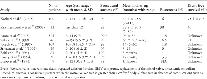
Percutaneous transvenous mitral dilation has become an established method of treatment for mitral stenosis in adults, with several series reporting the intermediate and long-term results.38–42 There is paucity of information, however, regarding the long-term efficacy of the procedure in young children. While the majority of the reports available on the procedure as used for juvenile mitral stenosis include patients of adolescent age, from 12 to 18 years,43–48 and in whom the results are quite comparable to adults (Table 1), there are only four studies of which we are currently aware describing the efficacy of the procedure in children aged less than 15 years.17, 20, 23, 48
Table 1. Immediate and follow-up results.

While Essop et al.48 first reported its feasibility and success exclusively in children aged less than 12 years, it was Krishnamoorthy et al.23 who undertook a study to determine whether the definition of mitral stenosis, and the recommendations for using Inoue balloons as in adults based on height, is applicable for children, and also to assess the effects of age and body surface area on the immediate outcome of the procedure. In their study, success was defined as achieving a mitral valvar area greater than 1.5 centimetre squared, with less than grade 2 mitral regurgitation, these criterions being similar to those used in adults. A stepwise dilation was carried out, at 1 millimetre increments, commencing with a balloon sized at 2 millimetres less than that calculated using the height-based nomogram, and going up to 2 millimetres above that obtained by the formula. Though the procedure was successful in over nine-tenths of the series, 2 children (15%) developed moderate mitral regurgitation, this being higher than the one-tenth incidence of this complication reported in the Inoue balloon registry obtained for adult patients.49 The authors attributed this problem to use of a height based nomogram in selecting the size of the balloon, and also in targetting a valvar area greater than 1.5 centimetre squared to define the procedure as successful. Similarly, when using the criterion of mitral valvar area less than 1.5 centimetre squared on follow up as the definition of restenosis, as is the case in adults, the authors reported a restenosis rate of almost two-fifths on follow up of 20 months, though none of the patients were symptomatic. Subsequently, Kothari et al.17 addressed the issues of procedural success and restenosis with respect to body surface area. They defined procedural success as achieving a mitral valvar area greater than 1 centimetre squared per metre squared body surface area in absence of complications such as death, tamponade, systemic embolism or severe mitral regurgitation. They considered restenosis as a decrease in the valvar area less than 0.8 centimetres squared per metre squared body surface area, or greater than or equal to half the loss in the initial gain.17 The initial size of balloon chosen was 2 to 4 millimetres less than the size as if calculated on the basis of height. The procedure was successful in 97 of 100 patients, and the maximum size of the balloon used was less than or equal to the recommended size. With this policy, the incidences of severe mitral regurgitation, at 4%, and restenosis at 12%, were both acceptable. The result of this study in children less than 12 years of age, with intermediate-term follow up at a mean of 34 months, reported echocardiographic restenosis in one-sixth of patients, which is comparable to our reported rate of restenosis in one-eighth of children aged less than 15 years and followed for over 96 months, and much less than that reported by Krishnamoorthy et al.,23 albeit that the latter group did not define restenosis on the basis of body surface area. Kothari et al.17 have also reported event-free survival at 100 months of 75% compared to 56% at similar duration of follow up in a large Western series in adults.41 It should also be remembered, nonetheless, that surgical procedures nowadays also carry a lower mortality, similar to that achieved using percutaneous transvenous dilation.
Conclusion and future directions
Though there are few guidelines about the optimal size of balloon to be used in children, it seems appropriate to follow the strategy of inserting an Inoue balloon sized at 2 to 4 millimetres less than the size recommended on the basis of height, or a Mansfield balloon sized at 21 to 22 millimetres per metre squared of body surface area. Procedural success should now be defined as achieving a mitral valvar area greater than 1 centimetre squared per metre squared body surface area, and restenosis as equal to or less than 0.8 centimetres squared per metre squared body surface area. There should be multi-centric trials conducted, exclusively involving children aged less than 15 years, as the current incidence of percutaneous transvenous mitral valvar dilation performed in children aged less than 15 years is only around 8%. Developing countries are long way off from achieving the eradication of rheumatic heart disease, as no single approach has been satisfactory or effective. Primary prevention of acute rheumatic fever by treating any sore throat is neither practical nor cost effective. Moreover, a sore throat is not clinically evident in nearly one-third to half of the patients, and yet the initial episode is still severe enough to produce cardiac disease. Secondary prophylaxis is generally practised in the majority of cases. A strategy of vaccination is expected to be the best alternative. One polyvalent M protein vaccine has been undergoing phase 2 clinical trials, while development of a vaccine using parts of the protein which are similar in all serotypes is in the developmental stages. Other approaches depending on the adherence capability of the streptococcal bacteria, like fibronectin binding protein or those targeted at virulence factor like 5a peptidases are in the laboratory phase.50 If trials of rheumatic vaccines prove to be successful, then the disease that affects so many children and young adults could one day be eradicated from the developing countries.