Although a great deal of the information required by our motor system for locomotion and survival-responses (fight and flight behavior) is encoded in our genes, throughout the course of life a large amount of motor information must be learned, remembered, and flexibly adapted to interact with the ever-changing conditions of our environment. The field of human motor cognition is broadly concerned with understanding action representations and associated higher mental processes. Fundamental questions for researchers are concerned with understanding how this information is encoded, assembled, stored, (re)activated, and executed, in other words, the building blocks of our purposeful interactions with the outside world, and how representations of action are organized in the brain.
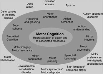
Some of the major components of motor cognition are illustrated in the darker circle of Figure 1, but are by no means complete. The figure is not intended to represent frameworks put forth by various theories (embodied cognition) and neurophysiological (mirror neurons) accounts of motor cognition. Rather, we aim to illustrate the wide range of topics in motor cognition that appear regularly in the scientific literature. Although this field of investigation is notably smaller than, for example, studies of language, memory, or executive functions, over the past decade there has been a steady rise in conference presentations and publications related to the neurobehavioral correlates of motor cognition, which this Special Issue highlights.
Aspects of motor cognition and associated clinical phenomena.

Neuroscientific approaches to unraveling the neural correlates of action representations have been particularly influential in the field, owing to the striking loss in specific facets of cognitive-motor abilities following acute brain damage and neurodegenerative disorders (Buxbaum, Kyle, Grossman, & Coslett, Reference Buxbaum, Kyle, Grossman and Coslett2007; Haaland, Harrington, & Knight, Reference Haaland, Harrington and Knight2000; Helmich, de Lange, Bloem, & Toni, Reference Helmich, de Lange, Bloem and Toni2007). Pioneering studies of patients with limb apraxia, visual agnosia, optic ataxia and brain injury due to stroke were particularly pivotal in motivating research into the neural mechanisms and cognitive facets of motor representations, including motor planning, visual coding, object recognition, and semantic representations (Goodale, Jakobson, & Servos, Reference Goodale, Jakobson and Servos1996; Jeannerod, Reference Jeannerod1997; Rizzolatti, Fogassi, & Gallese, Reference Rizzolatti, Fogassi and Gallese2001).
The diversity of clinical and behavioral manifestations that comprise contemporary studies into the neurobehavioral mechanisms underlying disturbances in motor cognition are shown in Figure 1 (border outside of the dark circle). Besides a more refined localization of brain damage and characterization of neurobehavioral disturbances in patients, the application of neuroimaging to the study of healthy individuals has notably accelerated our understanding of neurobehavioral mechanisms underlying normal motor cognition (Johnson-Frey, Reference Johnson-Frey2004; Lewis, Reference Lewis2006). This Special Issue of JINS selected eight manuscripts that relate to several topics of our figure. In addition to cutting-edge empirical investigations that characterize cognitive-motor dysfunctions in clinical groups, each manuscript also discusses fundamental research related to core questions about the representation of actions. Altogether, this collection of manuscripts advances our understanding of the nature and neuroanatomy of action representations and related concepts and, in some cases, empirical findings challenge current theoretical formations or show promise in contributing to earlier diagnosis of neurodegenerative conditions.
The first paper of this Special Issue is by Przybylski and Króliczak who investigated whether the praxis representation network of the left hemisphere supports the planning of tool grasping, otherwise known as transitive gestures, which is an important facet of action repertoires. The neurophysiological mechanisms of object interactions have long been of central interest in non-human primate research (Castiello, Reference Castiello2005; Gallese, Murata, Kaseda, Niki, & Sakata, Reference Gallese, Murata, Kaseda, Niki and Sakata1994; Jeannerod, Arbib, Rizzolatti, & Sakata, Reference Jeannerod, Arbib, Rizzolatti and Sakata1995), but now there are an increasing number of neurobehavioral studies of grasping in humans owing to the wide availability of neuroimaging technologies and more sophisticated recording devices (Vingerhoets, Reference Vingerhoets2014).
Using functional magnetic resonance imaging (fMRI), Przybylski and Króliczak had healthy participants plan grasps of tool versus non-tool objects, controlling for object complexity and hand-effector (i.e., dominant versus non-dominant hand performance). Whereas they found no difference of hand-effector on the activated network, planning functional grasps (tools) showed a significantly more asymmetrical activation of the left-hemisphere praxis representation network than did planning of non-functional grasps (non-tools) (Kroliczak & Frey, Reference Kroliczak and Frey2009). The differences in neural activity between the two object categories disappeared during grasp execution. Their findings underscore that the plan of a purposeful action is already present during the grasp component of a transitive action.
Our next three papers are related to the neurobehavioral mechanisms of limb apraxia, which has played a central role in the theoretical development and understanding of action representations. The writings of Hugo Karl Liepmann in the early 20th century were dominated by the ideas and research on apraxia that still resonate in many contemporary textbooks of neurology and cognitive neuroscience (Goldenberg, Reference Goldenberg2003a, Reference Goldenberg2013). In fact, the representation of action itself and the nature of action semantics remains a hotly debated topic (Binkofski & Buxbaum, Reference Binkofski and Buxbaum2013; Osiurak, Jarry, & Le Gall, Reference Osiurak, Jarry and Le Gall2011). One of the core diagnostic methods used to investigate the integrity of action representations in patients is the request to pantomime simple actions like hammering a nail or threading a needle. In absence of the tool object itself or a visual image of it, that is, without any tactile or visual sensory help, the patient must retrieve and execute correct grasp and gesture information. Failure to present an adequate pantomime is taken as a sign of apraxia (Hanna-Pladdy, Heilman, & Foundas, Reference Hanna-Pladdy, Heilman and Foundas2001).
Given the importance of pantomime for clinical diagnosis alongside other assessment strategies (e.g., imitation or actual tool use), a critical appraisal of the nature of pantomime is instructive to the informed clinician (Hermsdorfer, Terlinden, Muhlau, Goldenberg, & Wohlschlager, Reference Hermsdorfer, Terlinden, Muhlau, Goldenberg and Wohlschlager2007; Hoeren et al., Reference Hoeren, Kummerer, Bormann, Beume, Ludwig, Vry and Weiller2014). In this Special Issue, two papers focus on pantomime. In a critical review overviewing the complex relations between pantomime, imitation, and actual use, their respective lesion sites and their kinematic differences, Goldenberg comes to the conclusion that pantomime is not merely a replication of the motor programs of actual use, but rather that it is a different kind of gesture altogether. Goldenberg advances the provocative view that pantomime of tool use is a communicative gesture, constructed by a combination of features of tools and gestures that maximizes comprehensive demonstration, rather than simply copying the actual motor action. Given the different putative lesions underlying pantomime and imitation, the qualitative distinction between these types of behavior has implications for the interpretation of its disturbances (Goldenberg, Reference Goldenberg2003b).
A second inspiring paper on pantomime is an empirical study of Lesourd and colleagues who take the stance that defective pantomime of tool use not only depends on a loss of functional knowledge, but also on the degrading of mechanical knowledge (Osiurak, Reference Osiurak2014; Osiurak et al., Reference Osiurak, Jarry, Allain, Aubin, Etcharry-Bouyx, Richard and Le Gall2009). They investigated pantomime errors in patients with Alzheimer’s disease and semantic dementia, using separate tasks to assess functional, mechanical, and manipulation knowledge as predictors. A mechanical problem solving test assessing mechanical knowledge appeared to be a good predictor of the overall pantomime performance, whereas functional knowledge robustly predicted conceptual errors. As their results suggest a contribution of both functional and mechanical knowledge to pantomime, Lesourd et al. conclude that pantomime of tool use mimics a complex problem-solving task.
In a third study, Mutha and colleagues asked if apraxic patients can learn new cognitive-motor representations, despite left-hemisphere damage to frontal-parietal areas that are thought to support the internal representation of action. These investigators compare action learning in healthy adults and cohorts of left hemisphere stroke patients with and without ideomotor apraxia. In a motor adaptation task, participants were required to adapt their movements to a 30-degree visuomotor rotation. All groups showed an initial decrease in error rate, but only apraxic patients failed to show long-term improvement. Of interest, the late learning deficit was predicted by the degree of apraxia and by the volume of parietal damage. The authors interpreted the disrupted slow learning process of the apraxics as the failure to develop a durable action representation, the formation of which seemed to be associated with parietal integrity. These findings comport with the difficulties encountered in the treatment of apraxia, where strategies aimed at motor relearning meet with limitations in functional plasticity once the neural substrate underlying motor representation is damaged (Goldenberg, Daumuller, & Hagmann, Reference Goldenberg, Daumuller and Hagmann2001; Hartmann, Goldenberg, Daumuller, & Hermsdorfer, Reference Hartmann, Goldenberg, Daumuller and Hermsdorfer2005).
With the recently published Special Issue of JINS devoted to Preclinical Prediction (November, 2016), it pleases the editors that two studies in the present issue demonstrate the sensitivity of cognitive-motor measures for premanifest detection of two well-known neurodegenerative disorders of the basal ganglia that are characterized by dramatic motor disturbances: Parkinson’s disease and Huntington’s disease. The basal ganglia are central for planning and controlling hierarchically organized sequential motor patterns (Elsinger, Harrington, & Rao, Reference Elsinger, Harrington and Rao2006), which are also critical for carrying out homologous linguistic operations.
In the first study, García and colleagues explore whether language deficits are a precursor of Parkinson’s disease (PD). The investigators compared healthy controls to sporadic PD patients, genetic PD patients with parkin (PARK2) or dardarin (LRRK2) mutations, and asymptomatic first-degree relatives of the latter two cohorts with similar mutations. Participants’ performances on semantic, verb-production, and syntactic tasks were studied, controlling for individual differences in executive functioning. While the two clinical groups were impaired on all language measures, the asymptomatic mutation carriers showed a selective deficit on a syntactic comprehension test, in which participants chose which of four pictures best represented phrases that contained a verb and two nouns (e.g., touching the scissors with the comb). Thus, PARK2 and LRRK2 mutations appear to have disease-independent effects on syntactic operations that govern the structure of sequences, which may be a prodromal sign of focal basal ganglia deterioration and a risk factor for clinical PD.
In a second study, Misiura and colleagues sought to identify the relationships between preclinical phenotype variables in prodromal Huntington’s disease (prHD) and basal ganglia atrophy, which is the most sensitive imaging marker of early neuropathology in the prodromal phase. The analyses were based on a dataset of almost a thousand prHD individuals with volumetric measures of five subcortical brain regions and 34 cognitive, motor, psychiatric, and functional variables known to be associated with an emerging HD phenotype (Paulsen et al., Reference Paulsen, Long, Ross, Harrington, Erwin and Williams2014). Cluster analyses of the behavioral measures revealed five distinct clusters that were regressed against the subcortical volumes, controlling for genetic burden. The main results showed that smaller caudate and putamen volumes were related to clusters representing motor symptoms, cognitive control, and verbal learning, indicating that cognitive control is sensitive to basal ganglia changes that remain uncaptured by examining only motor scores.
The last two studies of this special issue pertain to more theoretical questions with regard to motor cognition. The first study by de Wit and Buxbaum uses a neuropsychological approach to determine whether non-biological motion trajectories are predicted by the same mechanisms used to predict human actions, or whether they rely on different mechanisms (Schubotz, Reference Schubotz2007). Performances of healthy adults and left hemisphere stroke patients were examined on a visual occlusion task that required prediction of pantomimed tool use, real tool use, and non-biological motion. Prediction difficulties of human and non-biological motion were associated with the presence of limb apraxia and motor production deficits, but not with action recognition. Impaired prediction was associated with localized left frontal and parietal lesions regardless of motion type. The authors concluded that the prediction of both human and non-biological motion trajectories critically rely on the sensorimotor network.
It has long been known from early studies of non-human primates that the motor cortex is involved in cognitive operations related to motor functioning, including spatial transformations, the coding of serial order, and memory consolidation (Georgopoulos, Reference Georgopoulos2000; Georgopoulos, Taira, & Lukashin, Reference Georgopoulos, Taira and Lukashin1993; Pellizzer, Sargent, & Georgopoulos, Reference Pellizzer, Sargent and Georgopoulos1995; Smyrnis, Taira, Ashe, & Georgopoulos, Reference Smyrnis, Taira, Ashe and Georgopoulos1992; Wise, Moody, Blomstrom, & Mitz, Reference Wise, Moody, Blomstrom and Mitz1998). The last study addresses a long-standing issue in human motor cognition, namely whether the primary motor cortex takes part in the mental simulation of movement. This issue dates back to discussions about whether motor processes are involved in mental rotation and mental imagery (Kosslyn, Digirolamo, Thompson, & Alpert, Reference Kosslyn, Digirolamo, Thompson and Alpert1998; Parsons, Reference Parsons1994), and of the precise role of the primary motor cortex in mental imagery (Kosslyn, Thompson, Wraga, & Alpert, Reference Kosslyn, Thompson, Wraga and Alpert2001).
The use of transcranial magnetic stimulation (TMS) to address these questions is not new (Ganis, Keenan, Kosslyn, & Pascual-Leone, Reference Ganis, Keenan, Kosslyn and Pascual-Leone2000), but the approach taken by Hyde and colleagues combines a novel adaptation of the hand laterality task and TMS of the hand area of the left primary motor cortex. In mental imagery users, the authors found increased excitability over this region of primary motor cortex during the hand laterality task, in particular for more complex simulated hand movements at later latencies. These findings offer further support for the involvement of primary motor cortex in the mental simulation of movement.
The contributions bundled in this special issue reflect some of the current themes on motor cognition addressed by researchers worldwide. The richness of the field is evidenced by the scope of scientific inquiries that stand to unravel the brain mechanisms of action plans, object use, action semantics and understanding, motor imagery, and movement planning and coordination. This body of research has already brought about the development of treatment strategies for the rehabilitation of motor impairments, ranging from the use of motor imagery during gait recovery after cerebral injury to the use of neurostimulation to improve movements in acquired and degenerative brain disease (Kang, Summers, & Cauraugh, Reference Kang, Summers and Cauraugh2016; Oostra, Oomen, Vanderstraeten, & Vingerhoets, Reference Oostra, Oomen, Vanderstraeten and Vingerhoets2015).
Cutting-edge developments in the area of cognitive-based neuroprosthetics for paralyzed patients also stem from basic science research in motor cognition (Aflalo et al., Reference Aflalo, Kellis, Klaes, Lee, Shi, Pejsa and Andersen2015; Collinger et al., Reference Collinger, Wodlinger, Downey, Wang, Tyler-Kabara, Weber and Schwartz2013; Wang et al., Reference Wang, Collinger, Degenhart, Tyler-Kabara, Schwartz, Moran and Boninger2013). Although much progress has been made thanks to recent technical and methodological advances, many issues with regard to the representation of action and its neural correlates remain unresolved. We must continue to refine and sharpen our concepts and theories of motor phenomena, only some of which are highlighted in Figure 1, to further advance fundamental knowledge in the field and promote clinical applications. These challenges ensure that important gains are still to be made in this exciting field, waiting to be uncovered by researchers intrigued by the science of our bodily interactions with the environment.