





Introduction
Pinworms (Oxyuridae: Syphaciinae) are parasites of a wide range of animals, including humans (Adamson, Reference Adamson1994). The family consists of several subfamilies, of which Oxyurinae occur in all mammalian orders, while Syphaciinae are restricted to rodents and lagomorphs (Adamson, Reference Adamson1994). Members of Syphaciinae parasitize 19 families and 80 genera within the order Rodentia and the family Sciuridae, consisting of squirrels and chipmunks, is no exception (Hugot, Reference Hugot1990). The genus Syphatineria Chabaud and Biocca, 1955 (hereafter abbreviated as ‘St.’) is exclusively described from squirrels (Hugot, Reference Hugot1981; Petter and Quentin, Reference Petter, Quentin, Anderson, Chabaud and Willmott2009) and is distinct from Syphacia sensu stricto that is exclusive to the Muroidea (Hugot, Reference Hugot1988; Sorci et al., Reference Sorci, Morand and Hugot1997). In the genus Syphacia Seurat, 1916 (hereafter abbreviated as ‘Sp.’), male individuals have 3 mamelons as opposed to 2 found in Syphatineria (Baylis, Reference Baylis1928; Sandground, Reference Sandground1933; Hugot, Reference Hugot1980, Reference Hugot1981, Reference Hugot1988).
Syphatineria consists of ∼ 19 species that are separated into four subgenera: Africanoxys Hugot, 1981, Quentenora Hugot, 1981, Orientoxys Hugot, 1981 and Syphatineria. Africanoxys and Syphatineria are parasites of African squirrels, while Quentenora and Orientoxys infect squirrels in the Eastern regions of the world (Johnson, Reference Johnson1968; Schmidt and Kuntz, Reference Schmidt and Kuntz1968; Hugot, Reference Hugot1981). Based on the latter classification, the Syphatineria assemblage includes 3 species (Syphatineria antiqua Hugot, 1981, Syphatineria pallaryi Seurat, 1915 and Syphatineria transafricana Chabaud and Biocca, 1955) while the Africanoxys group includes 7 species (Syphatineria adami Quentin, 1971, Syphatineria cepapi Hugot, 1981, Syphatineria feeri Hugot, 1980, Syphatineria interjecta Hugot, 1980, Syphatineria ividensis Hugot, 1980, Syphatineria paraxeri Sandground, 1933 and Syphatineria pearsi Baylis, 1928) (Johnson, Reference Johnson1968; Schmidt and Kuntz, Reference Schmidt and Kuntz1968; Hugot, Reference Hugot1981).
Paraxerus cepapi Smith, 1836 (Smith’s bush squirrel) is 1 of 5 squirrel species confined to southern Africa (Kingdon, Reference Kingdon1997) and it occurs sympatrically with the red squirrel, Paraxerus palliatus Peters, 1852, in Mozambique (Banotai et al., Reference Banotai, Mazzamuto and Koprowski2023). Smith’s bush squirrel is commonly found in the mopane and Acacia woodland habitats in South Africa and its range extends into Namibia, Botswana, Zimbabwe and Mozambique (Thorington and Hoffmann, Reference Thorington, Hoffmann, Wilson and Reeder2005; Thorington et al., Reference Thorington, Koprowski, Steele and Whatton2012). Viljoen (Reference Viljoen1977a), while investigating the feeding habits of P. cepapi at 5 localities in Limpopo province, South Africa, recorded the presence of a parasitic nematode in the caecum and large intestine and identified it at the time as Syphacia paraxeri, a pinworm that was first described from P. palliatus in Zimbabwe (Sandground, Reference Sandground1933). Subsequently, Hugot (Reference Hugot1981), using limited samples that originated from a different South African locality than Viljoen (Reference Viljoen1977a), described Syphatineria (Africanoxys) cepapi from P. cepapi. Although male and female individuals were examined by Hugot (Reference Hugot1981), it appears that the material originated from a single host individual and from a single locality. This, in addition to the incomplete species description for St. paraxeri (Sandground, Reference Sandground1933) led Hugot (Reference Hugot1981) to state, “we believe we can classify our animals in a new species ‘Syphatineria (Africanoxys) cepapi’ n. sp.”
The aim of the present study is to re-investigate the taxonomic description of Syphatineria (Africanoxys) cepapi and specifically to contribute additional morphological and new genetic data for St. cepapi occurring in P. cepapi in South Africa. This was achieved by using a wider geographic sampling approach, complemented by larger sample sizes. Molecular characterization of St. cepapi included both the mitochondrial DNA barcode Cytochrome Oxidase I (COI) and 18S rRNA nuclear genes. The wider sampling approach also allowed for some novel baseline ecological data to be added for St. cepapi.
Materials and methods
Sample collection and processing
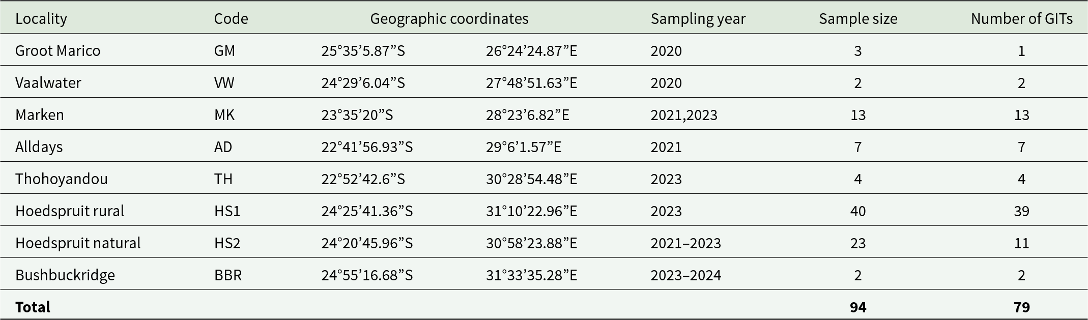
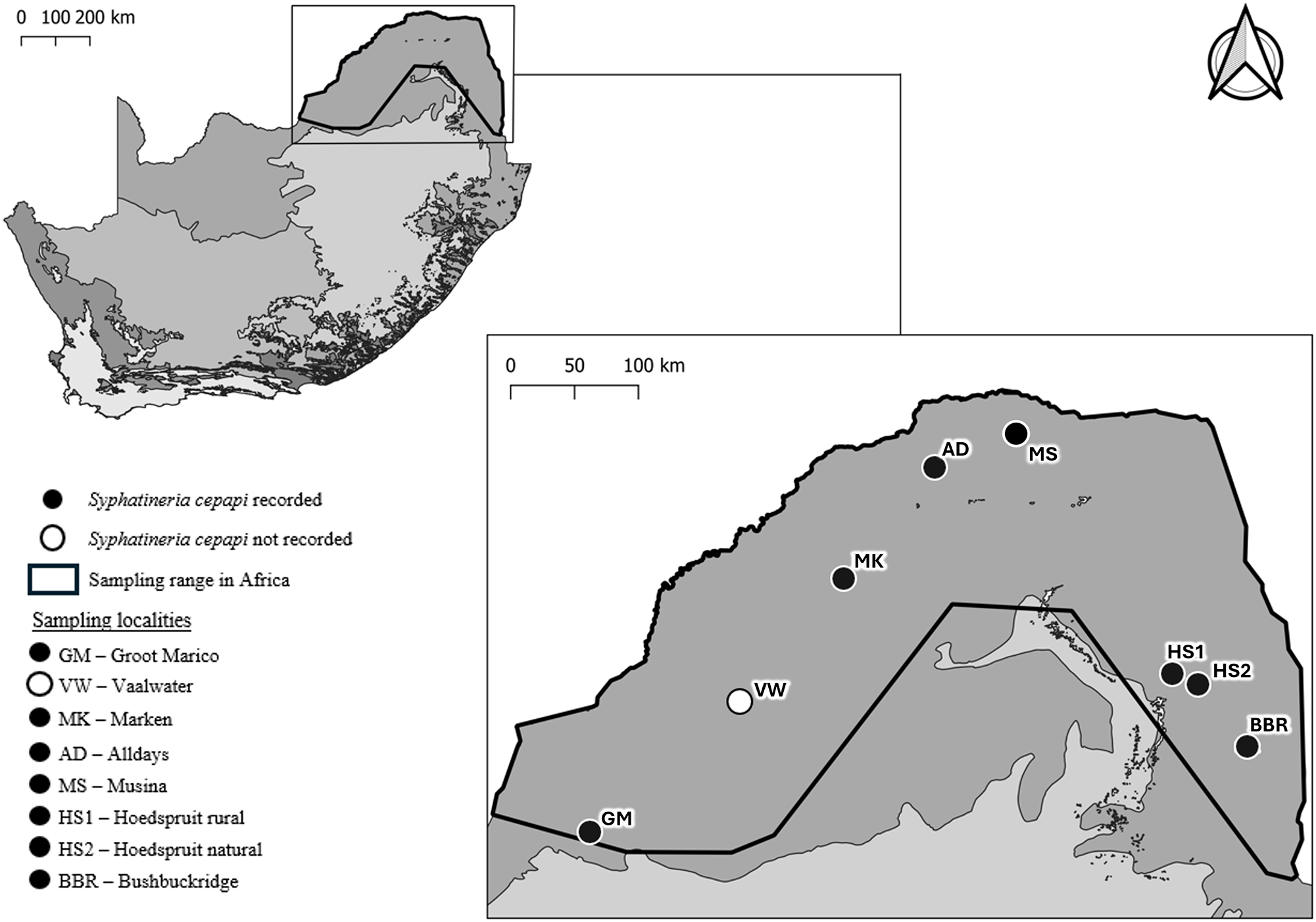
In total, 94 P. cepapi individuals were opportunistically obtained from 8 localities throughout the host’s distribution range in South Africa (Limpopo, Mpumalanga and the North West Provinces) (Table 1; Figure 1). The localities sampled included natural bushveld, farms and rural areas. The gastrointestinal tracts (GIT) of 79 squirrels were intact and available for examination. The GIT of each squirrel was removed and individually stored in labelled jars filled with 100% ethanol (Table 1).
Localities (n = 8), geographic coordinates, sampling year, number of Paraxerus cepapi individuals (n = 94) and the number of gastrointestinal tracts (GIT) included in the present study

Localities were abbreviated as follows: GM, Groot Marico; VW, Vaalwater; MK, Marken; AD, Alldays; TH, Thohoyandou; HS1, Hoedspruit rural; HS2, Hoedspruit natural; BBR, Bushbuckridge.
Localities from where Paraxerus cepapi (n = 8) and Syphatineria cepapi (n = 7) were obtained in the Savanna biome, South Africa (sampled between 2020 and 2024). Locality codes correspond to Table 1. The major vegetation types of South Africa (as represented by the biomes) are indicated in the inset map and indicated by the different shades of grey.

Figure 1 Long description
Map of South Africa with an inset showing the distribution and sampling sites of Syphatineria cepapi. Black circles indicate recorded sites: Groot Marico (GM), Marken (MK), Alldays (AD), Musina (MS), Hoedspruit rural (HS1), Hoedspruit natural (HS2), and Bushbuckridge (BBR). A white circle at Vaalwater (VW) indicates no record. The outlined area marks the sampling range in Africa. The map includes a scale bar and a legend explaining symbols and localities.
Helminth processing and morphological identification
Each GIT was separated into 4 sections: stomach, small intestine, large intestine and caecum. The content and tissue wall of each GIT section was placed in distilled water and individually and systematically examined for helminths (nematodes and cestodes) in a Petri dish using fine-point forceps and a Leica MZ75 stereomicroscope. All recovered helminths were recorded, separated according to taxonomic groups and stored in 100% ethanol. The nematodes were further identified to genus level (based on Petter and Quentin (Reference Petter, Quentin, Anderson, Chabaud and Willmott2009) and Gibbons (Reference Gibbons2010)) and all Syphatineria nematodes (based on the descriptions outlined by Sandground (Reference Sandground1933) and Hugot (Reference Hugot1981)) were stored in 100% ethanol. Ten Syphatineria males and 10 females from each of the 7 sampling localities were slide-mounted in lactophenol. The 140 mounted specimens were examined and measured using a Leica DM1000 light microscope fitted with a Zeiss Axiocam ERc 5s camera with accompanying ZEN 3.3 imaging software. Standard morphological characters, including, for example, total-, tail- and oesophagus-length, were photographed, measured and recorded. All measurements were taken in micrometres and converted to millimetres. The recorded measurements were compared to existing published data of 9 Syphatineria species (St. antiqua, St. cepapi, St. feeri, St. interjecta, St. ivindensis, St. pallaryi, St. paraxeri, St. pearsi and St. transafricana) that are associated with African squirrels (Baylis, Reference Baylis1928; Sandground, Reference Sandground1933; Hugot, Reference Hugot1980, Reference Hugot1981, Reference Hugot1988).
Molecular phylogenetic analysis
Genomic DNA was extracted following the protocol and using similar reagents as described by Subbotin et al. (Reference Subbotin, Toumi, Elekçioğlu, Waeyenberge and Maafi2018) from 2 female Syphatineria individuals per locality (n = 14). PCR amplification was performed using 2 primer sets: SyphaCOIF and SyphaCOIR (Okamoto et al., Reference Okamoto, Urushima, Iwasa and Hasegawa2007) for the COI gene and 5F and 7R (Giribet et al., Reference Giribet, Carranza, Baguna, Riutort and Ribera1996) for the 18S rRNA gene. All PCR reactions were carried out in a total volume of 25 µL. The reaction mixture per sample for 18S included 3 µL extracted DNA, 8.5 µL ddH₂O, 0.5 µL of 100 Mmol primers (forward and reverse) and 12.5 µL of Taq DNA Polymerase Master Mix Red (Ampliqon). The reaction mixture for COI included 3 µL of DNA, 7.5 µL ddH₂O, 1 µL of each primer at 100 Mmol concentration (forward and reverse) and 12.5 µL of Taq DNA Polymerase Master Mix Red (Ampliqon).
Amplification followed standard PCR protocols in a GeneAmp PCR system 2700 thermal cycler (Applied Biosystems) and included a negative control. Thermal cycling conditions included an initial denaturation of 5 minutes at 94 °C, followed by denaturation for 30 seconds at 94 °C, annealing for 45 seconds at 47 °C and extension for 45 seconds at 72 °C. This process was repeated for 35 cycles. A final extension of 72 °C for 5 minutes was performed. The PCR products, post-amplification, were visualized on a 1% agarose gel using electrophoresis in the presence of ethidium bromide. The positively amplified products were purified, sequenced using BigDye Chemistry and analysed on an ABI 3730 XL DNA Analyzer (Applied Biosystems). The chromatogram of each sequence was visually inspected for irregularities and edited/trimmed accordingly using BioEdit version 7.7.1 (Hall, Reference Hall2005).
Using the Basic Local Alignment Search Tool (BLAST) on GenBank (Nucleotide BLAST: Search nucleotide databases using a nucleotide query), 1 individual’s sequence from each gene was used to perform the search and Syphacia was specified to narrow the search parameters. From the BLASTN results, sequences from 2 to 5 Syphacia species (depending on the number of sequences that produced significant hits), were downloaded and combined with the newly generated sequences for each gene, respectively, using the MEGA X package (Kumar et al., Reference Kumar, Stecher, Li, Knyaz and Tamura2018). One Aspiculuris tetraptera Schulz, 1924 (Oxyuridae) pinworm sequence was included in the matrix as an outgroup (Feldman and Bowman, Reference Feldman and Bowman2007). The sequences were aligned using the ClustalW function (Thompson et al., Reference Thompson, Gibson and Higgins2003) available on MEGA X and trimmed at their ends to remove missing data. Optimal models for each gene were determined using the Bayesian Information Criterion in MEGA X.
The 18S rRNA tree was then constructed using the Maximum Likelihood method and the Kimura 2-parameter substitution model with a discrete Gamma distribution. For the COI gene, a Maximum Likelihood tree was constructed using the Tamura-Nei substitution model with a discrete Gamma distribution and Invariant sites. Bootstrap support for nodes was obtained by doing 1000 random samplings.
Results
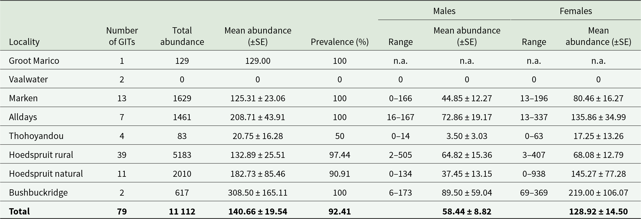
Syphatineria cepapi individuals were recorded from the caecum and large intestine of 79 P. cepapi squirrels (Table 2).
Localities, number of gastrointestinal tracts, total and mean abundance (±SE), prevalence and the range and mean (±SE) number of male and female Syphatineria cepapi recorded from Paraxerus cepapi

Table 2 Long description
The table has columns: Locality, Number of GITs, Total abundance, Mean abundance (plus–minus SE), Prevalence (percent), Males Mean abundance (plus-minus SE), Males Range, Females Mean abundance (plus–minus SE), Females Range. Row 1: Groot Marico, 1, 129, 129.00, 100, n.a., n.a., n.a., n.a. Row 2: Vaalwater, 2, 0, 0, 0, 0, 0, 0, 0. Row 3: Marken, 13, 1629, 125.31 plus–minus 23.06, 100, 44.85 plus–minus 12.27, 0 superscript subscript –166, 80.46 plus–minus 16.27, 13 superscript subscript –196. Row 4: Alldays, 7, 1461, 208.71 plus–minus 43.91, 100, 72.86 plus–minus 19.17, 16 superscript subscript –167, 135.86 plus-minus 34.99, 13 superscript subscript –337. Row 5: Thohoyandou, 4, 83, 20.75 plus–minus 16.28, 50, 3.50 plus-minus 3.03, 0 superscript subscript –14, 17.25 plus–minus 13.29, 0 superscript subscript –63. Row 6: Hoedspruit rural, 39, 5183, 132.89 plus–minus 25.51, 97.44, 64.82 plus–minus 15.36, 2 superscript subscript –505, 68.08 plus–minus 12.79, 3 superscript subscript –407. Row 7: Hoedspruit natural, 11, 2010, 182.73 plus–minus 85.46, 90.91, 37.45 plus-minus 13.15, 0 superscript subscript –134, 145.27 plus–minus 77.28, 0 superscript subscript –938. Row 8: Bushbuckridge, 2, 617, 308.50 plus–minus 165.11, 100, 89.50 plus–minus 59.04, 6 superscript subscript –173, 219.00 plus–minus 106.07, 69 superscript subscript –369. Total: 79, 11112, 140.66 plus–minus 19.54, 92.41, 58.44 plus-minus 8.82, 128.92 plus–minus 14.50.
Species description
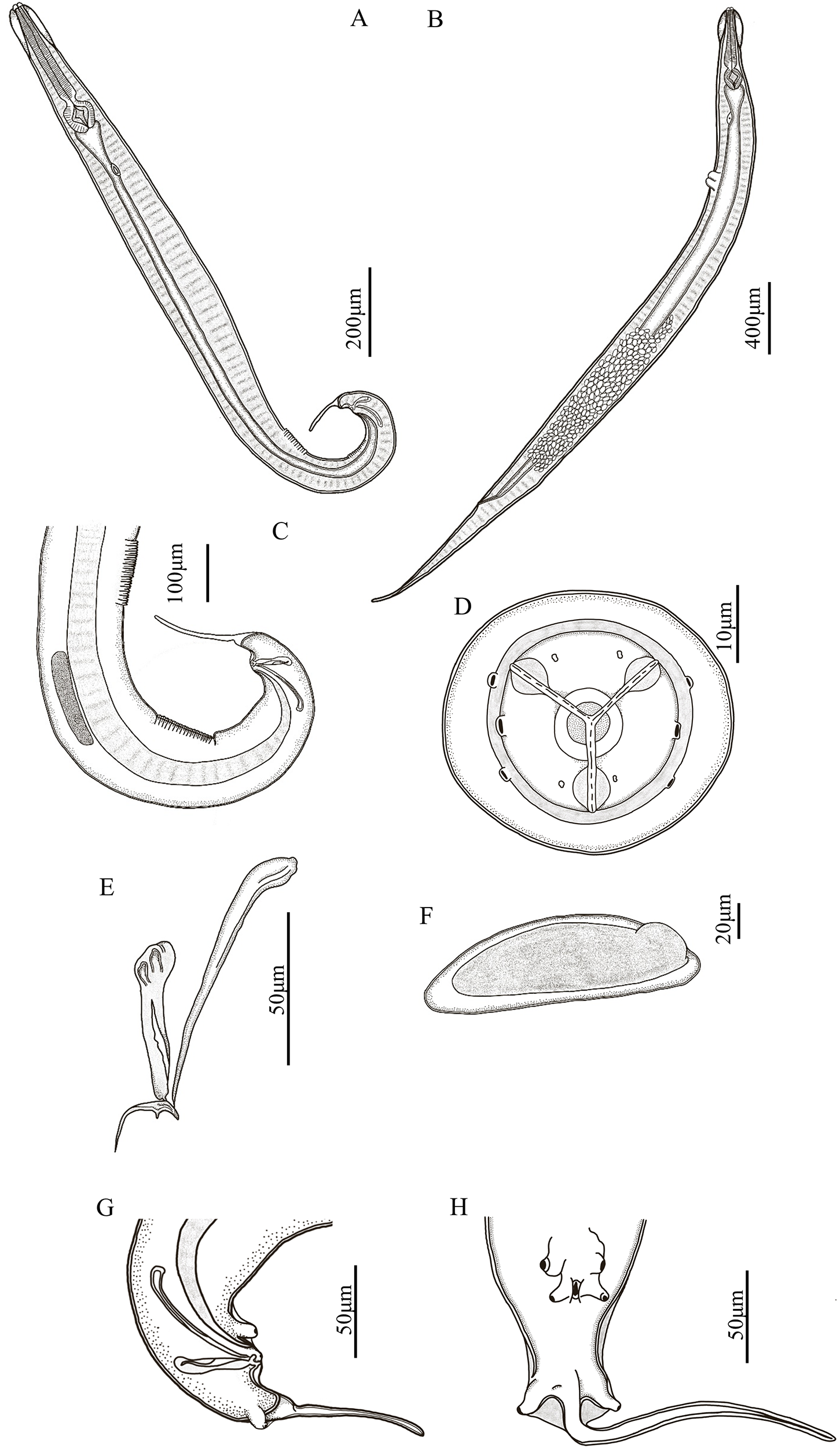
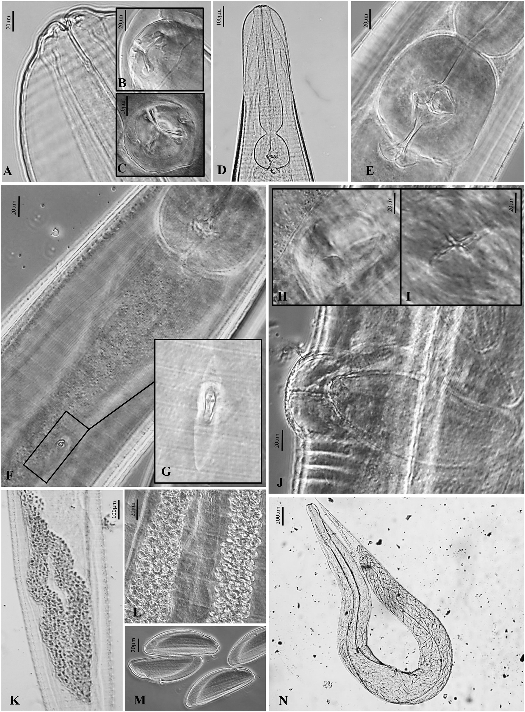
General. Specimens characterized by long, slender shape, mouth bounded by 3 lips, 2 laterally situated amphids and 4 cephalic papillae, 2 lateral to each amphid (Figure 2A,B,D). Cuticle transversally striated from anterior to posterior (Figure 3A,D). Lateral longitudinal alae visible from cephalic vesicle to near level of anus. Cephalic vesicle slightly inflated (Figure 3D). Oesophagus club-shaped, separated from the sub-globular oesophageal bulb by wide constriction (Figures 2A,B; 3D,E). Oesophageal bulb with distinct valvular apparatus (Figure 3E). Anterior end of intestine dilated with slight depression where the oesophageal bulb is positioned (Figure 3F). Intestine constricts slightly, continues at constant diameter to anus (Figures 2A,B; 3F). Nerve ring not clearly visible. Excretory pore small, located posterior to oesophago-intestinal junction (Figure 3F). Excretory pore connected to spindle-shaped cell (Figure 3G). Males were considerably smaller than females, with a coiled posterior extremity, while females end in a long narrow tip (Figure 2A,B). Males with 2 ventrally situated cuticular mamelons (Figure 2C).
Line drawings of Syphatineria cepapi from Paraxerus cepapi in South Africa. (A) Adult male; (B) adult female; (C) male posterior end; (D) en face view of the head; (E) male spicule, gubernaculum, accessory piece; (F) female egg; (G) male spicule, gubernaculum, accessory piece, 3 pairs of caudal papillae; (H) male caudal papillae.

Photomicrographs of Syphatineria cepapi from Paraxerus cepapi in South Africa. (A) Anterior end lateral view; (B) en face (angled) view of anterior end; (C) en face view; (D) anterior end; (E) Oesophageal bulb with denticular apparatus; (F) ventral view, oesophageal bulb, intestine, excretory pore; (G) excretory pore; (H-I) ventral view, 4 segment vulva; (J) lateral view, vulva and an egg; (K) female ovaries; (L) High magnification, female ovaries; (M) female egg; (N) adult female.

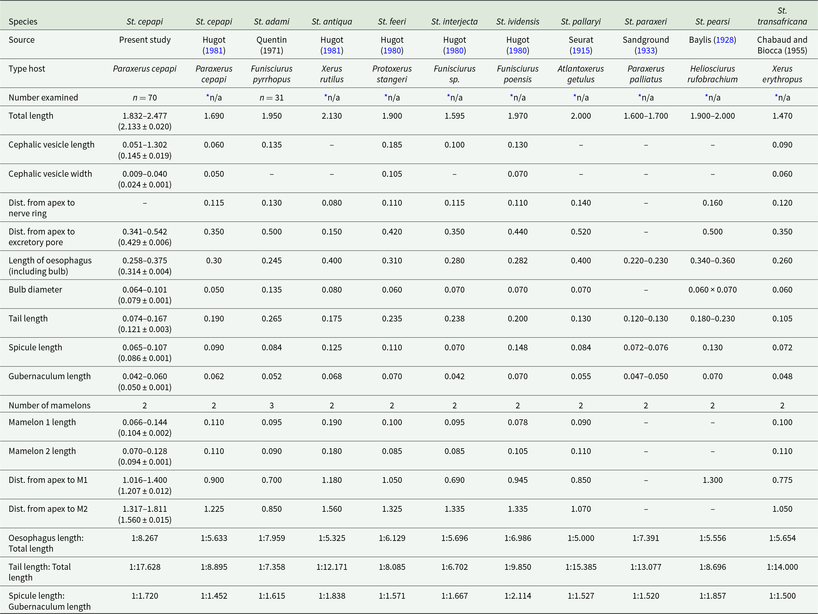
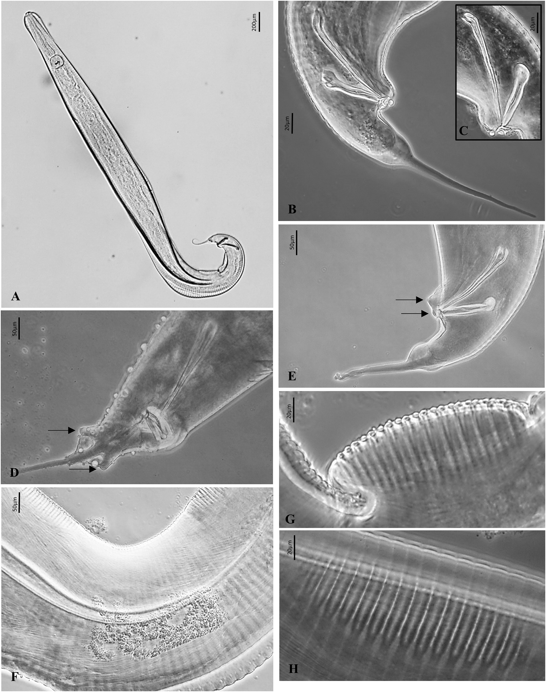
Males (n = 70). Dimensions of various morphological features are given in Table 3. Total body length 2.133 ± 0.020 mm. Body straight anteriorly, coiled posteriorly (Figure 2A). Oesophagus, including bulb, 0.314 ± 0.004 mm long. Two ventrally situated cuticular mamelons with prominent striae (Figures 4G,H). First mamelon 0.104 ± 0.002 mm long, second mamelon, nearest to tail, approximately 0.094 ± 0.001 mm long. Mamelons 0.174 ± 0.007 mm apart. Apex to first mamelon 1.207 ± 0.012 mm, to second mamelon 1.560 ± 0.015 mm. Testes are typically visible between first and second mamelon (Figure 4F). Some specimens, testes extend from first mamelon beyond posterior end of second mamelon. Spicule 0.086 ± 0.001 mm long, slightly curved, directed posteriorly (Figures 2C, 4B). Gubernaculum 0.050 ± 0.001 mm long, fused with a barbed accessory piece, tail-like structure directed posteriorly (Figures 2E, 4B). Three pairs of caudal papillae, 1 precloacal, 1 adcloacal, 1 postcloacal near base of tail (Figures 2G, H, 4D, E). Tail 0.121 ± 0.003 mm long.
Comparative measurements (in mm) of Syphatineria cepapi males (n = 70) recorded on Paraxerus cepapi to previously recorded Syphatineria species in Africa (from Sandground, Reference Sandground1933; Hugot, Reference Hugot1980, Reference Hugot1981). Where possible, range and mean values provided

Table 3 Long description
Morphometric data across 11 St. species. Columns represent species names: St. cepapi, St. cepapi, St. adami, St. antiqua, St. feeri, St. interjecta, St. ividensis, St. pallaryi, St. paraxeri, St. pearsi, St. transafricana. The leftmost column lists measurement categories and metadata. There are approximately 21 rows detailing sources, type hosts, sample sizes, and anatomical measurements. Measurements are likely in millimeters, with some entries as ranges and mean plus or minus standard deviation in parentheses. Key categories include total length, cephalic vesicle dimensions, distances from apex to nerve ring and excretory pore, oesophagus length, bulb diameter, tail length, spicule length, gubernaculum length, number of mamelons, mamelon lengths, and ratios of oesophagus, tail, and spicule lengths to total length or gubernaculum length. Notable points: Total length ranges from 1.470 to 2.477, with St. transafricana having the shortest and St. cepapi the longest. Tail length varies from 0.074 to 0.265, and spicule length from 0.065 to 0.148. St. adami is unique with three mamelons, while others have two. The table provides a comprehensive comparison of anatomical features across species, highlighting differences in size and structure.
* Not available as no exact count given.
Photomicrographs of Syphatineria cepapi from Paraxerus cepapi in South Africa. (A) Adult male; (B) lateral view, male posterior end; (C) high magnification lateral view, spicule, gubernaculum, accessory piece; (D) male posterior end, postcloacal papillae (arrows); (E) male posterior end, precloacal and adcloacal papillae (arrows); (F) lateral view, male testes; (G) lateral view, mamelon; (H) ventral view, mamelon.

Table 4 Long description
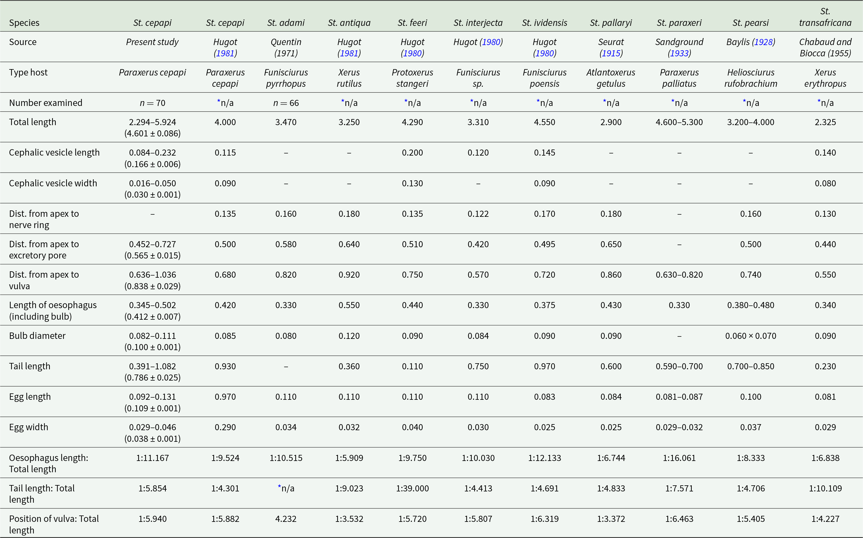
Morphological measurements across 11 species, with columns labeled: Species, Source, Type host, Number examined, Total length, Cephalic vesicle length, Cephalic vesicle width, Distance from apex to nerve ring, Distance from apex to excretory pore, Distance from apex to vulva, Length of oesophagus (including bulb), Bulb diameter, Tail length, Egg length, Egg width, Oesophagus length: Total length, Tail length: Total length, Position of vulva: Total length. Measurements are in millimeters. The table includes 18 rows of data. Notable patterns include St. paraxeri having the largest total length range (4.600 superscript subscript –5.300 mm) and St. transafricana having the smallest (2.325 mm). Tail length varies significantly, with St. cepapi showing a range of 0.391 superscript subscript –1.082 mm. Ratios like 'Oesophagus length: Total length' are expressed as 1:x, indicating proportional relationships. Blanks indicate data not reported. The table provides a comprehensive comparison of species morphology, aiding in understanding species differences.
Females (n = 70). Dimensions of various morphological features are given in Table 4. Total body length 4.601 ± 0.086 mm. No extreme curvature of body (Figure 2B). Oesophagus, including bulb, 0.412 ± 0.007 mm long. Vulva with protruding lips (Figure 3H, I). Apex to vulva approximately 0.838 ± 0.029 mm. Ovaries side by side in lower half of body (Figure 3K, L). Non-embryonated eggs 0.109 ± 0.001 mm long, 0.038 ± 0.001 mm wide. Eggs elongated, unilaterally flattened, numerous, occupy body from vulva to rectum (Figures 2F, 3M). Small operculum present on convex side of egg (Figure 2F). Tail 0.786 ± 0.025 mm long, gradually decreases in diameter (Figures 2B, 3N).
Comparative measurements (in mm) of Syphatineria cepapi females (n = 70) recorded on Paraxerus cepapi to previously recorded Syphatineria species in Africa (from Sandground, Reference Sandground1933; Hugot, Reference Hugot1980, Reference Hugot1981). Where possible range and mean values provided

* Not available as no exact count given.
Phylogenetic relationship
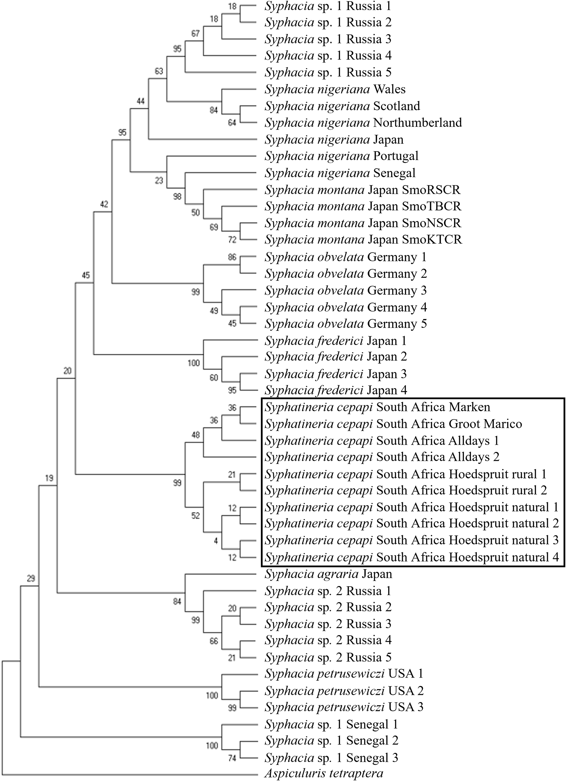
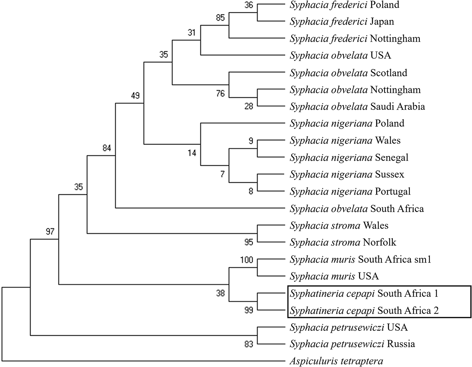
Two 18S rRNA (GenBank accession codes PQ790598-PQ790599) and 10 COI sequences (GenBank accession codes PZ435675-PZ435684) were generated. The fragment length, after being trimmed, was 600 bp for 18S and 203 bp for COI. BLAST analysis of the COI region showed 91.35% identity to Syphacia nigeriana Baylis, 1928 (OK272536.1), which was recovered from Mastomys huberti Wroughton, 1908, in Senegal, while the 18S rRNA sequence shared 96.35% identity with Syphacia obvelata (Rudolphi, 1802) (KY462826.1) recovered from Mastomys coucha (Smith, 1834) in South Africa. The phylogenetic analysis of the COI gene (Figure 5) clustered the 10 St. cepapi isolates into a monophyletic group with 99% support. Albeit based on a very short fragment sequenced, all specimens collected from localities in the North and West (Alldays, Marken and Groot Marico) clustered together with 48% support and all specimens collected from localities in the East (Hoedspruit rural and Hoedspruit natural) clustered together with 52% support. The Maximum Likelihood tree using 18S rRNA data (Figure 6) provides strong support for the monophyly of S. cepapi sampled herein and weak support for a sister taxon association with Syphacia muris Yamaguti, 1935 (Figure 6). Interestingly, Syphatineria cluster nested within a non-monophyletic Syphacia (Figure 5).
Maximum likelihood tree constructed using COI Syphatineria sequences generated in this study in addition to COI Syphacia species sequences available on GenBank. The COI sequences generated in this study are highlighted by the black rectangle.

Figure 5 Long description
The diagram is a maximum likelihood tree constructed using COI sequences, illustrating the phylogenetic relationships among various Syphacia species. The tree is oriented vertically, with branches extending from top to bottom. At the top, Syphacia sp. 1 from Russia is shown with five samples, forming a cluster supported by values ranging from 18 to 67. Below, Syphacia nigeriana is represented with samples from Wales, Scotland, Northumberland, Portugal, Senegal, and Japan, supported by values from 23 to 95. Syphacia montana from Japan is grouped next, with four samples supported by values from 50 to 86. Syphacia obvelata from Germany follows, with five samples supported by values from 45 to 99. Syphacia frederici from Japan is shown with four samples, supported by values from 36 to 100. Syphatineria cepapi from South Africa is highlighted within a black rectangle, showing samples from Marken, Groot Marico, Alldays, and Hoedspruit, supported by values from 12 to 99. Syphacia agraria from Japan is positioned below, followed by Syphacia sp. 2 from Russia and Senegal, supported by values from 20 to 99. Syphacia petrusewiczi from the USA is shown with three samples, supported by values from 19 to 100. At the bottom, Syphacia sp. 1 from Senegal and Aspiculuris tetraptera are displayed, supported by values from 74 to 100. The tree visually represents the evolutionary relationships and genetic distances among the species, with support values indicating the confidence level of each branch.
Maximum likelihood tree constructed using the 18S (SSU) Syphatineria sequences generated in this study, in addition to all 18S (SSU) Syphacia species sequences available on GenBank. Node validity was tested by applying 1000 bootstrap replicates. The 2 18S sequences generated in this study are highlighted by the black rectangle.

Figure 6 Long description
The phylogenetic tree displays evolutionary relationships among various Syphacia species, with branches labeled by geographic locations. The tree is oriented vertically, with branches extending from left to right. Bootstrap values are indicated at each node, representing the confidence level of the branching. Starting from the top, Syphacia frederici from Poland, Japan, and Nottingham form a cluster with bootstrap values of 85, 31, and 35. Syphacia obvelata from the USA, Scotland, and Nottingham follow, with values of 76 and 49. Syphacia obvelata from Saudi Arabia is next, branching with a value of 28. Syphacia nigeriana from Poland, Wales, Senegal, Sussex, and Portugal are grouped with values of 14, 9, and 8. Syphacia obvelata from South Africa branches separately. Syphacia stroma from Wales and Norfolk are grouped with a value of 95. Syphacia muris from South Africa and the USA are clustered with a value of 100. Syphatineria cepapi from South Africa 1 and 2 are highlighted within a black rectangle, branching with a value of 99. Syphacia petrusewiczi from the USA and Russia follow, with a value of 83. The tree concludes with Aspiculuris tetraptera at the base. The tree visually represents genetic relationships and geographic distribution among the species.
Nematode infestations
A total of 11 112 St. cepapi individuals were recorded (Table 2). Syphatineria cepapi was recorded at 7 of the 8 localities with an overall mean abundance of 140.66 ± 19.54 and an overall prevalence of 92.41% (Table 2). Males accounted for 38% of the collected individuals and females for 62% (Table 2). Overall and per locality female St. cepapi were more abundant compared to males (Table 2). Syphatineria cepapi had the highest mean abundance at Bushbuckridge (Table 2).
Discussion
The specimens described in this study were classified into the subfamily Syphaciinae, the genus Syphatineria and subgenus Africanoxys by the rounded cephalic plateau, cephalic papillae situated close to the amphids, well-developed cephalic vesicles and a triangular-shaped mouth with 3 individualized lips. More specifically, males possessed a simple spicule, elongated gubernaculum with a barbed accessory hook that ends ventrally and 3 pairs of caudal papillae. Additionally, male specimens have 2 instead of 3 cuticular mamelons. This feature is shared with 9 Syphatineria species that occur in African squirrels (Baylis, Reference Baylis1928; Sandground, Reference Sandground1933; Hugot, Reference Hugot1980, Reference Hugot1981, Reference Hugot1988) and other Syphacia species such as S. oceanica Schmidt and Kuntz, 1968, associated with the Southern Palawan tree squirrel in Indonesia and S. eutamii Tiner, 1948, found in the Least Chipmunk of North America. In females, non-embryonated, elongated, asymmetrical eggs with 1 concave and 1 convex side, and a small operculum are present (Hugot, Reference Hugot1981; Petter and Quentin, Reference Petter, Quentin, Anderson, Chabaud and Willmott2009). Furthermore, oxyurids of Sciurinae from Africa and the Oriental (Eastern) regions of the World are classified within the subgenus Africanoxys (Hugot, Reference Hugot1981).
The description and measurements of St. cepapi by Hugot (Reference Hugot1981), based on specimens from a single P. cepapi squirrel from a single locality, most likely resulted in narrow variations in measurements. When these measurements are compared to what is obtained for the taxon examined in the present study there is similarity in the length of some of the characteristics measured (Tables 3 and 4). For example, for St. cepapi in Hugot (Reference Hugot1981) males, the length of the spicule (0.090; 0.065–0.107, respectively), mamelon 1 (0.110; 0.066–0.144, respectively) and mamelon 2 (0.110; 0.070–0.128, respectively) (Table 3). There are, however, some differences also noted. For example, for St. cepapi males in Hugot (Reference Hugot1981) the gubernaculum is longer (0.062; 0.042–0.060, respectively) while the distance from the apex to mamelon 1 (0.900; 1.016–1.400, respectively) and mamelon 2 (1.225; 1.317–1.811, respectively) is shorter (Table 3). The most notable differences in the females presented by Hugot (Reference Hugot1981) and those measured herein are in the egg length (0.970; 0.092-0.131, respectively) and width (0.290; 0.029–0.046, respectively) (Table 4). However, the egg dimensions presented by Hugot (Reference Hugot1981) appear inconsistent compared to the dimensions presented herein and to the other species in Table 4 and possibly reflect an error. The morphological comparisons, in addition to the host species support the conclusion that the present specimens can be assigned to St. cepapi.
We regard the St. paraxeri classification by Viljoen (Reference Viljoen1977a) as an incorrect species delimitation for the pinworm that occurred in the gut and caecum of P. cepapi. The conclusion by Viljoen (Reference Viljoen1977a) was most likely influenced by the literature available at that time since the description of St. cepapi was only published in 1981 (Sandground, Reference Sandground1933; Hugot, Reference Hugot1981) and overlap in some of the characters for example, the spicule and gubernaculum length for males and the egg length and width for females between St. paraxeri and St. cepapi (Tables 3 and 4). However, it is less likely that St. paraxeri, described from P. palliatus, would also occur in P. cepapi given the level of host specificity of oxyurid nematodes (Behnke et al., Reference Behnke, Stewart, Smales, Cooper, Lowe, Kinsella, Bajer, Dwuznik-szarek, Herman, Fenn, Catalano, Diagne and Webster2022; Schlemmer et al., Reference Schlemmer, Junker, Matthee and Matthee2026) and the non-overlapping geographic ranges of P. palliatus and P. cepapi in South Africa (Monadjem et al., Reference Monadjem, Taylor, Denys and Cotterhill2015).
More compelling data are also provided by our study through comparing the morphometric data from St. cepapi (recorded in the present study), with St. paraxeri recorded in Zimbabwe, as well as 2 closely related species (St. pallaryi and St. pearsi; Sandground, 1933). Syphatineria cepapi males are differentiated from St. paraxeri by a longer total length and longer oesophagus, including the bulb. It is distinguished from St. pallaryi by a longer total length, shorter oesophagus including the bulb, longer distance between the apex and mamelon 1 and mamelon 2, and a shorter distance between the apex and the excretory pore, and from St. pearsi by a longer total length, shorter tail and spicule. Conversely, St. cepapi females are distinguished from St. paraxeri by a longer oesophagus including the bulb, and longer tail, from St. pallaryi by a longer total length, a longer tail and a shorter distance between the apex and the excretory pore, and from St. pearsi by a longer total length, larger oesophageal bulb and longer distance between the apex and the excretory pore and the apex and the vulva.
The noteworthy difference between minimum and maximum length of features measured in this study can be attributed to the size variation of the mounted nematodes. Previous studies on Syphacia species also recorded notable variation in the range of features. For example, Behnke et al. (Reference Behnke, Stewart, Smales, Cooper, Lowe, Kinsella, Bajer, Dwuznik-szarek, Herman, Fenn, Catalano, Diagne and Webster2022) recorded a relatively large range (3.4–5.4 mm) for the total length, distance from the anterior end to the vulva (0.363–0.700 mm) and tail length (0.410–0.710 mm) of Sp. nigeriana. Similarly, Stewart et al. (Reference Stewart, Lowe, Smales, Bajer, Bradley, Dwuznik, Franssen, Griffith, Stuart, Turner, Zalesney and Behnke2017) recorded great variation in the tail length, distance from the anterior to the vulva and distance from the anterior to the excretory pore for Sp. frederici Roman, 1945 and Sp. stroma (Linstow, 1884).
The nucleotide comparison of the generated sequences from St. cepapi to the nucleotide sequence database available on GenBank for Syphacia species (these are all Muridae-associated species, as no sequences are available for squirrel-associated Syphacia/Syphatineria species) indicated that the 18S rRNA generated sequence in the present study has a higher nucleotide identity to Sp. obvelata than the COI sequences had to Sp. nigeriana. The 3 species (Sp. obvelata, Sp. nigeriana and Sp. muris) are distinguished from St. cepapi by their tendency to only parasitize hosts in the family Muridae and the presence of 3 instead of 2 mamelons in male specimens. Interestingly, the phylogenetic clustering of Syphatineria based on 18S rRNA does not support the monophyly of Syphacia or the generic distinctness of Syphatineria. Accordingly, the generic taxonomy of Syphacia requires comprehensive revision based on broader taxon sampling, consistent with previous studies that have also highlighted its non-monophyletic nature and taxonomic instability (Okamoto et al., Reference Okamoto, Urushima, Iwasa and Hasegawa2007, Reference Okamoto, Urushima and Hasegawa2009). This further raises the possibility that additional genera remain to be recognized, or alternatively that Syphatineria merely represents a morphologically distinct group of Syphacia characterized by unique morphological apomorphies.
With the support from the morphological data and based on the distinct clustering of the novel 18S rRNA and COI sequences generated in this study, it is clear that putative St. cepapi is a unique species most likely occurring only in P. cepapi. Furthermore, the hint of geographic structure seen in the analyses of the COI sequences may support the close association between the parasite and host (Anderson, Reference Anderson2000; Schlemmer et al., Reference Schlemmer, Junker, Matthee and Matthee2026). However, our study contributes new sequence data and provides some preliminary evidence that St. cepapi is unique, a more definitive answer can only be obtained through the inclusion of all the potential sister species to St. cepapi.
In the present study, Syphatineria sensu stricto nematodes were highly abundant and prevalent across 7 of the 8 sampling localities and were primarily recovered from the caecum of P. cepapi squirrels. This is in agreement with other studies that also suggested that pinworms are highly prevalent in their respective rodent hosts (Froeschke and Matthee, Reference Froeschke and Matthee2014; Lutermann et al., Reference Lutermann, Medger and Junker2014; Spickett et al., Reference Spickett, Junker, Froeschke, Haukisalmi and Matthee2020; Schlemmer et al., Reference Schlemmer, Junker, Matthee and Matthee2026). Syphacia is predominantly transmitted when eggs are ingested from the perianal area through host grooming practices (Anderson, Reference Anderson1988, Reference Anderson2000) and the high abundance of this nematode in P. cepapi is most likely facilitated by allogrooming activities between family members (Viljoen, Reference Viljoen1977b). In addition to St. cepapi 5 nominal Syphatineria/Syphacia species and several unidentified Syphacia spp. have been recorded in South Africa to date. As mentioned above, the record of St. paraxeri in P. cepapi may represent a misidentification (Viljoen, Reference Viljoen1977a). Furthermore, Sp. muris (rat pinworm) was recorded in Rattus tanezumi (Temminck, 1844), Sp. obvelata (common mouse pinworm) was recorded in M. coucha and Rhabdomys dilectus (De Winton, 1897), Sp. nigeriana was recorded in Gerbilliscus brantsii (Smith, 1836) and Syphacia minuta (Schneider, 1866) in Acomys spinosissimus (Peters, 1852) (Lutermann et al., Reference Lutermann, Medger and Junker2014; Julius et al., Reference Julius, Schwan and Chimimba2017; Spickett et al., Reference Spickett, Junker, Froeschke, Haukisalmi and Matthee2020). Most of these records have been based on morphology alone and must be treated with caution, especially given the morphological difficulties in differentiating between Syphacia species (Behnke et al., Reference Behnke, Stewart, Smales, Cooper, Lowe, Kinsella, Bajer, Dwuznik-szarek, Herman, Fenn, Catalano, Diagne and Webster2022). Additional specimens, noted as Syphacia sp., were recorded in several other rodent species, including Rhabdomys pumilio (Sparrman, 1784) and Micaelamys namaquensis (Smith, 1834) in South Africa (Froeschke and Matthee, Reference Froeschke and Matthee2014; Spickett et al., Reference Spickett, Junker, Froeschke, Haukisalmi and Matthee2020; Schlemmer et al., Reference Schlemmer, Junker, Matthee and Matthee2026). As indicated by Schlemmer et al. (Reference Schlemmer, Junker, Matthee and Matthee2026), the unidentified Syphacia species recorded in R. pumilio and M. namaquensis that were trapped at the same localities and time represent distinct molecular clades and potentially distinct species. This reaffirms the need for future studies on both Syphatineria and Syphacia.
The present study offers insight into the morphological and molecular aspects and infestation patterns of St. cepapi through in-depth measurements, line drawings, photomicrographs and genetic sequences. Future studies of Syphatineria species associated with African squirrels should include sister species and follow an integrated morphological and molecular approach.
Data availability statement
All generated data for this study are included in this article. Additional data can be obtained from the corresponding author upon request.
Acknowledgements
We are grateful to all the farmers and landowners, and in particular Jan-Phillip Small, Reed van Zyl, Johan Jordaan, Sean du Toit, Giel Horn, Jeanette Wentzel and Lourens Swanepoel, who provided samples for this study. Fletcher Vincent assisted with the examination of the gastrointestinal tracts. Sarah Ackland assisted with the line drawings.
Author contributions
I.R. conducted the laboratory work, analysed the data, and wrote the first draft of the manuscript. C.A.M. acted as co-supervisor of I.R. and assisted with sample collection. S.M. conceived the study, provided funding, assisted with sample collection and supervised I.R. All authors contributed to the final version of the manuscript.
Financial support
I.R. received personal funding from the Department of Conservation Ecology and Entomology (Stellenbosch University). Stellenbosch University is thanked for its financial support.
Competing interests
The authors declare there are no conflicts of interest.
Ethical standards
The project was approved by Stellenbosch University Animal Ethics (ACU-2021-21609). All necessary permits were obtained for Mpumalanga province (MPB. 5698), Limpopo province (ZA/LP/116034) and North West province (NW 42843/08/2022) and Section 20 [12/11/1/7/5/2057(HP)] from the Department of Agriculture, Land Reform and Rural Development.

 Open access

