Introduction
In the span of 10 years, Huanglongbing (HLB) has devastated Florida's over the 10-billion-dollar citrus industry.[Reference Hodges and Spreen1] Recent works on greenhouse canker infiltration assays showed the local systemic activity of Zn-chelate[Reference Commerford, Gerberich, Rajasekaran, Young, Das, Graham, Santra and Johnson2]; however, there is no clear understanding of how the Zn-chelate moves in citrus trees. Although there are many analytical methods available for detecting Zn in aqueous solutions [e.g., inductively coupled plasma mass spectrometry and atomic absorption spectroscopy (AAS)], in situ monitoring of the movement of Zn-chelate in citrus trees has not been fully explored due to lack of experimental tools.[Reference Church, Armas, Patel, Chumbimuni-Torres and Lee3] There is an urgent need to develop a reliable Zn2+ monitoring tool, capable of tracking its systemic activity directly in plants. Successful in situ Zn2+ detection will lead to a better understanding of its potential fate in plants by estimating spatial and temporal phloem concentrations of Zn2+ between successive spray applications, which will benefit the assessment of spray rate and timings for effective HLB management.
Microelectrodes are needle-type electrochemical microsensors that have been used for in situ monitoring of chemical compounds of interest in biofilm and corrosion processes in drinking water distribution systems[Reference Lee, Wahman, Bishop and Pressman4–Reference Ma, Armas, Soliman, Lytle, Chumbimuni-Torres, Tetard and Lee6] and can be used to measure intracellular free-ion activities by inserting their tip into plant tissues. Previous studies have shown the usefulness of applying ion-selective microelectrodes to track the movement of H+, K+, NO3−, and Na+ in plants[Reference Miller, Cookson, Smith and Wells7–Reference Newman, Kochian, Grusak and Lucas10]; however, it was found that Zn2+ ion-selective electrodes show interference to Cu2+,[Reference Church, Armas, Patel, Chumbimuni-Torres and Lee3] a common micronutrient found in plants. Furthermore, ion-selective microelectrodes are difficult to manufacture and can be easily broken when the glass tip comes in contact with hard surfaces. The microelectrode tip must be microscale to impale the phloem, but durable enough to penetrate the plant without breaking. Miller et al.[Reference Miller, Cookson, Smith and Wells7] reported only 9% of the originally prepared triple-barreled electrodes working after plant impalement.
On the other hand, square-wave anodic stripping voltammetry (SWASV) has been recognized as a powerful tool for measuring trace metals for decades[Reference Wang, Lu, Hocevar, Farias and Ogorevc11] and can eliminate the need for fragile ion-selective membranes used in ion-selective electrodes. Metallic bismuth (Bi) electrodes have been used as a working electrode for the anodic stripping performance by forming amalgams with trace metals.[Reference Wang, Lu, Hocevar, Farias and Ogorevc11–Reference Wang, Lu, Kirgöz, Hocevar and Ogorevc14] SWASV using Bi films have shown a limit of detection (LOD) of zinc reaching 12 ppb.[Reference Hwang, Han, Park and Kang15] The enhanced sensitivity of SWASV, associated with its pre-concentration step, makes it an ideal tool for measuring trace metals in biologic matrices. However, conventional SWASV technique, using a three-electrode system, requires stirring during the deposition step, which is unsuitable for the measurement of Zn2+ within the vascular bundle of citrus plants. In addition, the conventional three-electrode system needs to be miniaturized for plant applications. Here, we propose the use of a novel two-step SWASV technique where the pre-concentration step is conducted in situ using a combined Bi/platinum (Pt) microelectrode system before being transferred to a stripping solution and then connected to a more sensitive three-electrode system. The use of microelectrodes in electroanalysis offers a number of advantages for in situ detection of Zn2+ in biologic samples including low background current, low IR drop, and enhanced mass transport rates.[Reference Bartlett, Denuault and Sousa16] The last property is particularly important since it could alleviate the need for stirring during the pre-concentration step and allows in situ measurements in plants where the stirring condition is not possible. Even with the many advantages described previously, SWASV-based Zn2+ detection using microelectrodes has not yet been explored.
This study is the first to develop a novel technique for in situ detection of zinc in citrus plants by exploring a solid type of microelectrode for Zn2+ measurement using SWASV. Two innovative concepts were introduced to overcome the challenges of in situ measurement of Zn2+ in plant. First, a low melting point Bi alloy (Belmont Alloy 2451: 44.7% Bi, 22.6% lead, 19.1% indium, 8.3% tin, and 5.3% cadmium, melting point 47 °C) was tested as a way to eliminate the need for electrochemical co-deposition of Bi traditionally used in the SWASV detection of Zn2+. Second, an in vivo pre-concentration step before anodic stripping analysis was evaluated to minimize the invasiveness of the method by only using a two-electrode system for pre-concentration before transferring to a more sensitive three-electrode system for anodic stripping analysis.
Methods
Metallic Bi microelectrode design and fabrication
Two different types of Bi microelectrodes were developed for Zn2+ detection using SWASV in this study. A single-barrel Bi microelectrode was used for testing the concept of two-step SWASV for detecting Zn2+, and a double-barrel microelectrode consisting of a Bi working electrode and a Pt reference electrode was used for real application to plant samples (Fig. S1). The fabrication steps for a single-barrel microelectrode can be found elsewhere.[Reference Lee, Lee, Choi, Hosni, Papautsky and Bishop17] Briefly, a micropipette (1B120-6, O.D. 1.2, I.D. 0.69, 150 mm, World Precision Instruments Inc., Sarasota, FL, USA) was pulled using a Flaming/Brown type micropipette puller (Model P-1000, Sutter Instrument Co., Novato, CA, USA). The tip of the micropipette was then broken using tweezers and tip diameter (6–85 µm) was verified under a microscope. A piece (3 cm) of Bi alloy wire (Belmont Alloy 2451: 44.7% Bi, 22.6% lead, 19.1% indium, 8.3% tin, and 5.3% cadmium, melting point 47°C) was then inserted into the pulled micropipette and pushed toward the tip using a metal plunger. The alloy was heated using a nichrome heating filament (Cat. No. 66258-066, VWR, Radnor, PA, USA). Pressure was applied using a plunger once the alloy began to melt to allow the alloy to pass through the tip of the micropipette. Any remnant of Bi alloy at the tip of the microelectrode was removed by a gentle shake. A copper wire (12 cm, S4828C, 26GA, Fisher Scientific, Hampton, NH, USA) was then fixed to the opposite end of the microelectrode by melting the alloy and inserting the wire. Next, the tip of the micropipette was beveled at a 45° angle to expose the alloy wire using a micropipette beveller (BV-10, Sutter Instrument Co.). The tip was then rinsed with deionized (DI) water, and the finished microelectrode was preserved in a pipette storage box (BX20, Sutter Instrument Co.) until its use.
A double-barrel microelectrode consisting of a Bi working electrode and a Pt reference microelectrode was fabricated using the same procedures described above but with a double-barrel micropipette (2BF150-86-10, Sutter Instruments Co.). A Pt wire (99.99%) with a diameter of 100 µm (#100-896, California Fine Wire Co., Grover Beach, CA, USA) was attached to a copper wire (12 cm, S4828C, 26GA, Fisher Scientific) using a silver conductive epoxy (8331-14G, MG chemicals, Ontario, Canada) and etched in 0.1 M KCN with 1.2 V for 25–30 s to taper the tip. The Pt wire was then inserted into one barrel of the double-barrel micropipette, and the micropipette was pulled using the micropipette puller to seal the wire within the glass. The tip of the second barrel was broken and the Bi alloy was formed at the tip of the microelectrode using the steps described for the single-barrel microelectrode fabrication.
Microelectrode performance evaluation using SWASV
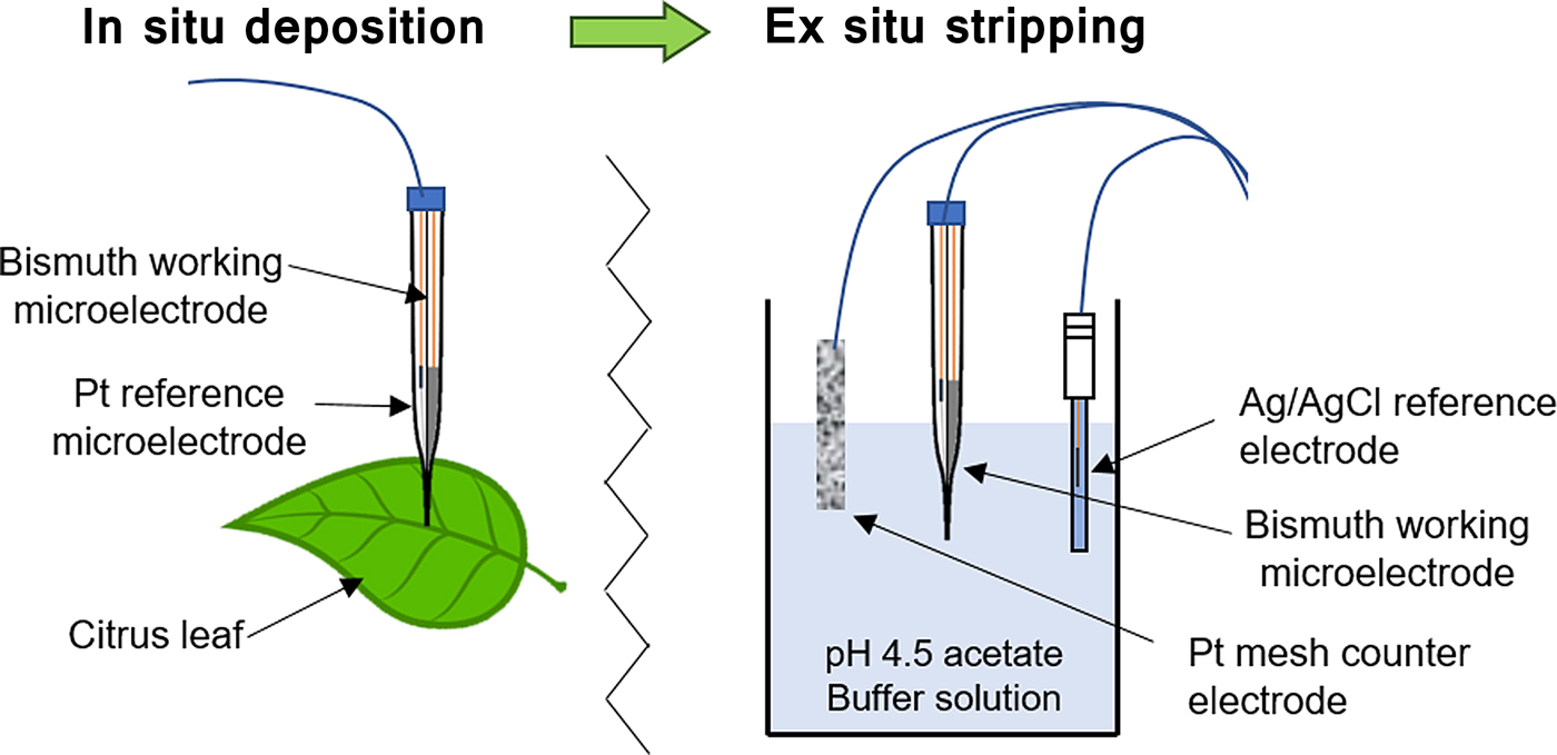
To apply the developed double-barrel microelectrode to the plant system, the traditional SWASV was modified with a two-step approach of in situ deposition and ex situ anodic stripping (Fig. 1). First, Zn2+ was deposited on the Bi microelectrode in a solution of Tris buffer (pH ranged from 5.0 to 7.5) with varying concentrations of ZnCl2 (0–300 ppm Zn2+) to represent the plant environment. Using a two-electrode system, a deposition potential of −1.4 V versus Pt reference electrode was applied to the Bi microelectrode for 180 s both under stirred and unstirred conditions. The microelectrodes were then removed, rinsed with DI water, and transferred to a stripping solution (unstirred) where a SWASV scan (scan, −1.4 to −0.4 V; step, 10 mV; amplitude, 40 mV; frequency, 5 Hz) was applied using a potentiometer (Palmsens3, Palmsens BV, The Netherlands) to determine Zn2+ concentration. During the square-wave scan, a Pt mesh was used as a counter electrode and an Ag/AgCl electrode (MI-401, Microelectrode Inc., Bedford, NH, USA) was used as a reference electrode. After the scan, the microelectrode was cleaned in the same solution by applying −0.4 V for 30 s.
A concept diagram of the two-step SWASV determination of Zn2+ in the vascular bundle of citrus plants.

In situ application to citrus leaves
Before applying the developed Bi/Pt microelectrode to the citrus leaves, sample leaves were collected from a sour orange seedling and introduced to a 1 mM solution of ZnCl2 by submerging the stem of the leaf into the solution for 6 h. Control samples were exposed to DI water for 6 h. The surfaces of all leaves were then rinsed with DI water and wiped dry with a Kimwipe. An automatic three-dimensional (3D) micromanipulator (UNISENSE A/S, Denmark) was used to position the tip of the combined Bi/Pt microelectrode 400 µm below the surface of the midrib for the in situ deposition of Zn2+. Preliminary results of pH microprofiles (Fig. S2) and cross-sectional images (Fig. S3) suggested that ~300–400 µm would be a typical depth for the phloem in citrus seedlings. Local in situ pH measurements were conducted using a pH microsensor (10 µm tip diameter, UNISENSE A/S) and the 3D micromanipulator (UNISENSE A/S).[Reference Ma, Armas, Soliman, Lytle, Chumbimuni-Torres, Tetard and Lee6] After Zn2+ deposition on Bi side of the Bi/Pt microelectrode, the microelectrode was removed, rinsed with DI water, and transferred using the automatic 3D manipulator to the anodic stripping solution (0.1 M acetate buffer at pH 4.5). A stereomicroscope with a CCD camera (World Precision Instruments Inc.) was used to monitor the location of the microelectrode tip during the movement steps. SWASV scans were produced using a potentiometer (Palmsens3, Palmsens BV) with the same parameters used in the calibration. All experiments were performed in a Faraday cage (81-334-04, Technical Manufacturing Co., Peabody, MA, USA) to avoid electrical interference. Pre- and post-calibrations were conducted for ensuring the sensor performance.
Results and discussion
Zn2+ detection using two-step SWASV
The remarkable sensitivity of anodic stripping analysis is attributed to the coupling of an efficient pre-concentration step with a sensitive voltammetric stripping measurement of accumulated metals. For conventional SWASV measurements, the pre-concentration and stripping steps occur in the same solution; however, this is not suitable for in situ detection in citrus trees. To minimize the impact of continuous SWASV measurements within the vascular bundle of citrus plants, the pre-concentration step needs to be conducted separately, in situ, using a combined two-electrode system before being transferred to a stripping solution and connected to a more sensitive three-electrode system. This two-step process was evaluated using a Bi microelectrode and compared with traditional SWASV measurements. For the conventional measurements, both the deposition step and stripping steps took place in a pH 4.5 acetate buffer solution (0.1 M) using a Pt mesh counter electrode and an Ag/AgCl reference electrode. For the modified two-step measurement used in this study, deposition was performed in a separate Zn2+ solution (Tris buffer + ZnCl2), simulating the plant system, before being transferred to a pH 4.5 acetate buffer where stripping occurred.
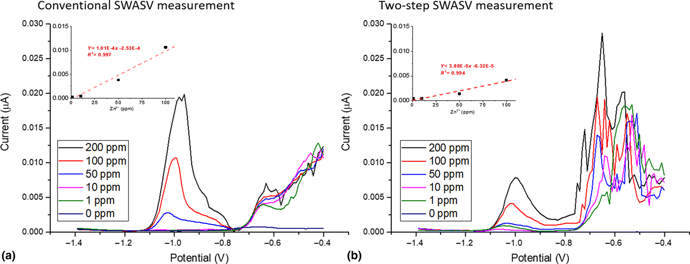
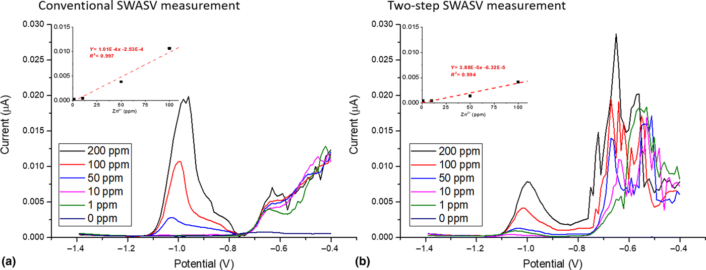
Figure 2 shows the difference in response between the two methods. For the conventional SWASV measurement, a Zn2+ peak height of 0.022 µA was observed for 200 ppm Zn2+. This is significantly larger than the 0.009 µA peak height for the two-step SWASV method using the same Bi microelectrode. However, it was found that both methods were able to demonstrate a linear response between 1 and 200 ppm Zn2+. While the conventional SWASV measurement showed higher sensitivity to Zn2+ (101 versus 38 pA/ppm), the two-step method was also able to measure Zn2+ concentrations of interest with an excellent linear response (R 2 = 0.990). This result is the first to show that the two-step SWASV method can work in plants systems.
SWASV responses of the developed Bi microelectrode (tip size: 6 µm in diameter) to various Zn2+ concentrations (1–200 ppm) between (a) a conventional method: deposition (stirred) and striping (unstirred) in a pH 4.5 acetate buffer and (b) a two-step SWASV method: deposition (unstirred) in ZnCl2 solution and stripping (unstirred) in a pH 4.5 acetate buffer at 23 °C.
Effect of plant pH on microelectrode response
The effect of plant pH was investigated over the pH range of 5.0–7.5, under a fixed concentration of 200 ppm Zn2+ in DI water, using the two-step SWASV method (Fig. S4). The pH was adjusted using HCl and NaOH. This pH range is typical for the vascular bundle of citrus plants[Reference Hijaz and Killiny18] and was also determined using a pH microsensor in this study (Fig. S5). The largest response was obtained at the lowest pH (5.0) with 1.78 µA; however, the responses between pH 5 and 6.5 were comparable and ranged from 1.40 to 1.78 µA. At pH 7.0, the response to Zn2+ decreased significantly by 63% with a 0.66 µA peak, and the response at pH 7.5 was <1% of the electrode signal for pH 5.0. This decrease in response with increasing pH is in accordance with the change of Zn2+ species depending on the pH in aqueous solution. Using a water chemistry modeling software (MINEQL), it was confirmed that the solubility of Zn2+ decreases as the pH increases above 7.2. Therefore, it is likely that the developed SWASV microelectrode is responding to the soluble concentration of Zn2+ across the tested pH range, and that when the pH was increased to 7.5, there was only a small amount of soluble Zn2+ to detect. Considering that the pH of the vascular bundle is expected to be below pH 7, it is likely that majority of the zinc would be detected by the developed two-step SWASV method using a Bi/Pt microelectrode.
Effect of tip size and mass transfer on SWASV response
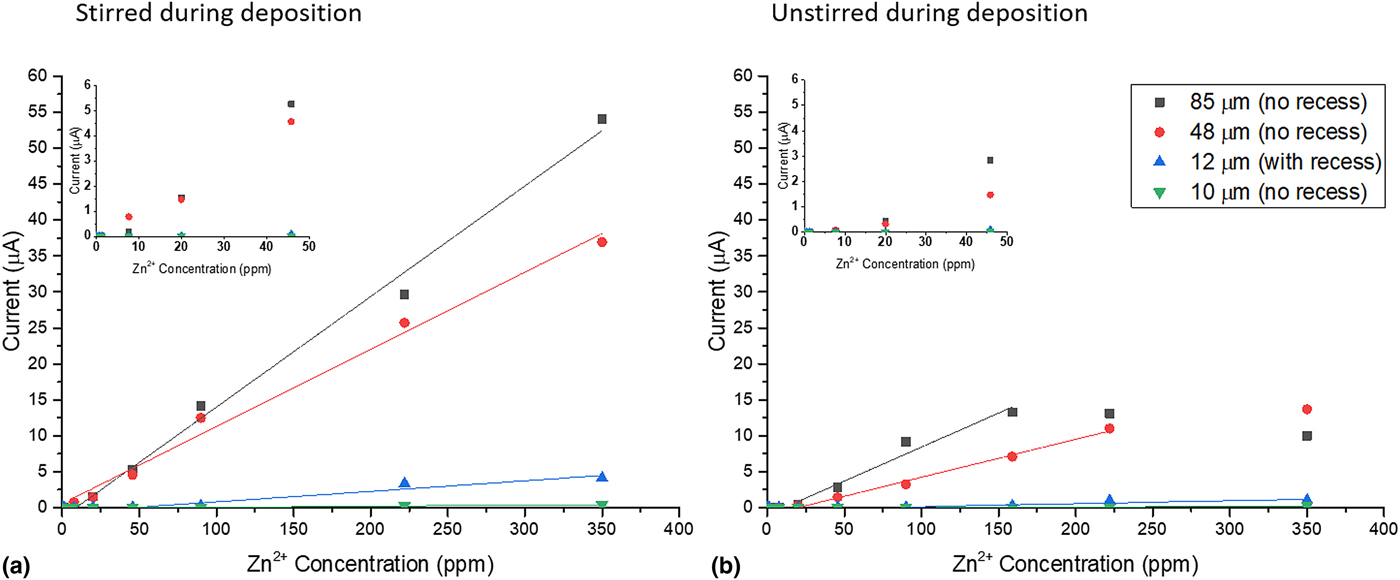
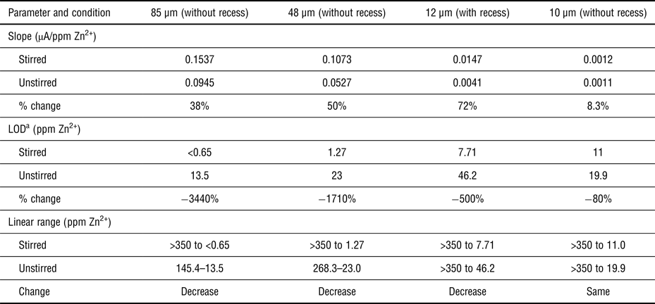
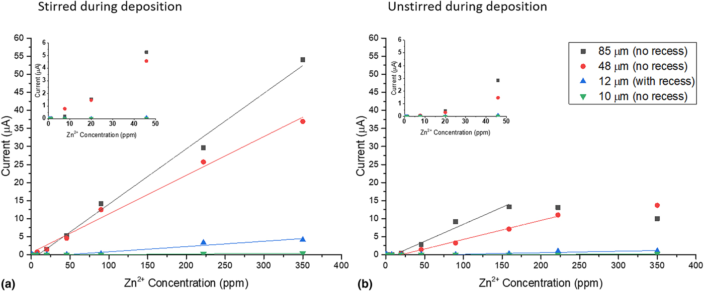
The response of SWASV to Zn2+ concentrations is partly related to the surface area of the working electrode. Therefore, the effect of the microelectrode tip size was investigated to optimize the developed Bi microelectrode for application to citrus plants. The ideal tip size would be small enough to penetrate the phloem, but large enough to produce distinguishable SWASV responses to different Zn2+ concentrations. Figure 3(a) shows the calibration curves of the developed Bi microelectrode with different tip sizes ranging from 10 to 85 µm (Fig. S6). As expected, larger tip sizes increase the sensitivity of the SWASV response. For example, the 85 µm tip size had an increased slope, 0.154 µA/ppm Zn2+, compared with the 10 µm tip size which only had a slope of 0.0012 µA/ppm Zn2+. The sensors displayed similar sensitivities per unit surface area (cm2) with an average normalized sensitivity of 1246 ± 285 µA/ppm Zn2+/cm. Under stirred conditions, the 85 µm tip displayed a LOD below 0.65 ppm Zn2+, while the smaller tip size (e.g., 10 µm) had a higher LOD (e.g., ~11 ppm Zn2+). The recess of the microelectrode shows no significant effect on the electrode response under stirred conditions (Table I).
Mass transfer effect on Zn2+ detection: Zn2+ SWASV calibration curves of various tip sizes of Bi microelectrodes under (a) stirred and (b) unstirred conditions during a deposition step. Insets: Expanded view of sensor response from 0 to 50 ppm Zn2+.
The effect of tip size on the mass transport interference of SWASV response to Zn2+ using the developed Bi microelectrode.

a Limit of detection.
The effect of mass transfer on SWASV response was also investigated (stirred versus unstirred). Conventional SWASV measurements use stirring during the deposition step to increase the mass transport of target heavy metal ions to the electrode; however, under unstirred conditions, which simulate inside the vascular bundle of citrus plants, the reduced mass transport may be a limiting factor during in situ deposition of Zn2+. Figure 4(b) shows the calibration curves of various microelectrode tip sizes under unstirred conditions. The slope and LOD are severely affected under unstirred conditions for the 85, 48, and 12 µm tip sizes. There was a 3440%, 1710%, 500%, and 80% increase in the LOD for the 85, 48, 12, and 10 µm tip sizes, respectively. Similarly, the linear range also decreased for the 85 and 48 µm tip sizes probably due to mass transport limitations but stayed the same for 12 and 10 µm tip sizes between stirred and unstirred conditions. Table I compares the slope, linear range, and LOD between stirred and unstirred conditions. These results show that there is the lesser effect of mass transport limitations on smaller sizes, compared with larger tip sizes. It is also clear that the recess enhances the effect of stirring on the sensor response. For example, the LOD of the 12 µm sensor with a recess was decreased by 500% when there was no stirring during the deposition, but a similarly sized sensor (10 µm) without a recess did not show a significant decrease in LOD. These findings agree with other literature[Reference Bartlett, Denuault and Sousa16] and is particularly important since microelectrodes with a smaller tip size could alleviate the need for stirring during the pre-concentration step and allow in situ measurements. Overall, a Bi tip size in the range of 45–90 µm (as a working electrode) was used for the biologic application to minimize mass transport interference and maximize LOD and slope.
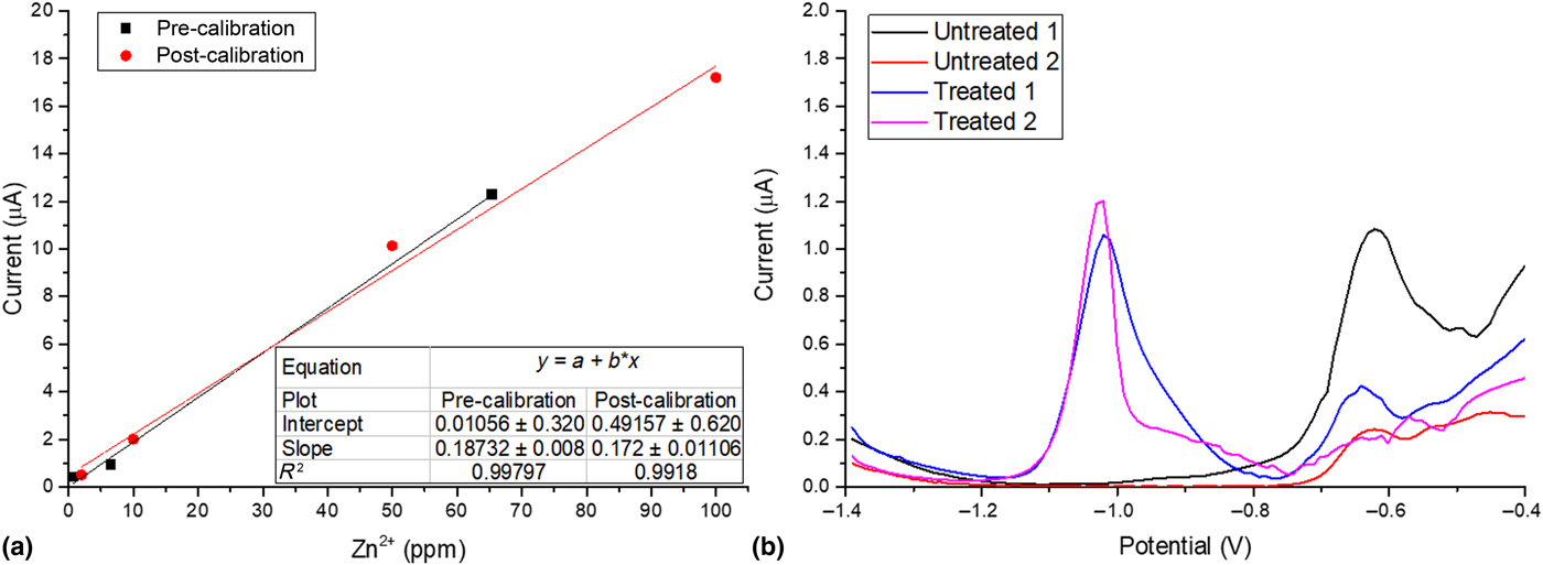
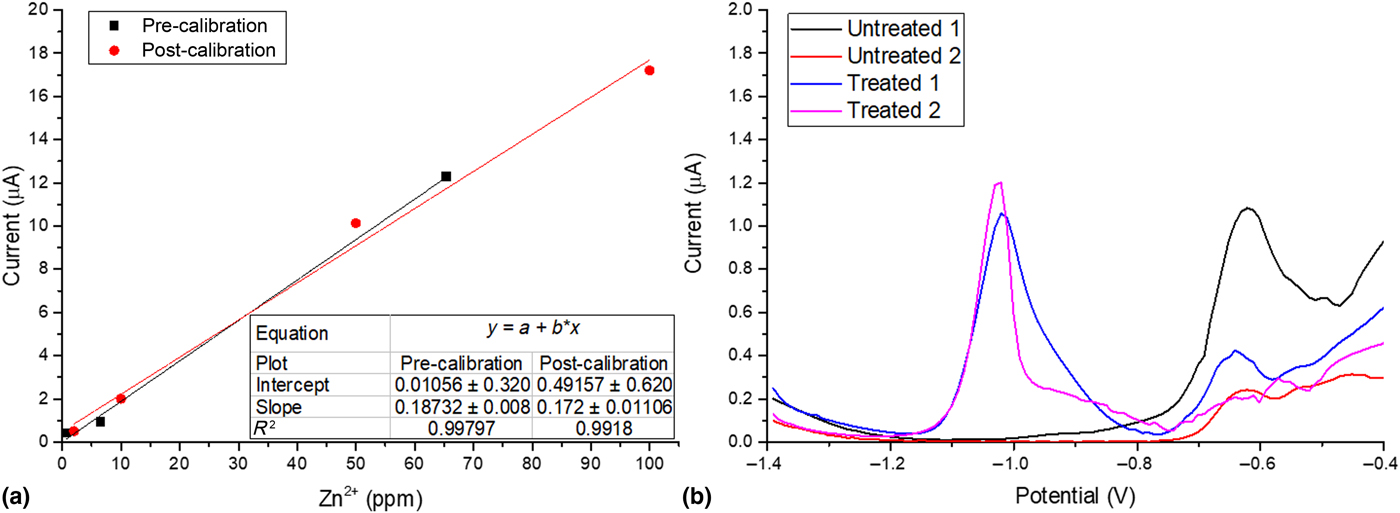
In situ application of the developed microelectrodes for plants. (a) Pre- and post-calibration curves of combined Bi/Pt reference microelectrode using a two-step SWASV method. (b) In situ detection of Zn2+ in untreated (DI water) and treated (1 mM ZnCl2) citrus leaves using the double-barrel Bi/Pt microelectrode.
In situ detection of Zn2+ using SWASV in the vascular bundle of citrus plants
The combined Bi/Pt microelectrode was applied for Zn2+ detection in the vascular bundle of citrus plants. The configuration of the double-barrel microelectrode allowed for simultaneous penetration of the working and reference electrodes into the vascular bundle of the citrus leaves for SWASV determination of Zn2+. The combined Bi/Pt microelectrode showed improved durability over the single-barrel Bi microelectrode and was not damaged by the leaf surface. The double-barrel Bi/Pt microelectrode had an overall tip diameter of 110 µm and a working Bi diameter of 70 µm. A calibration curve showed an excellent sensitivity toward Zn2+ with 0.172 µA/ppm Zn2+ slope and an R 2 value of 0.996 [Fig. 4(a)]. The calibration curve after application to the plant showed a response of 0.187 µA/ppm Zn2+ and demonstrated that the microelectrode was not damaged during the penetration. By microscopic observation, it was also found that the direct contact with plant tissue had no impact on the microelectrode tip. The pre- and post-calibration curves [Fig. 4(a)] also showed a LOD of 0.92 ppm with similar calibration curves (<10% difference in slope), and a lifetime study showed that the sensor's response does not change significantly over 18 uses (Fig. S7), which is acceptable for the purpose of detection of Zn2+ in plants.
Figure 4(b) shows the microelectrode's response to Zn2+ concentrations in the midrib of treated (1 mM ZnCl2 for 6 h) and untreated (DI for 6 h) citrus leaves. The two separate treated leaves (1 and 2) produced a SWASV response of 1.2 µA (treated 1) and 1.4 µA (treated 2) and the two separate untreated leaves showed a negligible response (0.06 and 0.003 µA) to Zn2+ compared with the treated leaves. Using the pre- and post-calibration curves [Fig. 4(a)], it was determined that the treated leaf contained 5.73 ± 0.88 ppm Zn2+, while the untreated leaf contained undetectable amounts of Zn2+. While the increase in Zn2+ concentration was expected in the treated leaf, the measured values were difficult to validate with existing literature due to the use of different analytical methods. Most studies primarily detect Zn2+ using inductively coupled plasma atomic emission spectroscopy or AAS for which samples need to be dried, grounded, and digested before analysis.[Reference Kalra19] This causes zinc concentrations being reported in mg/kg dry weight. A study using a more direct Zn measurement, micro x-ray fluorescence (μ-XRF), reported Zn2+ concentrations in mg/kg dry weight (34.6–42.4 mg/kg in sour orange plants).[Reference Tian, Lu, Labavitch, Webb, Yang, Brown and He20] Considering μ-XRF determines total zinc concentration and there will be a difference between dried (mg Zn/kg biomass) and in situ measurements (mg Zn2+/L), the measurement using in situ SWASV seems reasonable.
While the benefits of using a SWASV-based microelectrode for monitoring Zn2+ concentrations in citrus plants are clear, there are still limitations that need to be addressed. In particular, Cu–Zn intermetallic species are known to develop during the deposition step and interfere with the sensor response.[Reference Sanna, Pilo, Piu, Tapparo and Seeber21] The quantification of the interference from intermetallic Cu–Zn compounds is beyond the scope of this paper, as it is assumed that Cu2+ concentrations in the citrus leaves will be significantly lower than Zn2+.[Reference Graham, Timmer and Fardelmann22] However, for other applications, where both Zn2+ and Cu2+ exist in high concentrations, we suggest that one possible solution for determining the amount of copper in a sample would be to use a different deposition potential (e.g., −0.6 V) that is more anodic than that required for the reduction of Zn2+ to determine if the formation of Cu–Zn compounds may be a problem for sensor responses. With more validation regarding reproducibility and representativeness, it is expected that this novel approach of using two-step SWASV would provide a simple, fast, and reproducible detection of Zn2+ in plants, capable of distinguishing between treated and untreated samples. Overall, this study demonstrates the feasibility of using a SWASV-based microelectrode technique as a direct and rapid method for monitoring Zn2+ concentration in phloem tissue in a minimally invasive way.
Conclusions
A metallic Bi-based microelectrode was constructed and tested for concept proof of two-step SWASV for detecting Zn2+. We were able to demonstrate the feasibility of a two-step SWASV measure using in situ deposition of Zn2+ and ex situ stripping. It was also found that the tip size can be optimized to reduce mass transport limitation and improve LOD. The Bi microelectrode was then combined with a Pt microelectrode in a double-barrel microelectrode configuration for direct measurement of Zn2+ in plants. Our results showed that the solid metal tip (110 µm in diameter) of the Bi/Pt microelectrode is durable enough to penetrate the thick skin of the citrus leaves. Overall, the developed Bi/Pt microelectrode was able to respond to Zn2+ in the midrib of the citrus leaves and distinguish the Zn2+ concentrations between Zn2+-treated and -untreated samples with high resolution. This novel in situ detection method, capable of tracking its systemic Zn2+ activity directly in plants, will lead to better understanding of its potential fate in plants for effective HLB management in the agriculture industry.
Supplementary material
The supplementary material for this article can be found at https://doi.org/10.1557/mrc.2018.61
Acknowledgments
This work is supported by Citrus Disease Research and Extension (CDRE) (grant no. 2016-70016-24828/project accusation no. 1008984) from the USDA National Institute of Food and Agriculture. Any opinions, findings, conclusions, or recommendations expressed in this publication are those of the author(s) and do not necessarily reflect the view of the US Department of Agriculture.