Introduction
For decades, cancer has been recognized as a significant cause of mortality.Reference Liu, Chen, Feng and Liu1,Reference Denkova, de Kruijff and Serra-Crespo2 Conventional cancer treatment strategies, including surgery, radiotherapy, and chemotherapy, are widely used in clinical settings. However, these treatments have many problems, such as high cancer recurrence rates, systemic side effects, and cumulative radiation dose.Reference Lucky, Soo and Zhang3 A safe, potent, and cost-effective treatment modality needs to be developed and is highly desired.
Photodynamic therapy (PDT) has been considered as a promising strategy for cancer treatment due to its noninvasive nature, high therapeutic efficiency, and low systemic toxicity.Reference Park, Jiang, Feng, Mao and Zhou4,Reference Liang, Li, Jing, Yue and Dai5 In general, PDT contains three functional elements: photosensitizers, visible light, and oxygen. Photosensitizers effectively kill tumor cells by absorbing light and converting oxygen (O2) to cytotoxic singlet oxygen (1O2).Reference Lu, He, Guo, Chan, Ni, Weichselbaum and Lin6 Porphyrin photosensitizers, which are chromophores that exist in nature, are widely used in biomedical applications, especially for PDT of tumors due to their well-known photosensitizing properties. Nevertheless, low aqueous solubility and photostability, and lack of tumor targeting limit their in vivo bioapplications.
Over the past few decades, the rapid development of nanotechnology has resulted in new opportunities for improving the in vivo applications of porphyrin-based PDT. Nanotechnology not only improves water solubility and photostability of porphyrin, but also significantly increases accumulation in the tumor by passive (enhanced permeability and retention effect [EPR]) or active targeting (antibody or targeted peptide modification) strategies.Reference Zhou, Liang and Da7
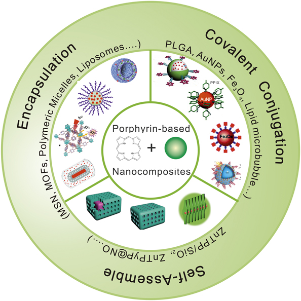
In this article, we first summarize the strategies for porphyrin delivery, including encapsulation, covalent conjugation, and self-assembly for PDT (Figure 1).Reference Zhao, Wu, Yao, Xu and Xu8–Reference Wang, Niu, Qiao, Cheng, Wang, Zhong, Bai, Wang and Fan17 Next, we describe the characterization methods for singlet oxygen (1O2) and the latest developments in multifunctional porphyrin-based nanoparticles for improving cancer PDT efficacy, such as increasing the tissue penetration depth of light, tumor hypoxia relief, and multimodal imaging guidance. Finally, the challenges and prospects of porphyrin-based nanocomposites in PDT of tumors are discussed.
Nanoparticle-mediated porphyrin delivery strategies, including encapsulation (mesoporous silica nanoshells,[MSNs],Reference Zhao, Wu, Yao, Xu and Xu8 metal–organic frameworks [MOFs],Reference Lu, He and Lin9 polymeric micelles,Reference Yan, Miller, Yuan, Liu, Busch, Tsourkas and Cheng10 and liposomesReference Feng, Wang, Song, Zhuo, Wang, Liao, Feng, Wei, Chen, Yang and Yang11), covalent conjugation (poly(lactic-co-glycolic acid) [PLGA]),Reference Jing, Liang, Li, Lin, Yang, Yue and Dai12 AuNPs,Reference Oo, Yang, Hu, Gomez, Du and Wang13 Fe3O4,Reference Gu, Xu, Yang, Chang and Xu14 and lipid microbubblesReference You, Liang, Yin, Chen, Qiu, Gao, Wang, Mao, Qu, Dai and Zheng15), and self-assembly (ZnTPP/SiO2Reference Wang, Zhong, Wang, Yang, Bai, Zhang, Alarid, Bian and Fan16 and ZnTPyP@NOReference Wang, Niu, Qiao, Cheng, Wang, Zhong, Bai, Wang and Fan17). Note: ZnTPP, zinc-tetra (4-pyridyl) porphyrin; ZnTPyP, zinc meso-tetra(4-pyridyl)porphyrin; NO, nitrous oxide; PPIX, protoporphyrin IX; NP, nanoparticle; 1O2, singlet oxygen.

Delivery strategies for nanoparticle-mediated porphyrin photosensitizers
Encapsulation
For inorganic nanoparticles, porphyrin photosensitizers can be effectively delivered to tumor tissues by using their specific mesoporous or hollow structures. For instance, Zhao et al.Reference Zhao, Wu, Yao, Xu and Xu8 synthesized porphyrin-doped mesoporous silica-coated gold nanorods and found these nanocomposites could be easily dispersed in an aqueous solution. This unique design facilitates the generation and release of 1O2 from the matrix, enhancing production efficiency to greater than that for pure porphyrin.Reference Zhao, Wu, Yao, Xu and Xu8 Lin et al.Reference Lu, He and Lin9 used nanoscale metal–organic frameworks (NMOFs) to deliver a new porphyrin derivative, 5,15-di(p-benzoato)porphyrin (H2DBP), for PDT to improve the hydrophilicity and tumor selectivity of the photosensitizer. NMOFs possess a variety of beneficial features not found in other reported nanocarriers, such as high porosity and tunable chemical composition.Reference Lu, He and Lin9 In the case of using organic nanoparticles, porphyrin photosensitizers can be encapsulated by nanoparticles formed into self-assembled polymeric micelle structures and delivered to tumor tissues. Roby et al.Reference Roby, Erdogan and Torchilin18 prepared meso-tetraphenylporphine-loaded polymeric micelles (polyethylene glycol-phosphatidylethanolamine conjugate [PEG-PE]), followed by modification with tumor-targeted monoclonal 2C5 antibody for PDT of murine cancer, including carcinoma of the lung and melanoma, which significantly improved the solubility of the photosensitizers and in vivo PDT effects for the cancer cells.
Covalent conjugation
In addition to encapsulation, porphyrin photosensitizers can be conjugated with other nanoparticles via covalent bonds, which can also enhance their in vivo stability. Fakayode et al. synthesized nanodrugs consisting of PEGylated gold coated superparamagnetic iron oxide nanoparticles and meso-tetrakis(4-hydroxyphenyl) porphyrin, forming magnetic porphyrin nano-drug conjugate, for killing MCF-7 cells (human breast adenocarcinoma cell line) under laser irradiation and external magnetic field (magnetic target). Both the cellular uptake and PDT efficacy were found to greatly increase in contrast to the control group that underwent nonmagnetic field-driven PDT.Reference Fakayode, Kruger, Songca, Abrahamse and Oluwafemi19
Self-assembly
Porphyrin nanoparticles were prepared, depending on the ordered arrays (hydrogen bonding and π−π stacking), through the self-assembly of molecular building blocks. Fan and Bai developed a new method of synthesizing self-assembled porphyrin−silica nanoparticles with ordered porphyrin networks as their cores surrounded by amorphous silica as the shell. The pH condition was then found to be the determining factor for tuning the final nanostructures. Low pH conditions promote porphyrin noncovalent self-assembly, forming core−shell particles. However, high pH conditions are favorable for silicate sol−gel process, where solid porphyrin−silica mesophase particles form.Reference Fakayode, Kruger, Songca, Abrahamse and Oluwafemi19 Subsequently, they reported the synthesis of noncovalent self-assembled porphyrin nanoparticle photosensitizers, followed by sorption with nitric oxide (NO) coordinates, which can produce singlet oxygen (1O2) and release NO simultaneously under light irradiation, leading to significantly enhanced antibacterial photodynamic therapy (APDT) activity. The APDT was determined by detecting the optical density at 600 nm with a multifunctional microplate reader.Reference Wang, Niu, Qiao, Cheng, Wang, Zhong, Bai, Wang and Fan17
Characterization methods of singlet oxygen (1O2)
Luminescence detection
Niedrel et al. reported that singlet oxygen can be directly detected by monitoring its near-infrared (NIR) luminescence at 1270 nm.Reference Niedre, Patterson and Wilson20 This method provides the most direct evidence of 1O2 generation, however, the short lifetime (μs) and low quantum yield of 1O2 in the NIR region, which have to be magnified by using other techniques, limited wide application at the subcellular level, such as fluorescence cell imaging and 1O2 detection in cells.Reference Ping, Peng, Qin, You, Wang, Chen and Song21
Electron spin resonance spectroscopy
Lion et al. originally set up the electron spin-resonance (ESR) method for the detection of 1O2 in 1976.Reference Lion, Delmelle and Vandevorst22 In this process, ESR signals can be detected after 1O2 reacts with a spin singlet oxygen scavenger, such as TEMP (2,2,6,6-tetramethyl-4-piperidine), yielding a stable paramagnetic product (TEMPO). Ge et al. used an ESR technique to detect the generation of 1O2 using graphene quantum dots (GQDs) under laser irradiation; they observed the characteristic 1O2-induced signal from 2,2,6,6-tetramethylpiperidine-1-oxyl, in the ESR spectra.Reference Ge, Lan, Zhou, Liu, Guo, Wang, Jia, Niu, Huang, Zhou, Meng, Wang, Lee, Zhang and Han23 The ESR method has high sensitivity and good selectivity, however the measured ESR signal is also affected by coexisting ions and solvents, which makes the quantitative detection error larger. At the same time, the instrument is expensive, and the operation procedure is relatively complicated.
Spectrophotometry
Generally, this method yields a fluorescence and an absorption spectrum. A variety of small-molecule probes was developed for 1O2 detection. The most widely used fluorescent probe is singlet oxygen sensor green (SOSG), which can react with 1O2 to yield SOSG endoperoxide (SOSG-EP) with a fluorescence peak at 528 nm under 485 nm excitation. DPBF(1,3-diphenylisobenzofuran) is a type of indicator that could react with 1O2 and cause a decrease in the absorption intensity of the DPBF band centered at 400 nm.Reference Fan, Bu, Shen, He, Cui, Liu, Zheng, Zhao and Shi24 Even though these photometric probes have been widely used for 1O2 detection, there are still a few inevitable shortcomings, especially stability of DPBF in the presence of light and oxygen in air. In addition, the sensitivity of the method also needs to be improved.
Chemiluminescence
Chemiluminescence probes can react with singlet oxygen, producing higher energy compounds. These products rapidly decompose and release energy in the form of light. CLA (2-methyl-6-phenyl-3,7-dihydroimidazo[1,2-a]pyrazin-3-one) is a typical chemiluminescence probe that only reacts specifically with singlet oxygen, rather than reacting with hydroxyl radicals or hydrogen peroxide (emission wavelength: 380 nm).Reference Nakano, Sugioka, Ushijima and Goto25 Subsequently, to improve the luminescence efficiency and extend the emission wavelength, a fluoresceinylcypridina luciferin analogue (FCLA) probe, a derivative of CLA, was synthesized. The luminescence efficiency of FCLA is nearly 10 times greater than that of CLA, with the emission peak centered at 532 nm. Xing et al. successfully used FCLA to detect 1O2 generated using upconversion nanoparticle-(UCNP)-pyropheophorbide (Ppa)-cyclo (Arg-Gly-Asp-d-Tyr-Lys) (c(RGDyK) (where Arg = arginine, Gly = glycine, Asp = aspartic acid, Tyr = tyrosine, Lys = lysine, c = cyclic; for RGDyK, where R = arginine, G = glycine, D = aspartic acid, y = tyrosine, K = lysine) nanocomposites after NIR light irradiation.Reference Zhou, Wei, Wu, Chen and Xing26
Improvement of the efficacy of PDT by porphyrin-based nanocomposites
Improving tissue penetration depth of light
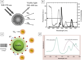
Currently, most conventional photosensitizers can only be excited by ultraviolet–visible (UV–vis) light, which is inefficient for deep tumors due to the short tissue penetration depth of UV–vis light. Thanks to the fluorescence resonance energy transfer (FRET) from photoconverting nanoparticles such as UCNPs can convert NIR light to UV–vis light, followed by transfer to photosensitizers for PDT. Shan et al. synthesized nanocomposites consisting of a prophine photosensitizer (meso-tetraphenylporphine, TPP) and UCNPs (NaYF4:Yb3+,Er3+). The NIR light (978 nm) was effectively converted to visible light (500–600 nm) for TPP excitation due to good overlap of fluorescence emission and absorption spectra (Figure 2a–b). Seventy-five percent of the HeLa cancer cells were killed after incubation with these nanocomposites and laser irradiation.Reference Shan, Budijono, Hu, Yao, Kang, Ju and Prud’homme27
Porphyrin-based photoconversion nanoparticles for enhanced photodynamic therapy. (a) Schematic illustration of porphyrin-based upconversion nanoparticles (UCNPs) for singlet oxygen (1O2) generation by converting near-infrared (NIR) light to visible light. (b) Absorbance spectrum of meso-tetraphenylporphine (TPP) and fluorescence spectrum of UCNPs irradiated by NIR light (978 nm). The NIR light was effectively converted to visible light (500–600 nm) for TPP excitation due to overlap of fluorescence emission and absorption spectra.Reference Shan, Budijono, Hu, Yao, Kang, Ju and Prud’homme27 (c) Schematic illustration of porphyrin-based nanoscintillator for 1O2 generation by converting x-rays to visible light.Reference Kamkaew, Feng, Zhan, Majewski and Cai29 (d) Absorbance spectrum of PPIX and fluorescence spectrum of LaF3:Ce/PLGA nanoscintillator irradiated by x-rays. The x-rays were successfully converted to green light (emission peak: 520 nm) for PPIX excitation due to overlap of fluorescence emission and absorption spectra.Reference Zou, Yao, Ma, Hossu, Han, Juzenas and Chen30 Note: PLGA, poly(lactic-co-glycolic acid); ET, excitation; PPIX, protoporphyrin IX; ScNPs, scintillating nanoparticles.
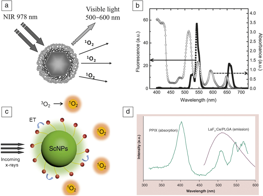
According to previous reports, the tissue penetration depth of NIR light is only 1.5 cm, which still cannot be used in deep tumor therapy (5–10 cm for human pancreatic cancer).Reference Hu, Tang, Elmenoufy, Xu, Cheng and Yang28 To further increase the depth of tissue penetration, x-rays, which have a greater penetration depth, have been broadly used in the clinic. The x-rays were introduced and converted by a nanoscintillator to visible light for tumor PDT (Figure 2c).Reference Kamkaew, Feng, Zhan, Majewski and Cai29 Chen et al. synthesized Ce-doped LaF3 nanoparticles (LaF3:Ce), then encapsulated protoporphyrin IX (PPIX) into poly(lactic-co-glycolic acid) microspheres, establishing an efficient FRET system. During x-ray irradiation (90 kV), the LaF3:Ce nanoparticles emitted significant green light (emission peak: 520 nm) that overlapped with the absorption spectrum of PPIX (Figure 2d). Thus, the x-rays were successfully converted into light that irradiated a photosensitizer for 1O2 generation to perform PDT of tumors.Reference Zou, Yao, Ma, Hossu, Han, Juzenas and Chen30
Tumor hypoxia relief
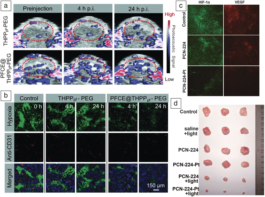
Oxygen is one of the three essential elements in PDT. However, the tumor microenvironment is hypoxic, thus weakening the PDT effect. Consequently, increasing the oxygen concentration in the tumor tissue is another promising strategy for enhancing PDT efficacy. Liu et al. constructed a tumor hypoxia relief-based nanoplatform for PDT that contained nano-covalent organic polymers, porphyrin photosensitizer, perfluoro-15-crown-5-ether (PFCE), and perfluorosebacic acid. Molecular oxygen was delivered to tumor tissues by PFCE, a type of perfluorocarbon.Reference Tao, Feng, Chao, Liang, Song, Wang, Yang and Liu31 Both the photoacoustic images (Figure 3a) under the oxy-hemo mode (hypoxia assessment within the tumor microenvironment) and immunofluorescence staining images of tumor slices (Figure 3b) revealed that the O2 content was significantly increased, mitigating the hypoxia of the tumor environment.
Enhancing photodynamic therapy (PDT) efficacy by relieving tumor hypoxia, including (a, b) O2 delivery and (c, d) O2 generation in situ. (a) Photoacoustic (PA) images of THPPpf-PEG and PFCE@THPPpf-PEG under the oxy-hemo mode. The red dashed circle indicates the region of interest for quantitative analysis of PA signal intensity. The tumor oxygenation of the (lower row) mice given an intravenous injection of PFCE@THPPpf–PEG (4 h p.i. and 24 h p.i.) was significantly increased compared to (upper row) the group given the THPPpf-PEG injection. Note: p.i., post injection; color bar (high and low), photoacoustic signal intensity. (b) Immunofluorescence staining images of tumor slices after intravenous injection of THPPpf-PEG and PFCE@THPPpf-PEG. The hypoxia areas and blood vessels were stained with (top) antipimonidazole antibody (green) and (middle) anti-CD31 antibody (red). (Bottom) Combination of the two images. Tumor hypoxia signals of the mice with intravenous injection of PFCE@THPPpf-PEG (4 h p.i. and 24 h p.i.) were significantly attenuated compared to the group given THPPpf-PEG.Reference Tao, Feng, Chao, Liang, Song, Wang, Yang and Liu31 (c) Immunofluorescence staining images (HIF-1α and VEGF) of tumor tissues after intravenous injection of PCN-224 and PCN-224-Pt. The tumors injected with PCN-224-Pt showed significantly weakened tumor hypoxia compared to those that received the PCN-224 injection, which is evidenced by the greatly decreased immonufluorescence intensity of HIF-1α and VEGF. (d) Tumor dissection images of different groups (control, saline injection + light irradiation, PCN-224 injection, PCN-224-Pt injection, PCN-224 injection + light irradiation, and PCN-224-Pt injection + light irradiation). The steel ruler was used to identify the tumor size. The tumor growth was completely inhibited with the injection of PCN-224-Pt after laser irradiation treatment due to O2-enhanced PDT.Reference Zhang, Wang, Liu, Wang, Kang, Huang, Dong, Ren and Qu32 Note: THPPpf, meso-5,10,15,20-tetra (4-hydroxylphenyl)porphyrin (THPP) + perfluorosebacic acid; PEG, poly(ethylene glycol); PFCE, perfluoro-15-crown-5-ether; HIF, hypoxia inducible factor; VEGF, vascular endothelial growth factor; PCN, porous coordination network.
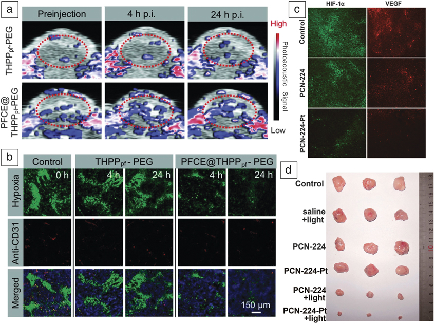
Tumor hypoxia can also be indicated by producing O2 through the reaction between nanoparticles and molecular markers with high expression in the tumor microenvironment. Qu et al. reported a simple yet versatile strategy for enhancing PDT based on the platinum nanozymes (platinum nanoparticles having an enzyme-like property)-decorated MOFs nanoplatform. The MOFs were synthesized by mixing ZrOCl2 • 8H2O and porphyrin photosensitizers (tetrakis (4-carboxyphenyl) porphyrin, TCPP) in the presence of N,N-dimethylformamide at 90°C. After Pt nanozymes decoration, the resulting porous coordination network (PCN)-224-Pt MOFs could decompose H2O2 in tumor microenvironment to O2 with the potent catalase-like activity of Pt. According to the final immunofluorescence staining images (hypoxia inducible factor (HIF)-1α and vascular endothelial growth factor) of the tumor slices (Figure 3c), the decreased fluorescence signals indicated the mitigation of tumor hypoxia. As expected, the PDT efficacy of PCN-224-Pt under laser irradiation was more enhanced versus other control groups (Figure 3d).Reference Zhang, Wang, Liu, Wang, Kang, Huang, Dong, Ren and Qu32
Multimodal imaging guidance
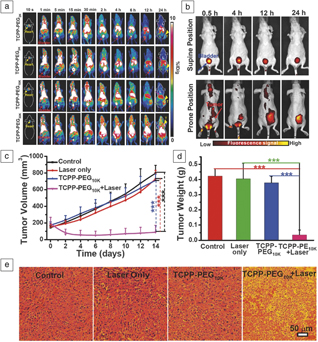
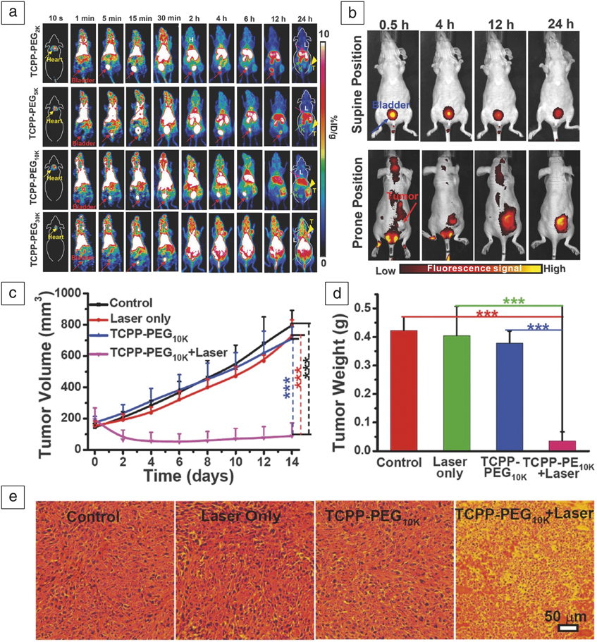
Molecular imaging allows for the detection and visualization of biomarkers in tumors.Reference Weissleder33,Reference Pysz, Gambhir and Willmann34 It can enable accurate detection of tumors, facilitating the use of molecular signatures to tailor effective and personalized therapies for improved patient survival. In particular, multimodal imaging techniques can replace the shortcomings of a single imaging mode and guide subsequent PDT of tumors. Srivatsan et al. established a “see and treat” approach by incorporating photosensitizer (3-devinyl-3-(1′-hexyloxyethyl)pyropheophorbide [HPPH]) with gold nanocages via hydrophobic interaction for dual-modal imaging capability-guided PDT. Fluorescence from HPPH and the photoacoustic signal from gold nanocages were able to guide the release of photosensitizer and the accumulation of nanocomposites in the tumor, which facilitated the PDT of the tumor.Reference Srivatsan, Jenkins, Jeon, Wu, Kim and Chen35 Cheng et al. synthesized meso-tetra(4-carboxyphenyl)porphyrin (TCPP)-PEG nanoparticles using different molecular weights of PEG. After 64Cu labeling, the in vivo metabolism behavior could be monitored by positron emission tomography imaging. It was found that TCPP-PEG nanoparticles with higher molecular weights showed higher tumor uptake efficacy due to the EPR effect, while those of lower molecular weight tended to be better for renal clearance (Figure 4a). The in vivo fluorescence imaging after injection of TCPP-PEG10K aids in further investigating the behaviors of clearance and tumor uptake efficiency (Figure 4b). TCPP-PEG10K nanoparticles were then used for further PDT of the tumor. It was found that the tumor treated with nanoparticles plus laser was significantly inhibited (Figure 4c–d). As shown in the hematoxylin-eosin staining images (Figure 4e), most of the tumor cells were killed by 1O2 generated by TCPP-PEG10K nanoparticles exposed to a laser.Reference Cheng, Jiang, Kamkaew, Valdovinos, Im, Feng, England, Goel, Barnhart and Liu36
(a) In vivo positron emission tomography (PET) imaging of 4T1 tumor-bearing mice at different points in time after intravenous injection of 64Cu-TCPP-PEG2K,TCPP-PEG5K,TCPP-PEG10K, and TCPP-PEG30K). TCPP-PEG nanoparticles with higher molecular weights (TCPP-PEG10K and TCPP-PEG30K) showed higher tumor uptake efficacy. (b) In vivo fluorescence imaging of 4T1 tumor-bearing mice at different points in time (0.5 h, 4 h, 12 h, and 24 h) after intravenous injection of 64Cu-TCPP-PEG10K. TCPP-PEG10K nanoparticles were able to be cleared and the prominent tumor uptake time is 24 h after injection. (c) Tumor growth curves, (d) tumor weight, and (e) hematoxylin-eosin staining images of mice in different groups (control, laser only, 64Cu-TCPP-PEG10K only, and 64Cu-TCPP-PEG10K+laser).Reference Cheng, Jiang, Kamkaew, Valdovinos, Im, Feng, England, Goel, Barnhart and Liu36 The tumor growth of 64Cu-TCPP-PEG10K+laser group was significantly inhibited, and tumor cells were killed and cell apoptosis was found. Note: TCPP, tetrakis (4-carboxyphenyl) porphyrin; PEG, poly(ethylene glycol); %ID, percentage injected dose.
Summary
In this article, we have summarized the characteristics of porphyrin-based nanocomposites and their applications for tumor PDT, including strategies for porphyrin delivery, characterization methods of 1O2, and the latest achievements in the development of multifunctional porphyrin-based nanoparticles for improving cancer PDT efficacy. Research into the combination of porphyrins and nanotechnology has shown this combination to be successful as a means of overcoming the deficiencies of porphyrins in PDT, such as photostability, water solubility, and target ability.
There still remain issues that need to be resolved: (1) the tumor-specific PDT of porphyrin-based nanocomposites is currently not sufficiently developed; (2) a simple and precise method of characterizing 1O2 has not yet been developed; and (3) strategies for improving the efficacy of PDT lack integration in one nanocomposite.
Correspondingly, the following actions are expected to be taken in the next step to overcome these obstacles: (1) nanoparticle design should proceed intelligently, according to the characteristics of the tumor microenvironment (weak acidity, overexpression of glutathione, enzymes, H2O2); (2) all of the 1O2 characterizing methods need to be further compared and developed; and (3) multifunctional nanocomposites must be designed to overcome the defects of the three elements (light, photosensitizer, and oxygen) in PDT. The development of porphyrin-based nanocomposites should thus move in the direction of multifunctionality.
Acknowledgments
This work was supported by the National Natural Science Foundation of China (81871399, 81801823, and 81571742) and the project was funded by the China Postdoctoral Science Foundation (1500229020).

Weitao Yang is a postdoctoral fellow in the School of Medicine at Tongji University, China. He obtained his PhD degree in the School of Materials Science and Engineering at Tianjin University, China, in 2017. His current research interests focus on designing and synthesizing smart protein-based biomimetic nanoprobes for cancer multimodal imaging (MRI, photoacoustic, fluorescence, and computed tomography) and phototherapy (photothermal and photodynamic therapy). He has co-authored more than 20 publications. Yang can be reached by email at weitaoyang@tongji.edu.cn.

Bingbo Zhang has been a full professor in the School of Medicine at Tongji University, China, since 2018. He received his PhD degree from Tianjin University, China, in 2009. He became an assistant professor at The Institute for Biomedical Engineering and Nano Science at Tongji University in 2009. His research focuses on biomaterials and photomedicine. He has authored or co-authored more than 50 papers on biomedical materials. Zhang can be reached by email at bingbozhang@tongji.edu.cn.