INTRODUCTION
Insomnia symptoms span the spectrum of difficulty falling asleep, difficulty staying asleep, waking up earlier than desired, or a subjective feeling that sleep is not restorative. Insomnia is a common health problem that is experienced across the lifespan, affecting up to 50% of the general population on an occasional basis and up to 10% on a chronic or more persistent basis (Ohayon, Reference Ohayon2002; Schutte-Rodin, Broch, Buysse, Dorsey, & Sateia, Reference Schutte-Rodin, Broch, Buysse, Dorsey and Sateia2008). These difficulties can emerge as a primary sleep disorder and/or symptoms associated with other medical conditions. Unlike some other sleep disorders, insomnia may be diagnosed on the basis of self-reported clinical history rather than objective laboratory measures (Bastien, Reference Bastien2011; Sateia, Doghramji, Hauri, & Morin, Reference Sateia, Doghramji, Hauri and Morin2000). Symptoms reach clinical significance when they result in considerable distress or interfere with social or occupational functioning (American Psychiatric Association, 2013). Disruptions in functioning are often due to associated sequelae, such as mood disturbances (Bastien, Reference Bastien2011). In addition, although not required for diagnosis, neuropsychological impairment is also frequently observed among individuals with insomnia in the domains of vigilance, attention, processing speed, and executive function (Bastien, Reference Bastien2011; Hauri, Reference Hauri1997).
Circadian rhythms and the 24-hr sleep-wake cycle are profoundly affected by exposure to both naturally occurring and artificially sourced light. The human eye contains retinal ganglion photoreceptor cells that uniquely project to the “circadian clock,” or the suprachiasmatic nucleus of the hypothalamus (Brainard et al., Reference Brainard, Hanifin, Greeson, Byrne, Glickman, Gerner and Rollag2001; Thapan, Arendt, & Skene, Reference Thapan, Arendt and Skene2001). These photoreceptors facilitate the synchronization of the circadian clock to the day–night cycle and are especially sensitive to light wavelengths in the blue to blue–green range of the visible light spectrum, around 460 nm (Phelps, Reference Phelps2008). The timing of exposure to blue light is a critical consideration as it may result in either beneficial or deleterious outcomes. For example, artificial manipulation of blue light exposure in the morning has reliable benefits to mood, such as the use of light boxes to effectively alleviate symptoms of seasonal affective disorder (Phelps, Reference Phelps2008). However, exposure to environmental blue light in the evening during hours that are naturally “dark” can postpone the onset of sleep, likely due to suppression and delay of the release of the pineal hormone melatonin, resulting in symptoms of insomnia (Cajochen, Krauchi, & Wirz-Justice, Reference Cajochen, Krauchi and Wirz-Justice2003; Fossum, Nordnes, Storemark, Bjorvatn, & Pallesen, Reference Fossum, Nordnes, Storemark, Bjorvatn and Pallesen2014). This is a critical issue in modern day society as individuals increasingly use electronic devices (e.g., computers, televisions, smartphones), common emitters of blue light, in the hours immediately preceding bedtime (Cajochen et al., Reference Cajochen, Frey, Anders, Spati, Bues, Pross and Stefani2011; Chang, Aeschbach, Duffy, & Czeisler, Reference Chang, Aeschbach, Duffy and Czeisler2015). In fact, a recent study indicated that up to 90% of individuals reported using an electronic device in the hour before bedtime (Gradisar et al., Reference Gradisar, Wolfson, Harvey, Hale, Rosenberg and Czeisler2013). While individuals often exhibit difficulty adhering to clinical recommendations to reduce exposure to nocturnal blue light (Phelps, Reference Phelps2008), evidence suggests that wearing amber-tinted blue light blocking lenses before bedtime reduces melatonin suppression and improves sleep quality (Gamble, May, Besing, Tankersly, & Fargason, Reference Gamble, May, Besing, Tankersly and Fargason2013; Kayumov et al., Reference Kayumov, Casper, Hawa, Perelman, Chung, Sokalsky and Shapiro2005; Phelps, Reference Phelps2008; Sasseville, Paquet, Sevigny, & Hebert, Reference Sasseville, Paquet, Sevigny and Hebert2006).
Although correlational studies show that neuropsychological dysfunction is associated with insomnia symptoms and that exposure to blue light regulates the circadian sleep cycle, little is known about how exposure to nocturnal blue light affects neuropsychological function in individuals with symptoms of insomnia. Establishment of causality in this context can be challenging and cross-sectional observational studies are inherently unable to address such issues. Therefore, the goal of this pilot study was to experimentally examine the impact of an intervention to block nocturnal blue light exposure across several neuropsychological functions (processing speed, working memory, attention, and executive function) that are thought to be particularly vulnerable to insomnia using a randomized, placebo-controlled crossover design. We hypothesized that individuals with insomnia symptoms would demonstrate an improvement in neuropsychological function across all domains of interest following blockage of nocturnal blue light but not following a control condition.
METHOD
Participants
Participants were recruited using flyers and electronic advertisements. The primary inclusion criterion for the study was age between 18 and 65 years and the report of chronic insomnia symptoms lasting for greater than 3 months. Insomnia symptom identification was achieved with the Insomnia Symptoms Questionnaire (ISQ), which is a self-report questionnaire guided by established diagnostic criteria (Okun et al., 2009). The ISQ is a 13-item self-report measure that provides a dichotomous outcome (“present” or “absent”) of the definition of insomnia symptoms based on diagnostic criteria from the American Psychiatric Association’s Diagnostic and Statistical Manual of Mental Disorders (DSM), Fourth Edition, and is also consistent with the American Academy of Sleep Medicine Research Diagnostic Criteria. To be considered as positive for insomnia symptoms (“present”), participants must indicate, as occurring over the 3-month duration, a frequent (3–4 times per week) or always (5–7 times per week) occurrence of at least one of the following sleep symptoms: difficulty falling asleep, difficulty staying asleep, or feeling that your sleep is unrefreshing. In addition to this, participants must also report being affected “quite a bit” or “extremely” by at least one of the listed daytime impairment symptoms (e.g., being bothered by sleep problems, sleep difficulties affecting work/social life, sleep difficulties causing feeling of irritability, trouble concentrating, sleepiness, fatigue).
Exclusion criteria included diagnosis of obstructive sleep apnea; history of deep vein thrombosis; working nights or rotating shifts; travel across time zones in the 2 weeks before the start of the study; diagnosis of a psychiatric disorder; current smoking, use of beta-blockers, consumption of excessive caffeine (>400 mg/day); having a child at home <1-year-old; or being a pregnant or breastfeeding woman. Participants were not permitted to use hypnotics/sedatives in the 2 weeks before the start of the study or during the duration of the study. Diagnosis of a psychiatric disorder, having a child at home <1 year old, current pregnancy, and current breastfeeding were exclusionary because they are frequently associated with disruptions in sleep quality due to mechanisms unrelated to evening blue light exposure, and, therefore, unlikely to be affected by the intervention. Current smoking and use of beta-blockers were exclusionary because these can decrease melatonin release (Ozguner, Koyu, & Cesur, 2005; Stoschitzky et al., 1999). Presence of exclusion criteria were ascertained by participant self-report, medical history, and physical conducted by a physician or nurse practitioner. Participants received $100 for their participation in these procedures. This study was approved by the Columbia University Medical Center (CUMC) Institutional Review Board, all procedures were in accordance with the Declaration of Helsinki, and all participants provided informed written consent. Data were collected at the Clinical Research Resource of the Irving Institute for Clinical and Translational Research at CUMC.
Trial Design
The study used a randomized, placebo-controlled crossover design. The measures used in the current analyses were part of a broader parent project that examined ambulatory sleep in response to the intervention (Shechter, Kim, St-Onge, & Westwood, Reference Shechter, Kim, St-Onge and Westwood2018). After enrollment, participants were randomized via simple randomization with a computer-generated random numbers generator into an intervention sequence (clear condition followed by amber condition for even numbers; amber condition followed by clear condition for odd numbers). Participants were randomly assigned to a 1-week intervention with amber lenses (filters out blue wavelength light with a blue-light absorption of 65% and visible light transmission of 90%; Uvex amber lenses, Bandit wraparound frames, Honeywell Safety, Smithfield, RI) or clear lenses (control; 90% transmission of blue-wavelength light and visible light transmission of 92%; Uvex clear lenses, Bandit wraparound frames, Honeywell Safety, Smithfield, RI). Both lenses were placed in identical wraparound frames that resemble sunglasses and can be worn over contact lenses or glasses. Participants were instructed to wear the lenses in the 2 hr preceding bedtime (removing frames at lights-out) each night for a 1-week period for each of the two intervention conditions. Participants were also instructed to wear the frames during any nighttime awakenings where they turned on lights or electronic devices. Intervention conditions were separated by a 4-week washout period to reduce carry-over effects.
During the 1-week intervention conditions, participants were freely dwelling and maintained habitual sleep routines and exposure to light levels. Neuropsychological function was uniformly assessed in-clinic at baseline and at the conclusion of each intervention phase between 6 p.m. and 7 p.m. Adherence to the clear and amber lenses intervention was assessed by requiring participants complete a sleep log entry that included the time of wearing lenses each night. Participants were not aware of the sequence of intervention type allocation. At enrollment in the study, participants were told that two different types of lenses would be tested, but were not told specifically what the lenses were, and they remained unaware of the lenses type until the start of each intervention phase. Outcomes assessors were aware of the sequence of intervention type allocation.
We designed the study to have a treatment period of 1 week based on the acute effects of blue light on sleep as well as the fast acting effects of blue light blockage on sleep. A 1-week treatment period would allow us to conduct a crossover trial with low attrition. Indeed, we achieved 93% completion in the study. This duration would also allow us to explore sleep changes over a series of work days and work-free days. The 1-month washout period was expected to be sufficient duration based on the aforementioned acute effects of blue light on sleep. We also aimed to account for the menstrual cycle in ovulating women by scheduling study phases approximately 4 weeks apart.
The CONSORT diagram illustrating flow of participants through each stage of the trial was reported previously (Shechter et al., Reference Shechter, Kim, St-Onge and Westwood2018). Briefly, 59 participants were assessed for eligibility, and 44 were excluded (n = 35 did not meet inclusion criteria and n = 9 declined to participate). A total of 15 participants were randomized. Eight participants were assigned to the clear lenses condition first and amber condition second, while seven participants were assigned to the amber lenses condition first and clear condition second. One participant randomized to the amber condition first declined to complete the second phase.
Sample size calculations were made for the improvements in sleep parameters. Sample size estimates were for paired-samples, two-tailed level of significance at 0.05 and 80% power for the primary outcomes in the parent study (Shechter et al., Reference Shechter, Kim, St-Onge and Westwood2018). We expected to be able to detect an effect size (Cohen’s d) of 0.95 for significantly improved subjective sleep quality in amber versus placebo lenses with n = 11, and to be able to detect an effect size of 0.92 for significantly decreased subjective sleep onset latency in amber versus clear lenses with n = 12. Assuming a 20% drop-out rate, we aimed to recruit n = 15 to have power to detect statistically significant improvements in sleep with amber lenses.
Subjective and Objective Sleep Assessment
Upon awakening during the intervention periods, participants noted their bedtime and waketime and time of wearing lenses on a sleep-log. During each 1-week intervention, participants also wore a non-dominant wrist-mounted accelerometer (wGT3X-BT Actigraph monitor; ActiLife LLC, Pensacola, FL). Measures of total sleep time (TST), sleep onset latency (SOL), wake after sleep onset (WASO), and sleep efficiency (SE) were obtained using ActiLife 6 data analysis software following Cole-Kripke scoring criteria (Cole, Kripke, Gruen, Mullaney, & Gillin, Reference Cole, Kripke, Gruen, Mullaney and Gillin1992).
The severity of insomnia symptoms during both study phases was assessed with the Pittsburgh Insomnia Rating Scale (PIRS), a 65-item 4-point scale that considers the impact of sleep on domains of nighttime/daytime symptom distress, sleep parameters, and quality of life (Moul, Pilkonis, Miewald, Carey, & Buysse, Reference Moul, Pilkonis, Miewald, Carey and Buysse2002; Moul, Hall, Pilkonis, & Buysse, Reference Moul, Hall, Pilkonis and Buysse2004). In addition to being designed as an instrument to determine broad-based clinical efficacy of sleep interventions for insomnia, the PIRS was used because it assesses sleep over the preceding 7-day period (asking participants to consider their sleep “over the past week”), coinciding with the current intervention period.
Neuropsychological Outcomes
Neuropsychological performance was measured with the NIH Toolbox Cognition Battery, which provides a computer-administered, reliable, and valid assessment of a comprehensive range of neuropsychological functions (Gershon et al., Reference Gershon, Cella, Fox, Havlik, Hendrie and Wagster2010; Heaton et al., Reference Heaton, Akshoomoff, Tulsky, Mungas, Weintraub, Dikmen and Gershon2014). Subtests have equivalent alternate forms and raw scores can be normed by age and education, permitting the use of this battery across multiple times points while minimizing practice effects. All tests are preceded by an introductory and practice session to ensure comprehension of task demands, and tests are supervised by a trained researcher to ensure task adherence and completion. Total time required to complete the battery was approximately 30 min.
Intelligence was estimated with the Picture Vocabulary (PV) test, a measure of receptive vocabulary that requires the participant to select a picture from an array of four pictures that most closely match the meaning of a target word. The PV test uses a computer-adaptive test administration in which the number and difficulty level of items a participant is exposed to varies as a function of their performance. Most participants provided responses to approximately 25 items. Because performance on tests of vocabulary is relatively spared from brain dysfunction and is tightly linked to education and intellectual functioning, it is commonly used as an estimate of premorbid intelligence. Scores on this measure have a mean of 100 and standard deviation (SD) of 15.
Processing speed was measured with the Pattern Comparison Processing Speed (PCPS) test, in which participants were presented with two simple pictures and were asked to indicate whether they were the same or not the same. Participants respond to as many picture sets as possible in an 85-s interval.
Executive function and attention were measured using two tests: (1) the Dimensional Change Card Sort (DCCS) test, a measure of cognitive flexibility, which requires participants to sort bivalent pictures (e.g., yellow balls and blue trucks) that vary along two dimensions (e.g., either color or shape) to a target cue word, and (2) the Flanker Inhibitory Control and Attention (FICA) test, which requires participants to focus on a target stimulus while inhibiting attention to other stimuli that are nearby. In the DCCS test, participants complete 30 test items, and in the FICA test, participants complete one block of 20 trials.
Working memory was assessed using the List Sorting Working Memory (LSWM) test, which requires participants to remember and sequence a series of different stimuli to a criterion category. There are two conditions consisting of one list (either food or animals) or two lists (both food and animals).
Statistical Analysis
Descriptive statistics were generated for participants and compared between individuals who were randomized to the clear lenses condition first and those who were randomized to the amber lenses condition first with t tests for continuous variables and a chi-squared test for proportional variables. Performance on the Picture Vocabulary test was used to estimate participants’ intellectual functioning and to gauge the degree of neuropsychological impairment on baseline neuropsychological measures and the amount of improvement following the intervention.
To test the primary hypothesis of the study, that is, that intervention with amber lenses improves neuropsychological functioning, a series of within-subject general linear models contrasted performance on each of the four NIH Toolbox tests (PCPS, DCCS, FICA, LSWM) across the three conditions: baseline, after 1-week treatment with clear lenses, and after 1-week treatment with amber lenses. Once an omnibus effect was established, we examined pairwise differences with least significant differences post hoc tests. We report effect sizes in eta squared (η2) for all omnibus effects, 95% confidence intervals (CIs) for post hoc tests of significant omnibus effects, and Cohen’s d effect size estimates for within-subjects comparisons of neuropsychological test scores between each condition and baseline (Howell, Reference Howell2010). Note that the neuropsychological data are presented as standard scores with a normative mean and SD of 100 and 15, respectively, for all tests. Thus, effect sizes are readily appreciable through simple comparison of means.
To examine the extent to which neuropsychological performance is related to objective measures of sleep quality, we first reproduced findings from a prior publication (Shechter et al., Reference Shechter, Kim, St-Onge and Westwood2018) to examine differences between actigraphy measures (TST, SOL, SE, WASO) during the 1 week of treatment with clear lenses compared to the 1 week of treatment with amber lenses using within-subjects t tests. We then focused on neuropsychological tests that showed significant treatment-related omnibus effects and correlated performance on those tests after treatment with clear lenses and after treatment with amber lenses with actigraphy measures.
RESULTS
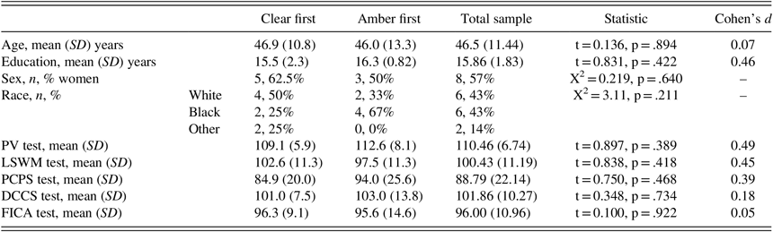
Fourteen participants with insomnia symptoms were included in the study analyses; 8 were randomized first to the clear lenses condition, and 6 were randomized first to the amber lenses condition. Demographic characteristics of the sample are presented in Table 1. Examination of performance on the Picture Vocabulary test indicated that participants had estimated intellectual functioning that was clinically in the “average” range and approximately 2/3 of an SD higher than normative expectations. Participants randomized to the clear lenses condition first did not differ from those who were randomized into the amber lenses condition first on any demographic feature or estimate of intellectual functioning (see Table 1). Mean (±SD) usage of lenses throughout the amber and clear conditions were 2.16±0.23 hr and 2.14±0.46 hr, respectively (p = .89).
Participant characteristics at baseline among participants who received the clear lens condition first, the amber condition lens condition first, and the total sample

Note. All neuropsychological test scores are presented as standard scores, which have a normative mean of 100 and SD of 15. The test statistic refers to the comparison between the two groups.
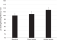
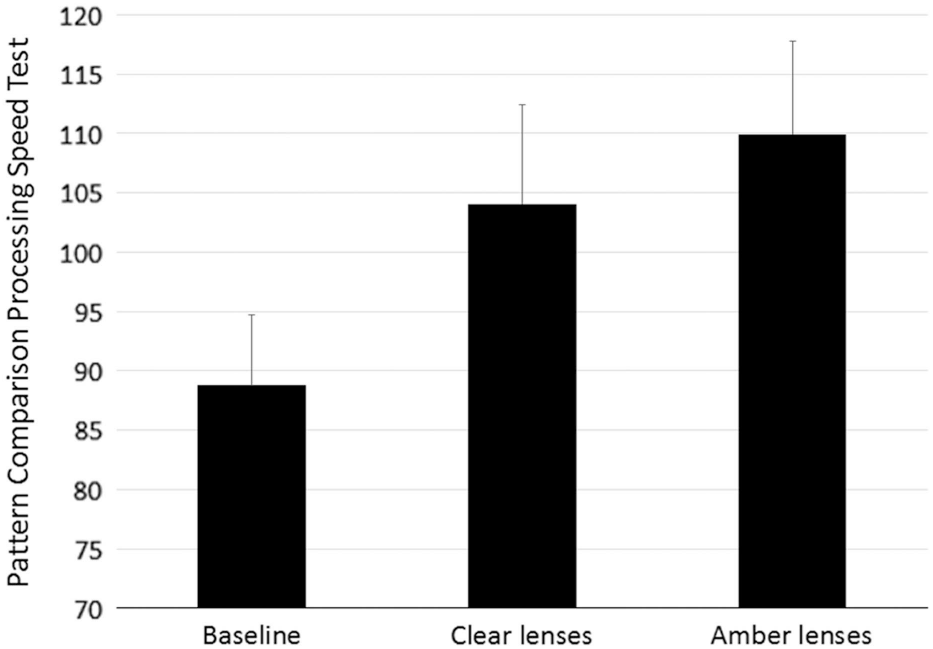
Compared with baseline, participants performed better on the LSWM test after treatment with amber lenses (p = .002; 95% CI [−11.81 to −3.58]; Cohen’s d = 0.66), which was not observed after treatment with the clear lenses (p = .419; 95% CI [−6.65 to 2.95]; Cohen’s d = 0.16; omnibus effect F = 5.16; p = .014; η2 = 0.301). Figure 1 displays mean scores on the LSWM test at the three study phases. A similar pattern emerged with the PCPS test, such that participants had the lowest performance at baseline, the highest performance following treatment with amber lenses, and intermediate performance after treatment with clear lenses (omnibus effect F = 7.65; p = .002; η2 = 0.370 see Figure 2). Here, performance was significantly greater than baseline after treatment with both clear (p = .03; 95% CI [−28.87 to −1.70]; Cohen’s d = 0.71) and amber (p = .003; 95% CI [−33.45 to −8.84]; Cohen’s d = 0.95) lenses.
Performance on the NIH Toolbox LSWM test at baseline and following treatment with clear lenses and treatment with amber lenses. Participants performed significantly better on the task after treatment with amber lenses than at baseline and after treatment with clear lenses. Performance did not differ significantly between the clear condition and baseline testing. Values are means; error bars are standard errors.

Performance on the NIH Toolbox PCPS test at baseline and following treatment with clear lenses and treatment with amber lenses. Participants performed significantly better on the task after treatment with amber and clear lenses than at baseline. Performance did not differ significantly between the amber and clear conditions. Values are means; error bars are standard errors.

Although performance after amber lenses treatment showed the largest effect, it did not differ significantly from performance following clear lenses treatment (p = .23; 95% CI [−15.88 to 4.16]). Of note, based on scores on the Picture Vocabulary test, treatment with amber lenses improved performance on the LSWM and the PCPS tests by approximately 0.5 SDs and 1.5 SDs, respectively, normalizing scores to the expected performance range. Performance did not differ across the three conditions for the FICA test (omnibus effect F = 2.70; p = .087; η2 = 0.184; Cohen’s d was 0.27 and 0.35 for, respectively, amber and clear conditions compared with baseline) or for the DCCS test (omnibus effect F = 0.063; p = .93; η2 = 0.005, Cohen’s d was 0.10 and 0.01 for, respectively, amber and clear conditions compared with baseline).
As reported previously (Shechter et al., Reference Shechter, Kim, St-Onge and Westwood2018), the most prevalent sleep complaints at baseline were the feeling that sleep is not refreshing or that sleep is not sound (78.6% and 71.4% reported these symptoms occurring 5–7 times per week). Sixty-four percent of participants reported difficulty staying asleep as occurring 5–7 times per week and 50% reported difficulty falling asleep 5–7 times/week. Also as previously reported (Shechter et al., Reference Shechter, Kim, St-Onge and Westwood2018), objective measures of sleep obtained using actigraphy showed numeric improvement during the 1-week treatment with amber lenses compared to the 1-week treatment with clear lenses across most measures, although with the exception of TST, differences in most actigraphy measures between the two conditions did not reach statistical significance.
Performance on the LSWM test after treatment with amber lenses was significantly related to several actigraphy measures recorded during the treatment week: TST, sleep onset latency (SOL), wakefulness after sleep onset (WASO), and sleep efficiency (SE) (Table 2). In all cases, better neuropsychological performance was associated with improved sleep characteristics. On the other hand, performance on the LSWM test after treatment with clear lenses was not associated with any actigraphy measures. Performance on the PCPS test after treatment with clear or amber lenses did not correlate systematically with actigraphy measures during their respective condition weeks (Table 2). Correlations between actigraphy measures and the DCCS and FICA tests were not performed because performance on those neuropsychological tests was unchanged by the intervention.
Correlations of performance on the LSWM test and the PCPS task after each condition with actigraphy-derived objective measures of sleep quality during the respective weeks of treatment (i.e., clear, amber)

Note. Note that we did not correct for multiple comparisons; only the correlation between TST and LSWM in the amber condition would have survived Bonferroni correction.
Assessment of intellectual ability indicated that treatment with amber lenses “normalized” performance on each test from approximately 1 SD below expected performance to expected performance. That is, on average, participants’ estimated intellectual functioning was approximately 2/3 of an SD higher than normative values while their baseline performance on the LSWM, PCPS, DCCS, and FICA tests was approximately 1 SD below normative values. After intervention with amber glasses, participants’ performance on these tests improved to the average range.
Insomnia symptom severity was improved with the active treatment, as illustrated by reductions in PIRS total scores after the amber versus clear lenses condition (mean±SD: 72.64±28.14 vs. 88.93±33.19; p = .023). Furthermore, objective measurement of TST was increased in the amber lenses versus clear lenses condition (358.80±66.29 min vs. 330.33±66.01 min; p = .035; Cohen’s d = 0.65). There were no statistically significant differences between conditions for SOL (amber: 11.27±12.23 min vs. 16.21±23.42 min; p = .221), SE (78.35±9.00 % versus 77.01±8.67%; p = .285), or WASO (88.43±38.45 min versus 83.95±35.53 min, p = .546). Self-reported time of going to bed, based on sleep-log, was unchanged between intervention conditions (amber: 23:27±01:46 vs. clear: 23:25±01:14; p = .833). Reported time of waking was significantly later in amber versus clear lenses condition (07:15±01:33 vs. 06:47±01:27; p = .033; Cohen’s d = 0.64).
DISCUSSION
Using a randomized, placebo-controlled crossover design, we demonstrated improvements in processing speed and working memory following a 1-week intervention that blocked blue light exposure for 2 hr before bedtime in individuals with insomnia symptoms. Improvements on the test of working memory were associated with objective actigraphic measures of sleep quality obtained during the same intervention period, suggesting that the improvement in cognition may be mediated partially by intervention-associated improvement in sleep. There was no change in neuropsychological performance from baseline during a 1-week control intervention period. These findings, while preliminary, show that some neuropsychological dysfunction experienced by individuals with insomnia symptoms is reversible with a short-term intervention. Furthermore, improvement in sleep appears to mediate the improvement in neuropsychological function on higher-order tasks that require holding and manipulating several concepts in mind at one time.
The neurocognitive model of insomnia (Perlis, Giles, Mendelson, Bootzin, & Wyatt, Reference Perlis, Giles, Mendelson, Bootzin and Wyatt1997) posits that individuals with insomnia experience cognitive and somatic hyperarousal that may predispose and perpetuate chronic sleep initiation and sleep maintenance symptoms (Bastien, Reference Bastien2011; Bastien, Vallieres, & Morin, Reference Bastien, Vallieres and Morin2004; Harvey, Reference Harvey2002). In this model, insomnia results from a conditioned arousal system emerging from learned associations between sleep stimuli and poor sleep response. Thus, individuals with insomnia symptoms may experience hyperarousal during the day and before bedtime that they are unable to inhibit during a typical sleep interval (Bastien, St-Jean, Morin, Turcotte, & Carrier, Reference Bastien, St-Jean, Morin, Turcotte and Carrier2008; Smith et al., Reference Smith, Perlis, Chengazi, Pennington, Soeffing, Ryan and Giles2002). Such a model of cognitive hyperarousal is consistent with the neuropsychological dysfunction frequently reported by individuals with insomnia. Chronic hyperarousal likely results in fatigue and impaired attention and concentration, which is consistent with our findings of decreased performance on a test of cognitive processing speed.
In addition, the inability to exert cognitive control is consistent with our findings of poor performance on a test of working memory and the ability to effectively hold multiple streams of information in mind at one time. While many studies find support for poor neuropsychological function in individuals with insomnia (Altena, Van Der Werf, Strijers, & Van Someren, Reference Altena, Van Der Werf, Strijers and Van Someren2008; Edinger, Means, Carney, & Krystal, Reference Edinger, Means, Carney and Krystal2008; Fortier-Brochu & Morin, Reference Fortier-Brochu and Morin2014; Hauri, Reference Hauri1997; Ling et al., Reference Ling, Lim, Gwee, Ho, Collinson and Ng2016; Schneider, Fulda, & Schulz, Reference Schneider, Fulda and Schulz2004), not all have found such relationships (Haimov, Hanuka, & Horowitz, Reference Haimov, Hanuka and Horowitz2008; Orff, Drummond, Nowakowski, & Perils, Reference Orff, Drummond, Nowakowski and Perils2007). Meta-analytic studies on this topic report largely equivocal findings (Riedel & Lichstein, Reference Riedel and Lichstein2000; Shekleton, Rogers, & Rajaratnam, Reference Shekleton, Rogers and Rajaratnam2010), although when present, deficits are apparent in the neuropsychological domains of attention, vigilance, memory, working memory, and executive function.
Our study, which used sensitive neuropsychological instruments developed with modern psychometric techniques (Gershon et al., Reference Gershon, Cella, Fox, Havlik, Hendrie and Wagster2010), confirmed neuropsychological dysfunction among individuals with insomnia. Our study design did not include a control group without insomnia symptoms, so we were not able to compare performance on neuropsychological tests between such groups. However, an estimate of intellectual function indicated that our participants’ neuropsychological performance levels should have been in the average range; yet, clinical examination of their scores fell below expected levels. Taken together, this indicates that individuals with insomnia symptoms in our sample evidenced neuropsychological dysfunction.
The primary finding of this study was that individuals with insomnia symptoms experienced improvement in working memory and processing speed when they engaged in a 1-week intervention that blocked blue light exposure 2 hr before bedtime, but not when they completed a control intervention. Although correlational data from the extant literature have suggested a relationship between blue light exposure and cognitive function (Alkozei, Smith, Dailey, Bajaj, & Killgore, Reference Alkozei, Smith, Dailey, Bajaj and Killgore2017; Souman, Tinga, Te Pas, van Ee, & Vlaskamp, Reference Souman, Tinga, Te Pas, van Ee and Vlaskamp2018), this study, to our knowledge, is the first to provide experimental evidence that reduction in nocturnal blue light exposure improves neuropsychological function in individuals with symptoms of insomnia.
Blue light exposure (i.e., wavelengths around 460 nm in the visible spectrum) reliably affects the 24-hr circadian clock, likely through an independent system of retinal ganglion cells that project directly to the hypothalamus (Brainard et al., Reference Brainard, Hanifin, Greeson, Byrne, Glickman, Gerner and Rollag2001; Thapan et al., Reference Thapan, Arendt and Skene2001). Blue light can be experienced in the natural environment as well as through electronic devices that use light emitting diodes, such as computers and smartphones. The timing of blue light exposure is critical, as focused exposure upon waking effectively alleviates the depressed mood symptoms associated with seasonal affective disorder and major depressive disorder (Glickman, Byrne, Pineda, Hauck, & Brainard, Reference Glickman, Byrne, Pineda, Hauck and Brainard2006). However, when experienced during the typical “dark” periods before bedtime, blue light functions as an alerting mechanism, delaying the release of the pineal hormone melatonin and the typical sleep onset response of somnolence. This may be a critical exacerbating factor for individuals with insomnia who are likely already managing somatic and cognitive hyperarousal responses (Perlis et al., Reference Perlis, Giles, Mendelson, Bootzin and Wyatt1997).
Our findings suggest that nocturnal blue light exposure may be negatively affecting neuropsychological abilities, which can be alleviated with an intervention to block nocturnal exposure to blue light. Correlations between working memory and objective sleep measures suggest that improvements in sleep quality may be mediating the cognitive response, although further examination with a larger sample is needed to test this hypothesis explicitly.
We did not observe a relationship between the improvement in processing speed and objective sleep measures, which raises several questions about potential mechanisms. We hypothesize that a relative reduction in blue-wavelength light exposure while wearing amber lenses improves sleep and insomnia symptoms via a lesser degree of melatonin suppression and/or higher nocturnal melatonin secretion during the hours leading up to bedtime. Unfortunately, we did not monitor melatonin secretion while participants were undergoing the intervention at home. Future work should measure ambulatory melatonin secretion in response to the amber lenses intervention. Furthermore, as we used wrist actigraphy to monitor sleep, we were also unable to determine if the intervention affected sleep architecture.
This study used a within-subject crossover design, which maximizes statistical power such that expected power is greater than a between subjects design with even twice the number of participants. Crossover designs boost power mostly because they control individual differences in relevant variables, such as age and sex, which may otherwise add error variance to between subjects designs. With smaller sample studies, internal validity that is based on a random assignment is not an issue in a crossover design studies but could be an unfortunate source of group differences in a between-subjects design. External validity is also maximized in a crossover design because the conditions are more of a reflection of natural behavior (e.g., individuals are more likely to wear amber colored glasses at various time points rather than choosing to wear amber colored glasses always).
With respect to statistical power per se the concern with a small sample study is that there will be insufficient power to detect otherwise meaningful effects (type 2 error). However, our results yielded statistically reliable results, suggesting that we indeed had sufficient power to detect effects. The other concern with small sample studies is that observed effects are spurious (type 1 error). We believe that this concern is also mitigated by the use of the crossover design, which reduces the likelihood of spurious findings because participants are essentially serving as their own controls. Crossover designs also have some disadvantages, including practice effects and differential carryover effects, which we attempted to mitigate by counterbalancing the conditions and introducing a washout period.
In addition, crossover designs may not be advantageous for individuals with conditions that have symptoms that are progressive or unstable in nature. We addressed this issue by recruiting only individuals within chronic insomnia symptoms that persisted for more than 3 months. Concerns about practice effects as a possible explanation of the changes in the follow-up condition rather than an actual therapeutic effect of the amber lenses is addressed by appreciating larger effect after the amber lens condition compared with the clear lens condition.
There are limitations to this study that warrant further consideration. First, this project was designed as a pilot study and the sample size was modest. However, we used an efficient crossover design, which maximizes statistical power with relatively small samples and reduces potential confounding relevant variables that can differ between groups in a parallel group design study. Replication with a larger sample is necessary to confirm the findings and to examine mediating mechanisms. In addition, our 1-week intervention period assumed nocturnal blue light exposure, but we did not objectively measure blue light exposure in the individual’s environment. Although we consider this naturalistic approach to have important ecological validity, it does not permit explorations of specific dose–response relationships. Furthermore, ambient light levels in the home environment during the intervention were not recorded. As a follow-up to current findings, habitual evening light levels via light intensity photometer, or more astutely, the levels of blue-wavelength light via spectral light meter, in the home environment should be recorded. The relatively large participant age range is important to consider, as a reduction in the transparency of the eyeball lens as occurs with age could influence the transmission of light through the retino-hypothalamic tract (Bloemendal et al., Reference Bloemendal, de Jong, Jaenicke, Lubsen, Slingsby and Tardieu2004). Visual impairments were not a specific exclusion criterion. However, based on self-report and medical history and physical conducted by a physician or nurse practitioner, no participant reported major visual impairment of light transmission, such as macular degeneration.
Relevant participant characteristics, such as age, sex, and other background variables, could also have an influence on the observed outcomes, such that certain subgroups may be more responsive to the intervention described here. The current study’s samples size was not large enough to explore these possibilities fully. However, we should note that we tested whether sex and age interacted with Visit for the primary neuropsychological outcomes observed and we did not see an effect of either variable (data not shown). It should also be noted that the crossover design of this study somewhat mitigates the concerns that differential distribution of demographic subgroups across condition might be confounding the observed results, as might be the case in small-sample between subjects designs.
Finally, insomnia can be quite heterogeneous across individuals, with various symptom subtypes, such as difficulty initiating sleep, difficulty maintaining sleep, and early morning awakening, and varying causes. In the current study, participants were enrolled based on symptom profiles associated with DSM and American Academy of Sleep Medicine diagnoses of insomnia (Okun, 2009). We did not have a large enough sample to conduct subgroup analyses, and it is important to note that blue blocker intervention may have differential effects on cognition across subtypes. Future studies should consider this possibility.
There are some important considerations regarding the choice of lenses for the intervention. As opposed to the amber lenses used here, orange-colored lenses would be the ideal choice to achieve a near-complete blockage of blue-wavelength light transmission (blue light transmission: <2%). However, orange-colored lenses also result in a substantial dimming of the overall light environment (visible light transmission: 45%). Conversely, although the amber lenses used here do not completely eliminate all transmission of blue-wavelength light (blue-light blockage: 65%), they also do not cause a dimming of the overall light environment (visible light transmission: 90%). The current objective was to test the effects of selectively blocking blue light, as opposed to testing the effects of an overall dimming of the ambient light environment. We, therefore, chose to use amber lenses, as they achieve a substantial degree of blue-light blocking while maintaining an overall visible light transmission (i.e., brightness) that is similar to clear lenses. Similarly, the choice of clear lenses may not be ideal as a placebo. We used clear lenses (visible light transmittance: 92%) as a sham, since any colored lenses would result in an overall dimming of the light environment, and we aimed to successfully match the level of visible light transmittance of the sham lenses to the amber lenses.
With the growing awareness of the adverse effects of nocturnal blue light exposure on sleep and discussion of the potential benefit of amber lenses in the media and lay literature, it is possible that participants were insufficiently blinded to experimental conditions. We did not assess participants’ knowledge of the lenses before the start of procedures, or query participants after study completion regarding their expectations of each condition. These should be done in a follow-up study, to confirm the integrity of blinding. Nevertheless, in this crossover study, participants received one colored lenses intervention at a time, and were not initially informed of each lenses color condition. Therefore, those receiving the clear lenses first were unaware of the amber lenses condition until the start of the second intervention phase, and vice versa. Participants were, therefore, blinded to the conditions, such that they were unaware if they were in an experimental condition or a control condition during the initial assessments. The 4-week washout period would also likely contribute to a reduction in demand characteristics.
Our findings have important implications for clinical practice. Individuals with insomnia symptoms may experience cognitive benefit from a short-term intervention to block blue light exposure with a tool, blue blocking lenses, that is relatively inexpensive, easy to obtain and integrate into one’s environment, and generally well-tolerated. Furthermore, the amount of improvement was both statistically significant and clinically meaningful, allowing individuals to experience a “return” to expected levels of neuropsychological function following the intervention.
ACKNOWLEDGMENTS
Sources of Financial Support: This project was supported by funding from a Focused-Project Award (144-FP-16) (PI: A. Shechter) from the American Sleep Medicine Foundation, a foundation of the American Academy of Sleep Medicine. This publication was also supported by the National Center for Advancing Translational Sciences, National Institutes of Health, through Grant Number UL1TR001873 (PI: M. Reilly). Conflicts of Interest: The authors have no conflicts of interest.