1. Introduction
Palaeoscolecids represent a class of early–mid-Palaeozoic marine nemathelminth worms characterized by a long, annulated, cylindrical body with a cuticle consisting of numerous, tiny, individual button-shaped plates and platelets. Initially described as compressed body fossils in shales from the Lower–Middle Ordovician (Ulrich, Reference Ulrich1878; Whittard, Reference Whittard1953), the disarticulated, isolated phosphatic sclerites of palaeoscolecids were subsequently found as common ‘problematic’ elements in early Palaeozoic (especially Cambrian) small shelly fossil assemblages (e.g. Bengtson, Reference Bengtson1977; Gedik, Reference Gedik1977; Wrona, Reference Wrona1982, Reference Wrona and Gaèdzicki1987, Reference Wrona2004; Peel & Larsen, Reference Peel and Larsen1984; Bendix-Almgreen & Peel, Reference Bendix-Almgreen and Peel1988; Märss, Reference Märss1988). Before articulated sclerite arrays were found, opinions about the affinity of the isolated sclerites were quite diverse; see Müller & Hinz-Schallreuter (Reference Müller and Hinz-Schallreuter1993) for a detailed historical review.
Over the last 20 years, recovery of unequivocal articulated sclerite arrays arranged in annulated bands has provided unequivocal evidence that the source of many of these sclerites can be attributed to palaeoscolecids (Kraft & Mergl, Reference Kraft and Mergl1989; van den Boogaard, 1989a, b; Hinz et al. Reference Hinz, Kraft, Mergl and Müller1990; Brock & Cooper, Reference Brock and Cooper1993; Müller & Hinz-Schallreuter, Reference Müller and Hinz-Schallreuter1993). Since their initial discovery over a century ago (Ulrich, Reference Ulrich1878), palaeoscolecids have been referred to a wide range of metazoan groups. Body fossils of palaeoscolecids have predominantly been referred to the Annelida (Miller & Faber, Reference Miller and Faber1892; Whittard, Reference Whittard1953; Conway Morris, Reference Conway Morris1977, Reference Conway Morris, Conway Morris, George, Gibson and Platt1985; Conway Morris, Pickerill & Harland, Reference Conway Morris, Pickerill and Harland1982; Kraft & Mergl, Reference Kraft and Mergl1989) or the Oligochaeta (Bather, Reference Bather1920; Reudemann, Reference Ruedemann1925a, Reference Ruedemannb). Current morphological evidence from well-preserved complete specimens supports an ecdysozoan systematic position for the palaeoscolecids within the Cycloneuralia (Budd, Reference Budd2001; Maas et al. Reference Maas, Huang, Chen, Waloszek and Braun2007a, Reference Maas, Waloszek, Haug and Müllerb), although their precise affinities with the Nematoida (including nematomorphs and nematodes) and the Scalidophora (including kinorhynchs, loriciferans and priapulids) remain unsettled (Maas et al. Reference Maas, Huang, Chen, Waloszek and Braun2007a, Reference Maas, Waloszek, Haug and Müllerb).
Isolated phosphatic sclerites have been recognized under various generic names since Ethington & Clark (Reference Ethington and Clark1965) first described specimens as ‘plate forms A and B’ recovered during routine acid etching for Lower Ordovician conodonts from Alberta, Canada. Due to the isolated nature of these sclerites, all names applied to the sclerites, including Hadimopanella, are here regarded as form taxa; more specifically, Hadimopanella is recognized as a form genus, and taxa within Hadimopanella are accordingly recognized as form species. The group of tiny, button-shaped sclerites (including the hadimopanellids) was independently described in near-synchronously issued publications from Turkey (Gedik, Reference Gedik1977) and east Siberia (Bengtson, Reference Bengtson1977). The form genus Lenargyrion (Bengtson, Reference Bengtson1977) described from east Siberia has since been considered a junior synonym of the form genus Hadimopanella Gedik, Reference Gedik1977 (Wrona, Reference Wrona1982; van den Boogaard, Reference Boogaard1983), a view that was recently challenged by Ivantsov & Zhuravlev (Reference Ivantsov, Zhuravlev and Ponomarenko2005). Individual sclerites of Hadimopanella are characterized by a smooth and slightly convex basal surface with a strongly raised, convex to flat upper surface ornamented by a series of nodes (Gedik, Reference Gedik1977; Bengtson, Reference Bengtson1977). A large number of similar sclerites have subsequently been reported from numerous localities and ages around the world (van den Boogaard, Reference Boogaard1983, Reference Boogaard1989a, Reference Boogaardb; Peel & Larsen, Reference Peel and Larsen1984; Wrona, Reference Wrona and Gaèdzicki1987, Reference Wrona2004; Hinz, Reference Hinz1987; Bendix-Almgreen & Peel, Reference Bendix-Almgreen and Peel1988; Märss, Reference Märss1988; Gedik, Reference Gedik1989; Wang, Reference Wang1990; Müller & Hinz-Schallreuter, Reference Müller and Hinz-Schallreuter1993; Wrona & Hamdi, Reference Wrona and Hamdi2001) with differentiation of species-level form taxa based on relatively minor variations in the spacing and number of nodes on the convex upper surface of the sclerites.
One of the more geographically widespread form species of Hadimopanella, H. apicata, was described by Wrona (Reference Wrona1982, pls 1–4) from the upper lower Cambrian (Bonnia–Olenellus Zone) of Spitsbergen; this form species is characterized by a single, centrally located, conical node with an undulate margin hosting radial striae. H. apicata has also been reported from lower Cambrian rocks in Antarctica (Wrona, Reference Wrona2004), Greenland (Peel & Larsen, Reference Peel and Larsen1984; Bendix-Almgreen & Peel, Reference Bendix-Almgreen and Peel1988; Skovsted, Reference Skovsted2006) and questionably, England (Hinz, Reference Hinz1987). A separate, but morphologically similar form species, H. antarctica Wrona, Reference Wrona and Gaèdzicki1987, previously thought to be endemic to the lower Cambrian of Antarctica, is characterized by having a large central node surrounded at the margin by numerous smaller, regularly spaced, conical tubercles (Wrona, Reference Wrona and Gaèdzicki1987, pls 5–8; Wrona, Reference Wrona2004, figs 17, 18A–D).
The vast majority of Hadimopanella form species are recognized only from isolated individual sclerites, with the exception of very poorly preserved, partly articulated phosphatized specimens of H. antarctica from glacial erratics on King George Island (Wrona, Reference Wrona and Gaèdzicki1987, pl. 8, fig. 1) and H. cf. oezgueli from the Middle Cambrian of Australia (Müller & Hinz-Schallreuter, Reference Müller and Hinz-Schallreuter1993, fig. 7D–G). However, sclerites of similar morphology are known from a number of shale-hosted palaeoscolecid species (Conway Morris, Reference Conway Morris1997; Ivantsov & Wrona, Reference Ivantsov and Wrona2004; Ivantsov & Zhuravlev, Reference Ivantsov, Zhuravlev and Ponomarenko2005). The aim of this paper is to describe and illustrate the morphology and details of the micro-ornament of articulated scleritome fragments recovered from the early Cambrian of South Australia and to elucidate the relationships of epidermal sclerites previously described as separate Hadimopanella form species.
2. Locality, stratigraphy and age
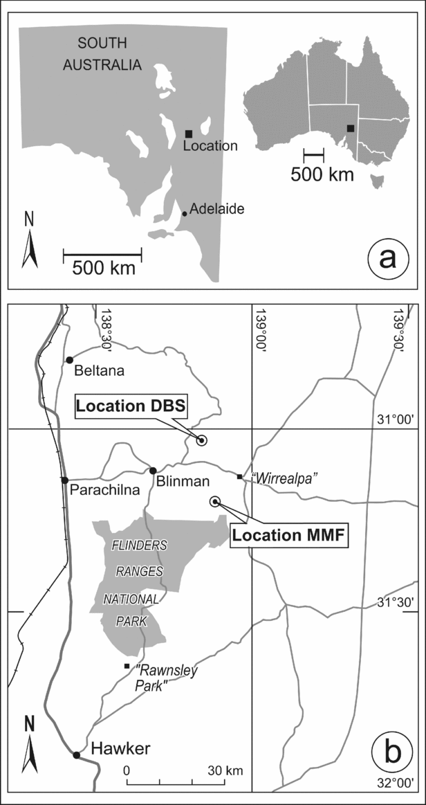
Two fragments of scleritome (specimens SAMP43299 and SAMP43301) were collected from a stratigraphic section (MMF) measured through the lower slope carbonates of the Mernmerna Formation cropping out along the eastern margin of the Bunkers Range on Angorichina Station in the central Flinders Ranges, South Australia. Both specimens come from sample horizon MMF/44.6, some 25.5 m true thickness above the base of the section (coordinates at base of MMF section: 31°11′38.4″ S, 138°52′28.7″ E; map datum: WGS84; see Fig. 1); other faunal elements that have been documented from this horizon include bradoriids and phosphatocopids (Skovsted, Brock & Paterson, Reference Skovsted, Brock and Paterson2006), and halkieriid sclerites (Paterson, Brock & Skovsted, Reference Paterson, Brock and Skovsted2009). A single, small scleritome fragment (specimen SAMP43300) was collected from sample horizon DBS250, some 216 m true thickness above the base of stratigraphic section DBS, measured through outcrops of the Mernmerna Formation on the eastern limb of the Donkey Bore Syncline, central Flinders Ranges (base of the DBS section is at 31°02′30.4″ S, 138°52′00.6″E; map datum: GDA94; see Fig. 1).
(a) Locality map showing position of the study area in South Australia. (b) Enlarged locality map (from Fig. 1a) of the study area and the position of the MMF and DBS sections.

The palaeontology, stratigraphy and sedimentology of the Mernmerna Formation of the MMF section have been documented in some detail by Brock & Paterson (Reference Brock and Paterson2004) and Paterson & Brock (Reference Paterson and Brock2007), and summarized by Skovsted, Brock & Paterson (Reference Skovsted, Brock and Paterson2006). Brock & Paterson (Reference Brock and Paterson2004) and Paterson & Brock (Reference Paterson and Brock2007) correlated the MMF section (above sample MMF/0.0) with the Third Plain Creek Member of the Mernmerna Formation at Wilkawillina Gorge type section in the Bunkers Graben based on similar lithologies, sedimentary structures and the occurrence of the index trilobite Pararaia bunyerooensis Jell (in Bengtson et al. Reference Bengtson, Conway Morris, Cooper, Jell and Runnegar1990). The sedimentology and lithology of the Mernmerna Formation at the DBS section have previously been documented in some detail by Topper et al. (Reference Topper, Skovsted, Brock and Paterson2007). The Linns Springs and Third Plain Creek members of the Mernmerna Formation at the Donkey Bore Syncline correlate with the Pararaia tatei and Pararaia bunyerooensis zones, respectively, based on the presence of the eponymous species. However, the dearth of identifiable trilobite specimens throughout the DBS stratigraphic section does not allow precise placement of the boundary between the two trilobite biozones in the Mernmerna Formation. SAMP43300 was collected from horizon DBS250 (216 m true thickness above the base of the section), a horizon that is situated between the two trilobite biozone boundaries, and consequently, precise biostratigraphic placement remains uncertain.
Paterson & Brock (Reference Paterson and Brock2007) suggested that the Pararaia tatei and Pararaia bunyerooensis zones correlate with the Eoredlichia–Wutingaspis Zone of the Qiongzhusian Stage, Yangtze Platform in China (Jago et al. Reference Jago, Zang, Sun, Brock, Paterson and Skovsted2006; Paterson & Brock, Reference Paterson and Brock2007). The presence of Eoredlichia, Wutingaspis and Yunnanocephalus in the Pararaia bunyerooensis Zone permits correlation with the Yunnanocephalus Assemblage subzone of the upper Eoredlichia–Wutingaspis Zone in China (Jago et al. Reference Jago, Zang, Sun, Brock, Paterson and Skovsted2006; Paterson & Brock, Reference Paterson and Brock2007). The upper Eoredlichia–Wutingaspis Zone correlates with the lower Botoman Bergeroniellus micmacciformis–Erbiella Zone of Siberia, based on the correlation of older (Abadiella huoi Zone equivalent) archaeocyaths (Paterson & Brock, Reference Paterson and Brock2007).
3. Morphological descriptions
The terminology used to describe the palaeoscolecid scleritome fragments largely follows that employed by Müller & Hinz-Schallreuter (Reference Müller and Hinz-Schallreuter1993), with some minor modifications. We use the term ‘interannular furrow', rather than ‘intercalation' (Müller & Hinz-Schallreuter, Reference Müller and Hinz-Schallreuter1993) or ‘intercalary zone' (Conway Morris, Reference Conway Morris1997). The terms ‘plates', ‘platelets' and ‘microplates' have never been formally defined in terms of dimensions, but differentiation can be assisted based on their position on the mineralized cuticle. Plates are typically circular to elongate in outline and are often preserved in two alternating rows on each annulus of the cuticle. Platelets are effectively miniaturized versions of plates, generally occupy the interannular furrow and are less commonly found separating plates on the annulus. Microplates generally do not display a resemblance to plates but instead tend to form in available space between plates and platelets, often resulting in an irregular morphology. Microplates are frequently distributed in the central region of each annulus as well as in the interannular furrow. Plates are essentially equivalent to the isolated cuticular sclerites previously described as form species of Hadimopanella. In this paper, plates specifically refer to cuticular elements associated with a scleritome, while sclerites refer specifically to isolated, cataphract elements mainly recovered from acid residues.
The lack of definitive posterior and anterior terminations in the specimens described here means that it is not possible to identify accurately the ‘posterior' or ‘anterior' and thus also ‘ventral' and ‘dorsal' orientations of the described fragments. As much as possible, descriptions and discussions avoid using terms related to orientation except when referring to figures. The orientation of the palaeoscolecid fragments in Figures 2–5, though consistent for each specimen, is arbitrary and may not reflect actual biological orientation.
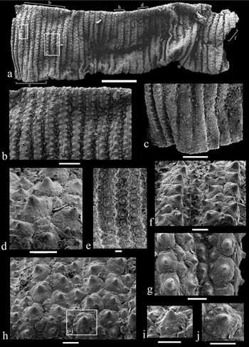
Early Cambrian palaeoscolecid scleritome fragment, SAMP43299. Specimen from sample horizon MMF/44.6; all scale bars 10 μm unless otherwise stated. Boxed and bracketed areas indicate the position of the relevant enlarged figure. (a) Lateral view of compressed trunk fragment; scale bar 200 μm. (b) Lateral view of upper (?dorsal) margin; scale bar 50 μm. (c) Lateral view of the lower (?ventral) margin; scale bar 50 μm. (d) Detail of plates referable to Hadimopanella antarctica Wrona, Reference Wrona and Gaèdzicki1987 situated on the upper (?dorsal) margin. (e) Detail of annuli, showing irregularly alternating plates. (f) Detail of annuli, showing pronounced interannular furrow occupied by platelets. (g) Detail of plates referable to Hadimopanella apicata Wrona, Reference Wrona1982. (h) Detail of the upper (?dorsal) margin, showing plates referable to H. antarctica. (i) Enlargement of (h), showing a plate referable to H. antarctica. (j) Enlargement of (h), showing a plate referable to H. antarctica.

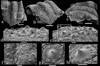
Early Cambrian palaeoscolecid scleritome fragment, SAMP43300. Specimen from sample horizon DBS/250. Boxed areas indicate the position of the relevant enlarged figure. (a) Lateral view of trunk fragment; scale bar 100 μm. (b) Oblique lateral view of trunk fragment; scale bar 100 μm. (c) Oblique lateral view of trunk fragment; scale bar 100 μm. (d) Detail of plates referable to Hadimopanella antarctica Wrona, Reference Wrona and Gaèdzicki1987; scale bar 10 μm. (e) Detail of plates referable to H. antarctica; scale bar 10 μm. (f) Detail of annuli, showing pronounced interannular furrow occupied by platelets; scale bar 20 μm. (g) Enlargement of (d) and (e), showing detail of a plate referable to H. antarctica; scale bar 10 μm. (h) Enlargement of (e), showing detail of a plate referable to H. antarctica; scale bar 10 μm.

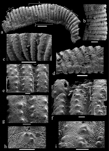
Early Cambrian palaeoscolecid scleritome fragment, SAMP43301. Specimen from sample horizon MMF/44.6; all scale bars 10 μm unless otherwise stated. Boxed and bracketed areas indicate the position of the relevant enlarged figure. (a) Lateral view of compressed trunk fragment; scale bar 100 μm. (b) Lateral view of trunk fragment; scale bar 50 μm. (c) Lateral view of upper (?dorsal) margin; scale bar 50 μm. (d) Oblique lateral view of lower (?ventral) margin; scale bar 50 μm. (e) Detail of annuli. (f) Detail of annuli situated on the upper (?dorsal) margin, platelets situated in the interannular furrow. (g) Enlargement of (e), showing irregularly alternating plates, separated by microplates, with microplates visible in the interannular furrow. (h) Enlargement of (f), showing detail of a plate with three median nodes, referable to H. apicata Wrona, Reference Wrona1982. (i) Enlargement of (f), showing detail of plates with two median nodes, interannular furrow occupied by platelets and microplates.

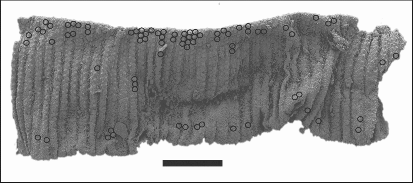
Early Cambrian palaeoscolecid scleritome fragment, SAMP43299. Circles indicate the position of Hadimopanella antarctica Wrona, Reference Wrona and Gaèdzicki1987 plates on the scleritome. Scale bar 200 μm.

Rather than add to the ever-increasing list of new palaeoscolecid genera and species described in recent years, it is our contention that formal taxonomic identification can only proceed when there is enough composite scleritome material (or enough complete specimens) to ensure all intraspecific and ontogenetic variation associated with palaeoscolecid cuticular morphology is adequately incorporated into the concept of the taxon. We thus prefer to leave the South Australian material under open nomenclature, and hope that the description of the cuticular fragments will not only provide the first step in documenting these well-preserved South Australian palaeoscolecids, but provide enough morphological detail to engender comparison with other taxa from this biogeographically widespread group. All specimens described and illustrated are housed in the palaeontological collection of the South Australian Museum (acronym: SAMP).
Palaeoscolecid cuticle fragment SAMP43299
Figures 2, 5
SAMP43299 is an incomplete, laterally compressed trunk fragment (1.3 mm in length, 493 μm maximum width) lacking oral and aboral terminations (Figs 2a, 5). The trunk fragment contains a total of about forty serially repeated, uniformly narrow annuli (average 25 μm in width) along the length of the entire trunk fragment. Lower (?ventral) margin (as figured) displays irregular ornamentation and appears to be slightly abraded (Fig. 2c). Cuticle ornamentation of each annulus consists of two rows of irregularly alternating plates (Fig. 2b); a few annuli display a variably developed, implanted third row (Fig. 2f). Plates are mostly in direct contact with each other, though in some regions they are separated intermittently by platelets (Fig. 2f, g). Plates are subcircular to slightly ovoid in outline with irregular margins and characterized by a single conical median node (Fig. 2d–g). Plates range in diameter from 12 to 18 μm (average diameter 14 μm) and vary widely in morphology, with a small proportion displaying a single median node surrounded by one to eight tiny nodes situated close to the plate margin (Fig. 2d, h–j). The majority of plates displaying this morphological variation appear to be situated along the upper (?dorsal) margin, though a few are developed in the central portion of the trunk fragment (Fig. 5). Interannular furrows are narrow but pronounced (5–7 μm in width), occupied in most cases by a single file of platelets (average 5 μm in diameter) (Fig. 2f, g). Each platelet is effectively a miniaturized version of a plate and displays a single median node (Fig. 2f, g).
Palaeoscolecid cuticle fragment SAMP43300
Figure 3
SAMP43300 is an incomplete trunk fragment (417 μm in length, 416 μm maximum width) lacking oral and aboral terminations (Fig. 3a–c). The trunk fragment consists of three irregular annulations, varying in width from a minimum of 85 μm to a maximum of 160 μm (average 139 μm). Cuticle ornamentation of each annulus consists of irregularly arranged plates in weakly defined rows, with the number of plate rows in each annulus varying from four to seven. Plates are mostly in direct contact with each other, though in some regions they are separated intermittently by platelets (Fig. 3f) and by small, nodular to polygonal microplates (Fig. 3d, e). Indifferent preservation has obscured most regions, preventing accurate description of plate relationships. Plates are subcircular in outline with irregular margins and characterized by a single prominent conical median node (Fig. 3f–h). Plates range in diameter from 22 μm to 32 μm (average 26 μm) and vary in morphology, with a small proportion displaying a single median node surrounded by four to eight tiny nodes situated close to the plate margin (Fig. 3d, e, g, h). Microplates are sporadically arranged but mostly occupy the central region of the annuli (Fig. 3d–f). The precise platelet arrangement within the interannular furrows has been obscured by poor preservation; however, in some regions a single file of platelets can be observed (Fig. 3f). Each platelet is effectively a miniaturized version of a plate, displaying a single median node with an average diameter of 17 μm.
Palaeoscolecid cuticle fragment SAMP43301
Figure 4
SAMP43301 is a laterally compressed, phosphatized trunk fragment (900 μm in length, 210 μm maximum width) lacking preserved oral and aboral terminations. The trunk fragment consists of thirty-two uniformly narrow (average 23 μm in width), serial annuli repeated along the length of the trunk fragment. Cuticle ornamentation predominantly consists of two rows of irregularly alternating plates positioned close to the borders of each annulus; a few annuli display a variably developed third row (Fig. 4e). Plates are subcircular in outline with irregular margins and commonly display a single prominent conical median node. Plates are sub-equal in size (average diameter 11 μm) but are variable in morphology with a minor proportion displaying 2–3 median nodes (Fig. 4h, i). Plates are generally isolated from one another and tend to be separated by a mosaic pattern of intervening microplates. Microplates are irregular in shape and size, varying from approximately hexagonal to completely irregular, typically exhibiting a raised marginal rim (Fig. 4i). Interannular furrows are narrow (8 μm in width), occupied by small, nodular to polygonal microplates with irregular margins. Platelets intermediate in size between the microplates and plates (average diameter 6–8 μm) can occasionally be found within the interannular furrows (Fig. 4f, i). Each platelet is effectively a miniaturized version of a plate and generally displays a single, median node.
4. Morphological comparisons
4.a. Morphological comparison of the South Australian specimens
The three scleritome fragments from South Australia all display the complex arrangement of plates and platelets in transverse annuli that are distinctive cuticular features of the Palaeoscolecida. SAMP43299 (Figs 2, 5) and SAMP43301 (Fig. 4) from MMF/44.6 display the common palaeoscolecid cuticular design with each annulus consisting of two rows of plates bounded on either side by interannular furrows. Both specimens are also dominated by subcircular plates characterized by a single conical median node referable to the form species Hadimopanella apicata Wrona, Reference Wrona1982. Annulus width and the diameter of individual plates and platelets are also similar. However, in SAMP43299 the plates are in direct contact with each other and there is seemingly a complete absence of microplates. Plates in SAMP43301 are isolated from one another and separated by a mosaic pattern of intervening microplates (Fig. 4e–i). Additional differences include the arrangement of platelets in the interannular furrow. The interannular furrow in SAMP43299 is occupied by a single row of platelets (Fig. 2g), and although platelets can occasionally be found in SAMP43301 (e.g. Fig. 4i), the interannular furrows are predominantly occupied by nodular to polygonal microplates (Fig. 4f, g). The morphological variation of individual plates also differs, with a small proportion of plates in the phosphatized cuticle of SAMP43299 displaying a single median node surrounded by numerous tiny nodes identical to the form species Hadimopanella antarctica Wrona, Reference Wrona and Gaèdzicki1987 (Fig. 2h–j), whereas a small proportion of plates in SAMP43301 display two to three median nodes (Fig. 4h, i) with no plates observed on the mineralized cuticle displaying the peripheral nodes diagnostic of the form species H. antarctica.
SAMP43300 (Fig. 3) from DBS/250 displays a more irregular plate and platelet arrangement with a high degree of variability in annulus width and the number of rows of plates occupying each annulus. The plates are generally in direct contact with each other, occasionally separated by platelets and microplates. Similarity with SAMP43299 is enhanced, as the majority of plates on SAMP43300 are characterized by a single median conical node with a small proportion displaying the peripheral nodes characteristic of the form species H. antarctica (Fig. 3g, h). SAMP43300 differs from SAMP43299 in the irregular arrangement of plates and the variable annulus width and the number of plate rows per annulus. Similarities between SAMP43300 and SAMP43301 are limited: both cuticles possess plates referable to the form species H. apicata as well as microplates, but otherwise the overall plate arrangement is vastly different. The width of the annuli in SAMP43300 is also much wider with an average of 139 μm, compared to 25 μm in SAMP43299 and 23 μm in SAMP43301. The individual plates in SAMP43300 are also larger in diameter with an average of 26 μm, compared to 14 μm in SAMP43299 and 11 μm in SAMP43301.
4.b. Comparison with other taxa
Excluding isolated phosphatic sclerites, there are at least 30 documented palaeoscolecid species where microstructural information regarding plates, platelets and microplates are discernable. The majority of palaeoscolecid taxa display a similar basic scleritome design, with each annulus typically consisting of two rows of marginal plates, separated by a median zone bounded on either side by interannular furrows (see for example, Ivantsov & Wrona, Reference Ivantsov and Wrona2004, fig. 11). However, differences in the mode of preservation can create difficulties when trying to compare palaeoscolecid taxa preserved as body fossils within fine-grained siliciclastics (e.g. Whittard, Reference Whittard1953; Robison, Reference Robison1969; Glaessner, Reference Glaessner1979; Conway Morris & Robison, Reference Conway Morris and Robison1986; Hou & Bergström, Reference Hou and Bergström1994; Ivantsov & Wrona, Reference Ivantsov and Wrona2004), with those derived as phosphatized cuticular structures recovered using acid-etching techniques (e.g. Hinz et al. Reference Hinz, Kraft, Mergl and Müller1990; Brock & Cooper, Reference Brock and Cooper1993; Müller & Hinz-Schallreuter, Reference Müller and Hinz-Schallreuter1993; Zhang & Pratt, Reference Zhang and Pratt1996). Compressed specimens on fine-grained siliciclastic bedding planes (typically in Konservat-Lagerstätten) often represent nearly complete individuals with variably preserved microstructural details, while the acid-etched phosphatized material, though invariably fragmentary, usually has better-preserved microstructure, especially ultra-fine features such as microplates.
It has been noted and illustrated by various authors that individual palaeoscolecid sclerites display wide intraspecific variability, with different types of sclerites occurring on the same palaeoscolecid scleritome (van den Boogaard, Reference Boogaard1989a, Reference Boogaardb; Müller & Hinz-Schallreuter, Reference Müller and Hinz-Schallreuter1993; Wrona & Hamdi, Reference Wrona and Hamdi2001; Ivantsov & Wrona, Reference Ivantsov and Wrona2004). Consequently, the formal taxonomic assignment of isolated phosphatic sclerites to specific palaeoscolecid families, genera or species may be very difficult. It is therefore essential that the morphological variation in the shape, size and arrangement of plates and platelets on the palaeoscolecid scleritome be as completely and precisely documented as possible, before these features can be used as valid systematic characters. On functional morphological grounds alone, one would expect the morphology of individual cuticle features to change depending on their position (and function) along the length of the vermiform scleritome. Although palaeoscolecid sclerites are considered to have biostratigraphic potential (Wrona, Reference Wrona1982; Gedik, Reference Gedik1989; Müller & Hinz-Schallreuter, Reference Müller and Hinz-Schallreuter1993; Zhang & Pratt, Reference Zhang and Pratt1996), their intraspecific variability and the difficulties with generic (let alone specific) assignment of isolated sclerites actually diminish the biostratigraphic utility of the group.
The arrangement of plates on scleritome fragment SAMP43299 is very distinctive, and unlike many palaeoscolecid taxa, the plates are in direct contact with each other and there is an absence of microplates. The closest cuticular similarity is with Schistoscolex angustosquamatus (Müller & Hinz-Schallreuter, Reference Müller and Hinz-Schallreuter1993, text-fig. 10A–C, E–G, I, L, M) from the Middle Cambrian Monastery Creek Formation, SW Queensland. Both scleritomes display two rows of irregularly alternating plates in close contact with each other. Interannular furrows are narrow and plates are comparable in size. However, S. angustosquamatus differs in that the platelets are only developed as filling of interspaces between the plates with their shape often quite irregular and seemingly strongly influenced by available space; the majority tend to be elongate to triangular in shape. Platelets in SAMP43299 are effectively miniaturized versions of plates and occupy the interannular furrow and are also sporadically placed between plates in the annuli. Plates themselves are substantially different, with S. angustosquamatus plates displaying four to five irregular conical nodes, differentiating into one larger node positioned towards the midline of the annulus and pointing towards the annulus border. The remaining smaller and irregular nodes are always positioned along the borders of annuli (Müller & Hinz-Schallreuter, Reference Müller and Hinz-Schallreuter1993, text-fig. 10G).
Scleritome fragment SAMP43300 displays a unique arrangement of plates, and unlike many palaeoscolecid taxa, the plates are mostly in direct contact with each other and the common basic cuticular plan of two rows of plates on each annulus is not followed. Overall, the best congruence in terms of the cuticular morphology of SAMP43300 is with Houscolex lepidotus Zhang & Pratt, Reference Zhang and Pratt1996, from the lower Cambrian Qiongzhusi Formation of China (Zhang & Pratt, Reference Zhang and Pratt1996, fig. 2.1–2.13). The cuticle of H. lepidotus is covered with tightly packed plates and platelets, with plates distributed within undulating rows, with the number of rows per annulus variable (Zhang & Pratt, Reference Zhang and Pratt1996). Although annuli are uniform in length in individual cuticular fragments of H. lepidotus, annuli widths vary considerably between specimens, with wider annuli bearing more plates and platelets than cuticular fragments with narrower annuli (Zhang & Pratt, Reference Zhang and Pratt1996). However, there is little similarity in the morphology of individual plates and platelets found in H. lepidotus. Plates in H. lepidotus are small (9–22 μm in diameter), concave projections with a smooth upper surface, surrounded by a fine circular furrow (Zhang & Pratt, Reference Zhang and Pratt1996, fig. 2.7–2.9). Platelets are generally small (2–5 μm) polygonal tubercles (Zhang & Pratt, Reference Zhang and Pratt1996, fig. 2.8). Plates are scattered among platelets and can be found occasionally separated by rows of platelets that display a hexagonal suture (Zhang & Pratt, Reference Zhang and Pratt1996, fig. 2.12). In addition, some annuli of H. lepidotus host distinctive nipple-like projections (Zhang & Pratt, Reference Zhang and Pratt1996, fig. 2.7–9, 13) which are absent in the South Australian material.
The overall disposition of plates on scleritome fragment SAMP43301 is somewhat similar to the cuticle arrangements found in some species of Schistoscolex Müller & Hinz-Schallreuter, Reference Müller and Hinz-Schallreuter1993 and Shergoldiscolex Müller & Hinz-Schallreuter, Reference Müller and Hinz-Schallreuter1993. The closest comparable scleritome is Schistoscolex umbilicatus Müller & Hinz-Schallreuter, Reference Müller and Hinz-Schallreuter1993 (text-figs 11, 12). Schistoscolex umbilicatus possesses annuli ornamented with two rows of plates separated by a mosaic pattern of microplates; annuli are bounded by a narrow interannular furrow consisting of nodular to elongate platelets with an irregular shape and chaotic arrangement. Similarity between SAMP43301 and S. umbilicatus is further enhanced, as the latter typically exhibits plates characterized by one or two median nodes (Müller & Hinz-Schallreuter, Reference Müller and Hinz-Schallreuter1993, text-figs 11F, 12B–F). However, S. umbilicatus can be differentiated from cuticle fragment SAMP43301 based on the greater variation in width of annuli and the fact that many annuli commonly display ?dorsal bi- or trifurcation. In addition, the conical median nodes of individual plates of S. umbilicatus are not as prominent as found in plates associated with cuticle fragment SAMP43301, though it is possible that such differences may be the result of ontogenetic variation and/or slight preservational differences.
Morphological similarities are also apparent between cuticle fragment SAMP43301 and the genus Shergoldiscolex Müller & Hinz-Schallreuter, Reference Müller and Hinz-Schallreuter1993 from the Middle Cambrian of Australia. In Shergoldiscolex polygonatus Müller & Hinz-Schallreuter, Reference Müller and Hinz-Schallreuter1993, the plates occur in two rows, separated by a mosaic pattern of polygonal platelets (Müller & Hinz-Schallreuter, Reference Müller and Hinz-Schallreuter1993, text-fig. 14A–F). Another similarity with cuticle fragment SAMP43301 is the morphology of individual plates; both specimens display plates with a number of centrally located conical nodes, although in S. polygonatus there are typically four nodes (Müller & Hinz-Schallreuter, Reference Müller and Hinz-Schallreuter1993, text-fig. 14C, F). S. polygonatus can be differentiated from SAMP43301 by the more complex arrangement of platelets in the interannular furrow. The type species of Shergoldiscolex, S. nodosus (Müller & Hinz-Schallreuter, Reference Müller and Hinz-Schallreuter1993, text-fig. 14G–M), is more easily differentiated. Although S. nodosus also displays annuli containing two rows of plates, separated by a mosaic pattern of polygonal platelets, the two rows of plates are not identical. In one row, plates typically have an elevated centre with four to six nodes surrounded by tubercles and in the opposite row, plates may lack a large central elevation; instead, many small nodes are developed (Müller & Hinz-Schallreuter, Reference Müller and Hinz-Schallreuter1993, text-fig. 14M). Annuli are also variable in width and interannular furrows contain a single row of irregularly ovoid, nodular microplates (Müller & Hinz-Schallreuter, Reference Müller and Hinz-Schallreuter1993, text-fig. 14M).
5. Discussion
There is an abundance of isolated phosphatic plates associated with palaeoscolecid scleritomes described in the literature, the large majority of which have been assigned to the form genera Hadimopanella Gedik, Reference Gedik1977, Kaimenella Märss, Reference Märss1988, Milaculum Müller, Reference Müller1973 and Utahphospha Repetski, Reference Repetski1981. The form genus Hadimopanella itself consists of 14 documented form species with differentiation based on relatively minor variations in the spacing and number of nodes on the convex upper surface of the sclerites. Although the South Australian material provides the first well-preserved articulated partial scleritome with attached Hadimopanella plates, plates similar to Hadimopanella have been observed in the cuticles of other palaeoscolecid taxa.
Plates similar in ornamentation and structure to the type species, Hadimopanella oezgueli Gedik, Reference Gedik1977, have been reported occurring in many different configurations (Müller & Hinz-Schallreuter, Reference Müller and Hinz-Schallreuter1993; Conway Morris, Reference Conway Morris1997; Wrona & Hamdi, Reference Wrona and Hamdi2001; Ivantsov & Wrona, Reference Ivantsov and Wrona2004). Ivantsov & Wrona (Reference Ivantsov and Wrona2004) suggested that the type species possesses a common morphology and may occur in the cuticles of a variety of different palaeoscolecid species and possibly genera, following observations of Hadimopanella-like plates in three species of Palaeoscolex Ivantsov & Wrona, Reference Ivantsov and Wrona2004 and Sahascolex labyrinthus Ivantsov & Wrona, Reference Ivantsov and Wrona2004 from the lower Cambrian of Siberia. The Siberian specimens of Palaeoscolex lubovae and P. spinosus have subsequently been reassigned to a new genus, Wronascolex Ivantsov & Zhuravlev, Reference Ivantsov, Zhuravlev and Ponomarenko2005, and S. labyrinthus has been reassigned as Corrallioscolex labyrinthus (Ivantsov & Wrona, Reference Ivantsov and Wrona2004). Müller & Hinz-Schallreuter (Reference Müller and Hinz-Schallreuter1993, p. 567) considered H. oezgueli sclerites to closely resemble the plates of Palaeoscolex sinensis Hou & Sun, Reference Hou and Sun1988, and Conway Morris (Reference Conway Morris1997) upon re-examination of Palaeoscolex piscatorum Whittard, Reference Whittard1953 also observed that a number of the plates closely resemble those of Hadimopanella sclerites, in particular, H. oezgueli Gedik, Reference Gedik1977 (pl. 5, figs 1–5), H. antarctica Wrona, Reference Wrona and Gaèdzicki1987 (pl. 5–8) and H. collaris Märss, Reference Märss1988 (pl. 2, figs 1–8, pl. 3, fig. 7, pl. 4, figs 1–3).
Variability within assemblages of isolated Hadimopanella sclerites is also not uncommon. Wrona & Hamdi (Reference Wrona and Hamdi2001) documented an assemblage of isolated phosphatic sclerites of H. oezgueli from the Upper Cambrian of Iran with the number of nodes on individual sclerites ranging from two (Wrona & Hamdi, Reference Wrona and Hamdi2001, pl. 1:1) to over 15 (Wrona & Hamdi, Reference Wrona and Hamdi2001, pl. 3:6). Wrona & Hamdi (Reference Wrona and Hamdi2001) recognized that sclerites with two to four central nodes appear reminiscent of H. apicata sclerites described from Greenland by Bendix-Almgreen & Peel (Reference Bendix-Almgreen and Peel1988, fig. 3A–E), who reported up to four central nodes in individual specimens, but the morphology and size differ markedly from the holotype H. apicata documented from Spitsbergen (Wrona, Reference Wrona1982, pl. 2:4). Sclerites with 15 or more nodes arranged in rows bear a close resemblance to sclerites described as species of Milaculum by Müller (Reference Müller1973); however, the dense basal mineralization and lack of vertical canals in the basal layers led Wrona & Hamdi (Reference Wrona and Hamdi2001) to assign the forms to Hadimopanella.
The large majority of plates on the cuticle fragments from South Australia display a single, central conical node and are clearly referable to the form species H. apicata. A number of plates on SAMP43301 display one to three central nodes (Fig. 4h, i), corresponding with the morphological variation in H. apicata sclerites reported from the lower Cambrian of Greenland (Bendix-Almgreen & Peel, Reference Bendix-Almgreen and Peel1988). A number of plates on SAMP43299 and SAMP43300 also display a single, central, conical node surrounded by the characteristic peripheral nodes of the form taxon H. antarctica. Although plates referable to H. antarctica do occur sporadically over the entire fragmentary cuticle of SAMP43299, they do appear to be concentrated along the flattened trunk margin (Fig. 5). Whether this represents some sort of functional differentiation of plates in the scleritome is difficult to ascertain. Of particular note is the lack of the diagnostic radial striae on the marginal brim of plates referable to H. apicata on all three of the scleritome fragments. The absence of this particular characteristic feature is probably the result of the small size of the articulated plates in comparison to recovered isolated sclerites of H. apicata (cf. Wrona, Reference Wrona1982, Reference Wrona2004; Peel & Larsen, Reference Peel and Larsen1984; Bendix-Almgreen & Peel, Reference Bendix-Almgreen and Peel1988; Skovsted, Reference Skovsted2006). With an average diameter ranging from approximately 11 μm (SAMP43301) to 26 μm (SAMP43300), the articulated Hadimopanella plates from South Australia are significantly smaller than the isolated holotype sclerite, with a diameter of 134 μm (Wrona, Reference Wrona1982, pl. 2:4), but are comparable in size to plates on the majority of articulated scleritome fragments described by Müller & Hinz-Schallreuter (Reference Müller and Hinz-Schallreuter1993) from the Middle Cambrian of Australia.
Current palaeoscolecid taxonomy is based on the shape, size, spacing and arrangement of cuticular structures, including the morphology of individual plates and platelets (Conway Morris & Robison, Reference Conway Morris and Robison1986; Kraft & Mergl, Reference Kraft and Mergl1989; Brock & Cooper, Reference Brock and Cooper1993; Müller & Hinz-Schallreuter, Reference Müller and Hinz-Schallreuter1993; Zhang & Pratt, Reference Zhang and Pratt1996; Ivantsov & Wrona, Reference Ivantsov and Wrona2004). However, most taxa have been established based on cuticular fragments and isolated sclerites either preserved as three-dimensional, phosphatic structures or as variably preserved (e.g. internal and external moulds and carbonized) remains in fine-grained siliciclastics. Due to their fragmentary nature, it is difficult to assess the extent of morphological variation in ornament along the entire trunk segment. While it is generally accepted that the Palaeoscolecida belong within the Cycloneuralia (Budd, Reference Budd2001; Maas et al. Reference Maas, Huang, Chen, Waloszek and Braun2007a, Reference Maas, Waloszek, Haug and Müllerb), very little is known about their moulting stages or how the arrangement of plates and platelets changes with ontogenetic development. Brock & Cooper (Reference Brock and Cooper1993) reported a gradual increase in the size and number of tubercles on each annulus in Kaimenella dailyi Brock & Cooper, Reference Brock and Cooper1993 from the Toyonian of South Australia and suggested that such a morphological series may reflect a sequential mode of development through successive ontogenetic stages. However, it remains uncertain whether there is a continued increase in the size of plates and platelets with growth. Zhang & Pratt (Reference Zhang and Pratt1996) documented possible growth stages in Houscolex lepidotus Zhang & Pratt, Reference Zhang and Pratt1996, based on variations in the length of annuli and the density of individual plates. Unlike K. dailyi, the plates and platelets of H. lepidotus increase only in density with growth, with their size remaining virtually the same (Zhang & Pratt, Reference Zhang and Pratt1996).
The three palaeoscolecid cuticles from South Australia also display variation in the length of annuli and in the size and density of individual plates and platelets. Although annuli are generally uniform in width in individual cuticular fragments, annulus width varies considerably between specimens, with wider annuli (e.g. SAMP43300) bearing more plates and platelets than cuticular fragments with narrower annuli (e.g. SAMP43299 and SAMP43301). The two cuticular fragments from section MMF (SAMP43299 and SAMP43301) are comparable in terms of average width of the annuli (25 μm in SAMP43299 and 23 μm in SAMP43301) and average diameter of individual plates (14 μm in SAMP43299 and 11 μm in SAMP43301). However, the cuticular fragment SAMP43300 from the Donkey Bore Syncline displays much wider and variable annuli (average 139 μm) and a larger average plate diameter (26 μm). Each annulus also displays a higher number of rows of plates per annulus, varying from four to seven. It is difficult, with the available material, to determine whether this morphological variation is ontogenetic or simply due to intraspecific differentiation among specimens. Such uncertainties regarding intraspecific morphological variation and ontogenetic development among palaeoscolecid genera emphasize the difficulties in assessing the affinities of cuticular fragments.
The plates and platelets of SAMP43301 display poorly defined margins in comparison to the other two cuticular fragments and may possibly represent the phosphatized organic template beneath the true phosphatic cuticle. Similar organic linings can be seen in phosphatized palaeoscolecid cuticles from the Middle Cambrian of Australia (see Müller & Hinz-Schallreuter, Reference Müller and Hinz-Schallreuter1993, text-fig. 4H).
While the South Australian material demonstrates that different Hadimopanella form species can appear in the same scleritome, it would be imprudent to assume that H. apicata and H. antarctica always occur in the same scleritome. Based on the distribution of individual sclerites of H. apicata and H. antarctica, this does not appear to be the case in Spitsbergen (Wrona, Reference Wrona1982) or Greenland (Bendix-Almgreen & Peel, Reference Bendix-Almgreen and Peel1988; Skovsted, Reference Skovsted2006), although a poorly preserved sclerite referred to H. apicata (Bendix-Almgreen & Peel, Reference Bendix-Almgreen and Peel1988, fig. 4) displays peripheral nodes reminiscent of H. antarctica. As isolated sclerites, both taxa have only been described co-occurring in early Cambrian glacial erratics on King George Island, Antarctica (Wrona, Reference Wrona2004), where Wrona (Reference Wrona2004) also recognized a third co-occurring Hadimopanella form species, Hadimopanella staurata Wrona, Reference Wrona2004.
6. Conclusions
Palaeoscolecid form taxa preserved as phosphatic, three-dimensional cuticular structures have the capacity to reveal in great detail the microstructural relationships between cuticular plates, platelets and microplates. Exceptional preservation of partial scleritome fragments from South Australia reveals a complex ornamentation of plates and platelets identical to widely reported isolated sclerites assigned to the form genus Hadimopanella. Hadimopanella plates on the three specimens display considerable morphological variation, with plates referable to both H. apicata and H. antarctica occurring as elements on the same palaeoscolecid scleritome. The overall scleritome arrangement of the specimens is similar to phosphatic palaeoscolecid specimens recovered from the Middle Cambrian of Australia (Müller & Hinz-Schallreuter, Reference Müller and Hinz-Schallreuter1993), however, individual Hadimopanella (or similar) sclerites have been reported from numerous different taxa, including Palaeoscolex and Wronascolex. Detailed microstructural comparison remains difficult because specimens of Palaeoscolex and Wronascolex are preserved as compressed body fossils in fine-grained siliciclastics. Such quandaries highlight the significance of morphological variation when dealing with fragmentary scleritomes and isolated sclerites. Minor disparities in the shape and arrangement of plate and platelets on the cuticle could reflect intraspecific morphological variability or even various growth stages in an ontogenetic series. While the preserved scleritome fragments from South Australia do not contribute any new data regarding the systematic position of palaeoscolecids, the fragments do elucidate the level of variation in the relationship between individual plates and platelets in palaeoscolecid cuticular arrays and highlight the problems associated with the taxonomic assignment of fragmentary scleritomes and isolated phosphatic plates.
Acknowledgements
Financial support towards fieldwork and laboratory costs comes from a Macquarie University Research Development Grant to GAB and a Macquarie University Postgraduate Research Fund grant to JRP. GAB thanks the Wenner Gren Foundation for financial support during his stay in Sweden during the final write-up of this paper. We extend warm thanks to Ian and Di Fargher (owners of Angorichina Station) for access to the field area and allowing us to stay on their property. We are indebted to P. Bell, P. Cockle, B. Jonak, R. Morgan, B. Pyemont, D. Smith, L. Strotz and N. Wilson for their assistance during the 2003 and 2004 field seasons when this material was collected. Initial picking of the MMF samples was completed by Phil Bell, Bo Jonak and Luke Strotz. We are indebted to Dean Oliver (Dean Oliver Graphics) for drafting Figure 1. The manuscript benefited from the constructive reviews of Andreas Maas and an anonymous reviewer.